Anatomy
Human Anatomy
A regional course built around real 3D specimens. Read a lesson with the specimen beside it, tap a term to find the structure, then practise pointing to it.
Available now · Head & Neck
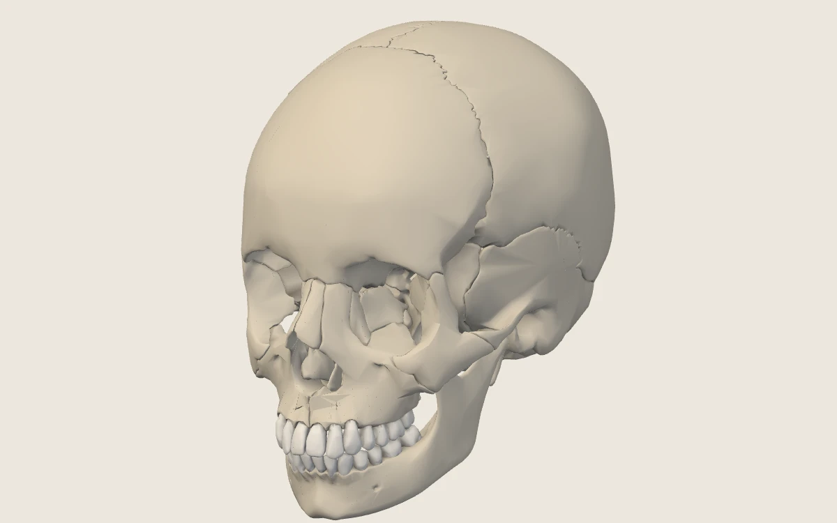
The adult skull
22 bones as separate pieces, six lessons in Deep Learning and High-Yield views, a searchable atlas and point-to-identify practice.
 Also available · Back and ThoraxThe vertebral column and thoracic cage24 vertebrae, the sacrum, 23 discs, the ribs and the sternum as separate pieces; four lessons and practice on the specimen.
4 topics available in 2 units, 70 topics planned. Planned topics have no lessons yet and are not counted in your progress.
Find a structure
Search by English name, Latin term or synonym. A result opens that structure in the atlas.
The subject map
- 1
Foundations
7 plannedThe language and the basic building blocks used in every region.
- Anatomical position, left and right, directional termsPlannedThe reference posture, the patient's left and right, and the words that describe position.
- Body planes, surfaces, cavities and membranesPlannedSagittal, coronal and axial planes; the body cavities and their linings.
- Bone, cartilage and skeletal landmarksPlannedKinds of bone and cartilage and the vocabulary of bony landmarks.
- Joint types and movementsPlannedFibrous, cartilaginous and synovial joints and the movements they allow.
- Muscle architecture and actionsPlannedHow fibre arrangement and attachments predict what a muscle does.
- Basic vascular and neural organisationPlannedArteries, veins and lymphatics; spinal nerves, plexuses and the autonomic outflow.
- Reading a cross-section and image orientationPlannedHow to orient an axial, coronal or sagittal image before naming anything on it.
- 2
Head & Neck
1 available · 13 plannedFrom the skull outward: spaces, joints, muscles, viscera, nerves and vessels.
- The adult skullAvailable22 bones on a 3D specimen: bone groups, each bone and its landmarks, sutures, the cranial base, the bony orbit and nasal cavity, and the openings with what passes through them.
- Cranial fossae and their contentsPlannedWhat lies in each fossa: lobes, cranial nerves, venous sinuses.The bony fossae and their boundaries are taught in the Skull unit.
- Facial skeletonPlannedThe face as a framework for the orbits, nose and jaws.The facial bones are taught in the Skull unit.
- OrbitPlannedWalls, apex, muscles, nerves and vessels of the eye socket.The bony orbit is taught in the Skull unit.
- Nasal cavity and paranasal sinusesPlannedConchae and meatuses, the septum, sinus drainage.The bony nasal cavity is taught in the Skull unit.
- Oral cavity and palatePlannedTongue, palate, teeth and salivary glands.
- Mandible and temporomandibular jointPlannedMovements of the jaw and the muscles that produce them.The mandible and the joint surfaces are taught in the Skull unit.
- Scalp and facePlannedLayers of the scalp; sensory and motor supply of the face.
- Muscles of mastication and facial expressionPlannedTwo muscle groups, two nerves (V3 and VII).
- EarPlannedExternal, middle and inner ear; the auditory ossicles.
- Pharynx and larynxPlannedThe shared airway and food passage and the voice box.
- Neck triangles and fascial spacesPlannedSurface triangles, deep fascia and the spaces infection travels in.
- Thyroid and parathyroidsPlannedPosition, blood supply and the nerves at risk in surgery.
- Nerves, vessels and lymphatic drainage of the head and neckPlannedCarotid and jugular systems, cranial nerves in the neck, lymph node groups.
- 3
Back & Vertebral Column
2 available · 4 plannedThe column, its joints and muscles, and the cord it protects.
- The vertebral column and its regionsAvailable24 vertebrae, the sacrum and 23 discs on a 3D specimen: the curves of the column, the plan of a vertebra, and what makes a cervical, thoracic, lumbar or sacral vertebra recognisable.
- Atlas and axisAvailableThe two vertebrae that carry and turn the skull: a ring without a body, and the dens it turns on.
- Intervertebral discs and facet jointsPlannedThe three-joint complex at each level.The discs (as pieces of the model), the facet joints and disc herniation are introduced in the Vertebral Column unit.
- Vertebral canal and ligamentsPlannedBoundaries of the canal and the ligaments that line it.The long ligaments are introduced in words in the Vertebral Column unit.
- Intrinsic back muscles and regional movementPlannedErector spinae and the deeper layers; movements by region.
- Spinal cord, meninges and nerve roots in the canalPlannedLevels, enlargements, cauda equina and where a needle goes.
- 4
Thorax
1 available · 7 plannedThe chest wall, pleura and lungs, mediastinum and heart.
- Ribs, sternum and costal cartilagesAvailableThe thoracic cage on a 3D specimen: the three parts of the sternum, true, false and floating ribs, the costal cartilages and how each rib meets the column.
- Intercostal spaces and the chest wallPlannedThe muscles of the wall and the neurovascular bundle in each space.The ribs, the costal groove and the sternum are taught in the Vertebral Column and Thoracic Cage unit.
- Diaphragm and its aperturesPlannedAttachments, openings and levels; the phrenic nerve.
- Pleura, lungs and hilaPlannedSurfaces, lobes, fissures and the structures at each hilum.
- MediastinumPlannedDivisions and what each contains.
- Pericardium and heart: chambers and valvesPlannedSurfaces, borders, chambers and valves.
- Coronary circulation and great vesselsPlannedCoronary arteries and veins; the aorta, venae cavae and pulmonary vessels.
- Trachea, oesophagus and thoracic lymphaticsPlannedThe tubes of the posterior chest and the thoracic duct.
- 5
Abdomen
9 plannedThe wall, the peritoneal cavity and the organs within and behind it.
- Surface regions and abdominal wallPlannedRegions and quadrants; the muscle layers and rectus sheath.
- Inguinal canalPlannedWalls, rings and contents; why hernias form here.
- Peritoneum and mesenteriesPlannedGreater and lesser sacs, mesenteries, intra- and retroperitoneal organs.
- Oesophagus at the hiatus and stomachPlannedParts, relations and blood supply of the stomach.
- Small and large intestinePlannedHow to tell the parts apart and what supplies each.
- Liver, porta hepatis and biliary treePlannedLobes and segments, the portal triad and the bile ducts.
- Pancreas and spleenPlannedParts and relations of the pancreas; the spleen and its vessels.
- Kidneys, suprarenal glands and uretersPlannedPosition, coverings, hilum and the course of the ureter.
- Abdominal vessels and autonomic plexusesPlannedAorta and its branches, the portal system, the vena cava and the plexuses.
- 6
Pelvis & Perineum
8 plannedThe bony ring, the floor, and the pelvic viscera with their supply.
- Bony pelvis, inlet and outletPlannedThe hip bones and sacrum; diameters and differences between reference specimens.The sacrum and coccyx are taught in the Vertebral Column unit.
- Pelvic floor, spaces and fasciaPlannedLevator ani and the supports of the pelvic organs.
- Bladder and urethraPlannedPosition, relations and the course of the urethra.
- Rectum and anal canalPlannedRelations, sphincters and the watershed of supply and drainage.
- Male pelvic and reproductive anatomyPlannedProstate, seminal glands, ductus deferens and external genitalia.
- Female pelvic and reproductive anatomyPlannedUterus, uterine tubes, ovaries, vagina and their supports.
- Perineal regionsPlannedUrogenital and anal triangles; the pudendal nerve and vessels.
- Pelvic vessels, nerves and lymphaticsPlannedInternal iliac branches, sacral plexus and lymph drainage.
- 7
Upper Limb
7 plannedFrom the shoulder girdle to the hand, compartment by compartment.
- Shoulder girdle and axillaPlannedClavicle, scapula and humerus; the walls and contents of the axilla.
- Arm and elbowPlannedCompartments of the arm and the elbow joint.
- ForearmPlannedFlexor and extensor compartments and their nerves.
- Wrist and handPlannedCarpal bones, joints and the intrinsic muscles.
- Brachial plexusPlannedRoots, trunks, divisions, cords and branches, and what each lesion looks like.
- Arteries and veins of the upper limbPlannedFrom the subclavian to the palmar arches; superficial veins.
- Cubital fossa and carpal tunnelPlannedTwo spaces where arrangement matters clinically.
- 8
Lower Limb
7 plannedFrom the hip to the foot, compartment by compartment.
- Gluteal region and hipPlannedHip bone, hip joint and the gluteal muscles; safe injection.
- Thigh and kneePlannedCompartments of the thigh; the knee joint and its ligaments.
- LegPlannedAnterior, lateral and posterior compartments.
- Ankle and footPlannedTarsal bones, joints and arches.
- Lumbosacral plexusPlannedFemoral, obturator and sciatic nerves and their territories.
- Arteries and veins of the lower limbPlannedFrom the femoral artery to the foot; the superficial and deep veins.
- Femoral triangle, popliteal fossa and tarsal tunnelPlannedThree spaces and the order of their contents.
- 9
Neuroanatomy
8 plannedThe central nervous system, its coverings, its blood supply and its connections.
- Brain surfaces and sectionsPlannedLobes, gyri and sulci; standard sections.
- Deep nucleiPlannedBasal nuclei, thalamus and the internal capsule.
- Brainstem and cerebellumPlannedExternal features, levels and cranial nerve nuclei.
- Spinal cord and levelsPlannedSegments, grey and white matter, vertebral versus cord levels.
- Meninges, ventricles and the CSF pathwayPlannedThree coverings, four ventricles and the route of the fluid.
- Arterial territories and venous sinusesPlannedCircle of Willis, cortical territories and the dural sinuses.
- Cranial nervesPlannedNuclei, course, skull exits and the effect of a lesion.The openings each nerve uses are taught in the Skull unit.
- Major tracts and functional connectionsPlannedCorticospinal, dorsal column and spinothalamic pathways.
A topic is marked available only when it has lessons, a structure list and practice. Everything else is planned.