Telling the regions apart: cervical, thoracic, lumbar, sacral
Every vertebra follows the same plan, but each region changes it for its own job. Three questions sort any vertebra: is there a hole in the transverse process, is there a facet for a rib, and how big is the body?
Cervical vertebrae: small, mobile, with a hole in each transverse process
The seven cervical vertebrae carry only the head, so their bodies are the smallest in the column. The feature that belongs to this region alone is the transverse foramen, an opening in each transverse process that transmits the vertebral vessels. If a vertebra has it, the vertebra is cervical.
A typical cervical vertebra, C3 to C6, also has a short spinous process that is usually bifid, forked at its tip. The last one breaks the pattern: the spinous process of C7 is much longer, which is why C7 is called the vertebra prominens. Its tip, the spinous process of C7, is the landmark from which the vertebrae are counted at the base of the neck.
Teaching schematic
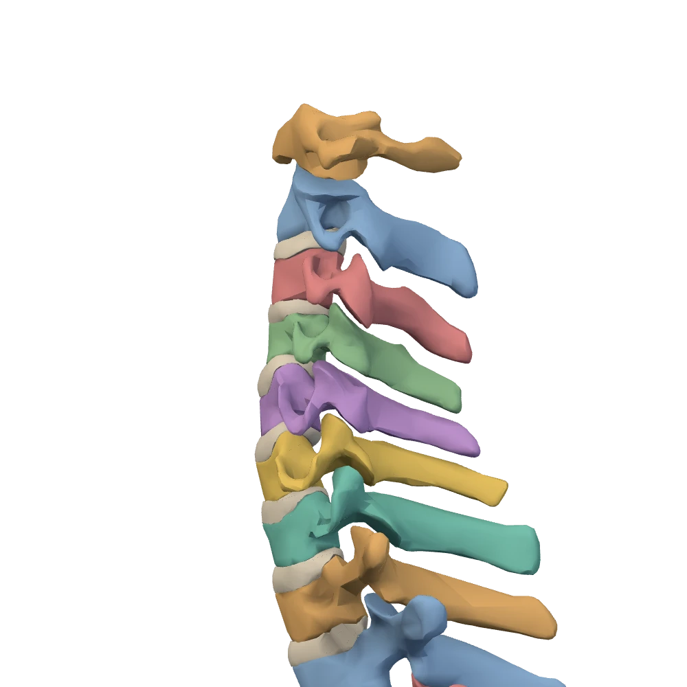
Column alone, left lateral view, close-up. Ribs, cartilages and sternum hidden. The patient's left side: the front of the body is toward your left.
Atlas and axis: the two vertebrae that carry and turn the head
The atlas (C1) does not look like a vertebra at all. It has no body and no spinous process: it is a ring made of an anterior arch and a posterior arch joined by two lateral masses. The upper surface of each lateral mass is a curved socket for an occipital condyle of the skull. The head rocks forward and back on these two joints.
The axis (C2) has the feature the atlas lacks. Its dens is a peg that rises from the body into the ring of the atlas, where it meets the back of the anterior arch. The atlas, carrying the skull, turns around this peg: this is the movement of shaking the head to say “no”.
Teaching schematic
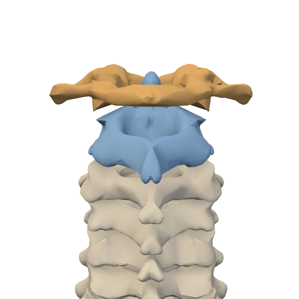
Posterior view, close-up. From behind: the patient's left is on your left.
Thoracic vertebrae: facets for ribs
The mark of a thoracic vertebra is the costal facet. The head of a rib sits against the side of the bodies, and the tubercle of the rib rests on a facet of the transverse process. On a typical thoracic vertebra the superior costal facet takes the head of the rib with the same number, and the inferior costal facet takes part of the head of the rib below.
The spinous process is long and slopes steeply downward, so that it overlaps the vertebra below; in the posterior view the tips form a close-set row. The joints with the ribs, and through them with the sternum, limit movement here: the thoracic region is the stiffest part of the column above the sacrum. In return it gives the cage a firm back wall.
Lumbar vertebrae: built for weight
The five lumbar vertebrae, L1 to L5, carry the weight of everything above them and have the largest, thickest bodies in the column. Their spinous processes are short and blunt and point straight backward. They are recognised as much by what they lack: no transverse foramen and no costal facet. Their transverse processes are for muscle attachment, and no rib meets them.
Teaching schematic
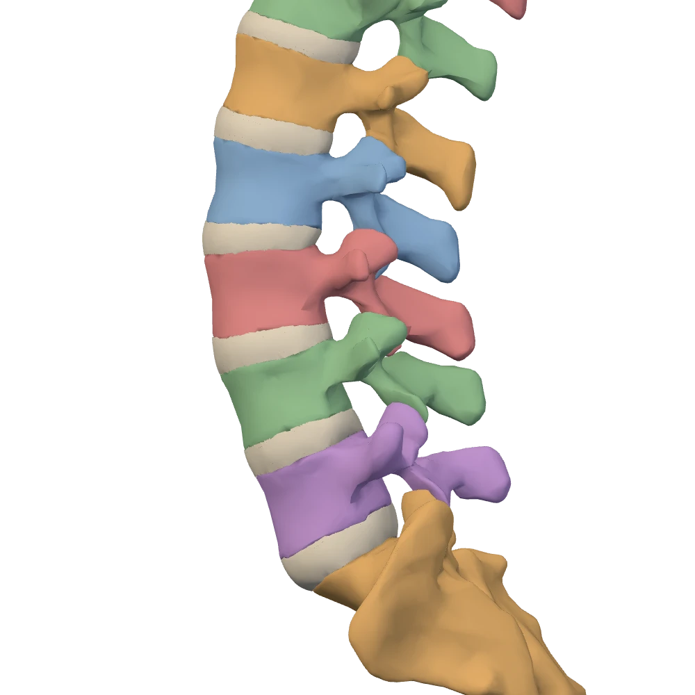
Column alone, left lateral view, close-up. Ribs, cartilages and sternum hidden. The patient's left side: the front of the body is toward your left.
Unlike the thoracic region, the lumbar region moves freely: flexion, extension, bending to the side and some rotation. A region that is both heavily loaded and mobile is where discs fail, as section 3 explains.
Sacrum and coccyx
The sacrum is five sacral vertebrae fused into one wedge. Its wide upper end, the base, takes the weight of the column through the L5–S1 disc and passes it sideways to the hip bones at the sacroiliac joints; the sacrum forms the back wall of the pelvis. The anterior lip of the base is the sacral promontory.
Fusion leaves traces. The fused spinous processes form the median sacral crest down the back of the bone. Where the intervertebral foramina would have been there are sacral foramina: four pairs on the front and four pairs on the back, for the ventral and dorsal rami of the sacral nerves. The vertebral canal continues as the sacral canal and ends at an opening near the tip, the sacral hiatus.
The coccyx is the small tail end of the column, made of four small segments. It carries no weight in standing, though it takes some in sitting, and a fall onto it can break it. On this specimen it is the narrow lower end of the sacrum piece.
Side by side, and practice
Three questions that sort a vertebraOpenStax 7.3Netter p.181–186
| Cervical | Thoracic | Lumbar | |
|---|---|---|---|
| Body | Small | Medium | Large and thick |
| Transverse foramen | Present | Absent | Absent |
| Costal facets | Absent | Present | Absent |
| Spinous process | Short, usually bifid (C7 long) | Long, sloping downward | Short, blunt, pointing backward |
| Movement | Considerable | Limited by the ribs | Considerable |
Body
- Cervical
- Small
- Thoracic
- Medium
- Lumbar
- Large and thick
Transverse foramen
- Cervical
- Present
- Thoracic
- Absent
- Lumbar
- Absent
Costal facets
- Cervical
- Absent
- Thoracic
- Present
- Lumbar
- Absent
Spinous process
- Cervical
- Short, usually bifid (C7 long)
- Thoracic
- Long, sloping downward
- Lumbar
- Short, blunt, pointing backward
Movement
- Cervical
- Considerable
- Thoracic
- Limited by the ribs
- Lumbar
- Considerable
Point to the vertebra
0 of 25All 24 vertebrae and the sacrum, asked by number. The prompt names one structure and states the starting view. Labels are hidden. Tap the structure on the specimen; you may rotate and zoom first. You get the recognition cue after a miss and can try again.
Knowledge check · 2
Answer, read why, and try again whenever you like. Answers are kept in both study modes.
- Q1Transverse foramenOpenStax 7.3Netter p.181–182
An isolated vertebra has an opening in each transverse process. To which region does it belong?
- Q2AtlasNetter p.31–32
Which statement about the atlas is correct?
- Q3Atlas and axis: movementsNetter p.31–32OpenStax 7.3
Turning the head from side to side, as in saying “no”, takes place mainly around which structure?
- Q4Counting cervical vertebraeBodyParts3D
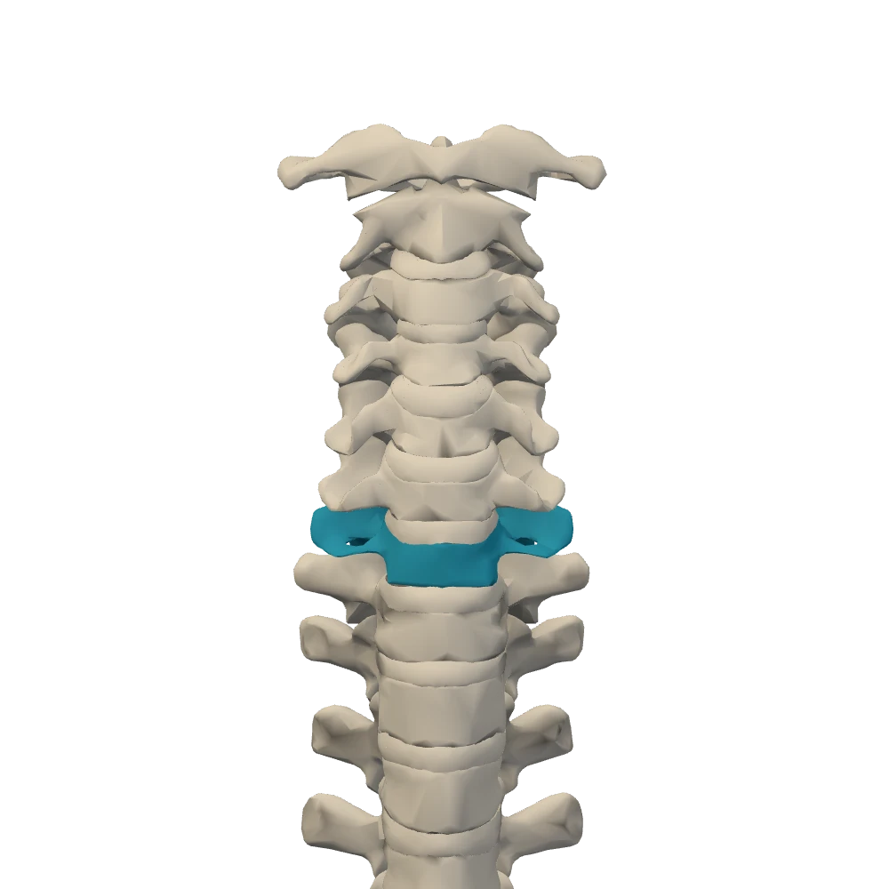
Anterior view, close-up. Facing the chest: the patient's left is on your right.
The picture shows the upper column from the front with the ribs and sternum hidden. Which vertebra is highlighted?
- Q5Costal facetsNetter p.183–184, 225–226
The superior costal facet on the body of a typical thoracic vertebra articulates with:
- Q6Lumbar vertebraOpenStax 7.3Netter p.185–186
Which description fits a lumbar vertebra?
- Q7SacrumOpenStax 7.3Netter p.191–192
What forms the median sacral crest?