The thoracic cage: sternum, ribs and costal cartilages
Twelve pairs of ribs leave the thoracic vertebrae, curve round the chest and, for most of them, reach the sternum through a bar of cartilage. How each rib ends in front is what classifies it.
The sternum: three parts and two landmarks
The sternum is the flat bone in the midline of the chest. It has three parts. The manubrium is the wide upper part. The body is the long middle part. The xiphoid process is the small tip at the lower end. On the specimen the three are separate pieces.
Teaching schematic
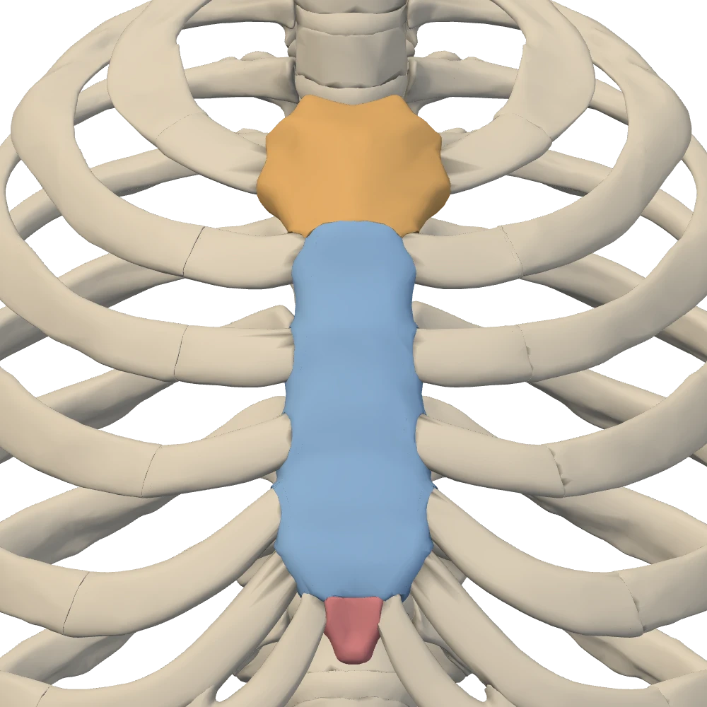
Anterior view, close-up. Facing the chest: the patient's left is on your right.
Two landmarks on it are used every day at the bedside. The jugular (suprasternal) notch is the dip in the top edge of the manubrium, with a clavicular notch for the clavicle on each side of it. The sternal angle is the junction of the manubrium and the body; the two do not lie in one flat plane, so the junction forms a slight bend that can be felt as a ridge.
True, false and floating ribs
There are twelve pairs of ribs, one pair for each thoracic vertebra. A rib does not reach the sternum itself: its front end continues as a bar of hyaline cartilage, the costal cartilage. Ribs are classified by what that cartilage does.
Classes of ribsOpenStax 7.4Netter p.223–224
| Ribs | Class | How the front end is attached |
|---|---|---|
| 1 to 7 | True (vertebrosternal) | Each has its own costal cartilage that joins the sternum directly |
| 8 to 10 | False (vertebrochondral) | The cartilage joins the cartilage of the rib above: 10 to 9, 9 to 8, 8 to 7 |
| 11 and 12 | Floating (vertebral); also counted as false ribs | No attachment in front: the short cartilage ends in the muscles of the abdominal wall |
- Class
- True (vertebrosternal)
- How the front end is attached
- Each has its own costal cartilage that joins the sternum directly
- Class
- False (vertebrochondral)
- How the front end is attached
- The cartilage joins the cartilage of the rib above: 10 to 9, 9 to 8, 8 to 7
- Class
- Floating (vertebral); also counted as false ribs
- How the front end is attached
- No attachment in front: the short cartilage ends in the muscles of the abdominal wall
Teaching schematic
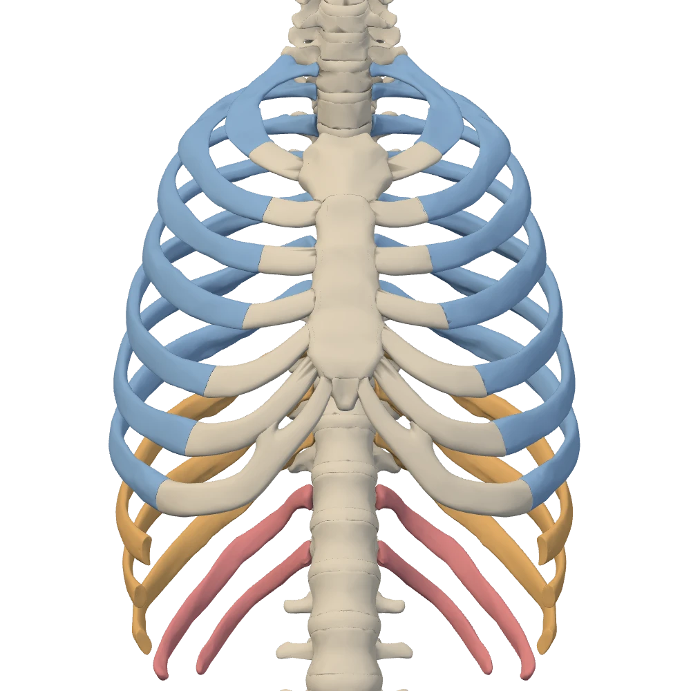
Anterior view, close-up. Facing the chest: the patient's left is on your right.
The joined cartilages of ribs 7 to 10 form the costal margin, the lower edge of the front of the cage that runs down and outward from the xiphoid process. “False ribs” therefore means ribs 8 to 12: all five fail to reach the sternum on their own, and the last two do not even reach another rib.
The parts of a rib and how it meets the column
A typical rib has a head at its posterior end, then a narrow neck, then a small bump on its back surface, the tubercle. Just beyond the tubercle the rib bends most sharply at its angle, and the long body (shaft) then curves forward to the costal cartilage. A shallow costal groove along the lower edge of the body shelters the vessels and nerve of the space below the rib.
Each rib makes two kinds of joint with the column, both plane synovial joints. At the costovertebral joint the head of the rib sits against the costal facets on the bodies: the superior facet of its own vertebra and the inferior facet of the vertebra above. At the costotransverse joint the tubercle rests on the transverse process of its own vertebra. Costotransverse joints are present for ribs 1 to 10; ribs 11 and 12 have none.
Teaching schematic
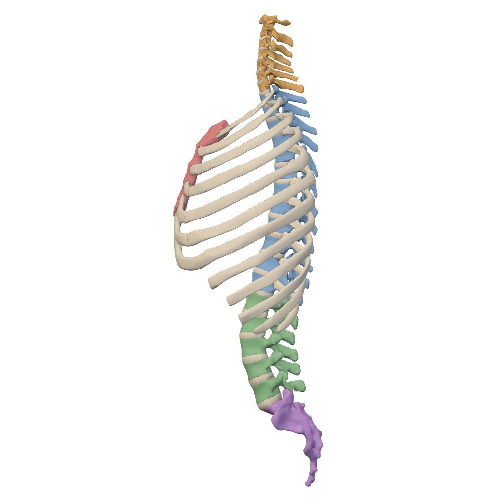
Left lateral view. The patient's left side: the sternum (the front) is toward your left.
The cage as a whole
The thoracic cage is the sternum, the twelve pairs of ribs with their costal cartilages, and the twelve thoracic vertebrae behind. It belongs to the axial skeleton, with the skull and the vertebral column. The clavicle and scapula lie against it but belong to the upper limb, and they are not part of this specimen.
The cage is open at both ends. The superior thoracic aperture is the small opening at the top, ringed by T1, the first ribs and the manubrium; look at it in the superior view. The inferior thoracic aperture is the wide opening at the bottom, which is closed in life by the diaphragm.
Practice on the specimen
Point to the rib or the part of the sternum
0 of 27The 24 ribs, left and right asked separately, and the three parts of the sternum. The prompt names one structure and states the starting view. Labels are hidden. Tap the structure on the specimen; you may rotate and zoom first. You get the recognition cue after a miss and can try again.
Left or right?
0 of 8A rib or a costal cartilage is highlighted. Decide whether it is the patient's left or right in that view. The prompt names one structure and states the starting view. Labels are hidden. Choose the side. You get the recognition cue after a miss and can try again.
Point to the landmark
0 of 4Only the surface points that were computed on this model and checked on a marked picture are asked. The prompt names one structure and states the starting view. Labels are hidden. Tap the structure on the specimen; you may rotate and zoom first. You get the recognition cue after a miss and can try again.
Knowledge check · 4
Answer, read why, and try again whenever you like. Answers are kept in both study modes.
- Q1Sternal angleOpenStax 7.4
The costal cartilage of which rib joins the sternum at the sternal angle?
- Q2Classes of ribsOpenStax 7.4Netter p.223–224
Which ribs are the true ribs?
- Q3False ribsOpenStax 7.4
How does the costal cartilage of the ninth rib end?
- Q4Costotransverse jointNetter p.225–226OpenStax 7.4
The tubercle of the sixth rib articulates with:
- Q5Second rib and sternal angleBodyParts3DOpenStax 7.4
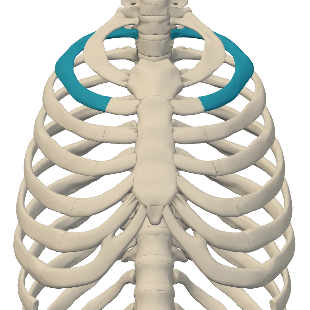
Anterior view, close-up. Facing the chest: the patient's left is on your right.
The picture shows the upper cage from the front with one pair of ribs highlighted. Which bedside landmark identifies this pair?
- Q6Costal grooveOpenStax 7.4
What runs in the costal groove on the lower edge of a rib?
- Q7Thoracic cage and pectoral girdleNetter p.223–224
Which of these is not part of the thoracic cage?
- Q8Thoracic aperturesNetter p.223–224BodyParts3D
Which structures ring the superior thoracic aperture?