The column as a whole: regions, curves and the plan of a vertebra
Count the pieces, learn the four curves, and learn the one plan that every vertebra follows. With those three things you can place any vertebra before you know its regional details.
Five regions, 24 separate vertebrae
The vertebral column develops as 33 vertebrae. In the adult, 24 of them stay separate: 7 cervical (C1 to C7) in the neck, 12 thoracic (T1 to T12) in the chest and 5 lumbar (L1 to L5) in the lower back. Below them, five sacral vertebrae have fused into one bone, the sacrum, and four small coccygeal vertebrae form the coccyx. So the adult column is 24 vertebrae plus the sacrum and the coccyx.
Each vertebra is named by the first letter of its region and its number from above. The first two have names of their own, the atlas (C1) and the axis (C2). The thoracic vertebrae are the ones that carry ribs, which gives you a fixed point on any specimen: T1 is the first vertebra with a rib and T12 the last. Everything above T1 is cervical; the five large vertebrae below T12 are lumbar.
Teaching schematic
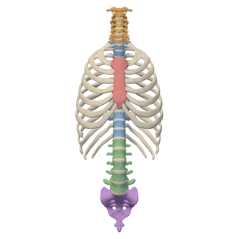
Anterior view. Facing the chest: the patient's left is on your right.
Four curves: two you are born with, two you acquire
Seen from the side, the adult column is not straight. It curves forward in the neck, backward in the chest, forward again in the lower back and backward in the sacrum. Open the Column alone, left lateral view to see all four at once.
Teaching schematic
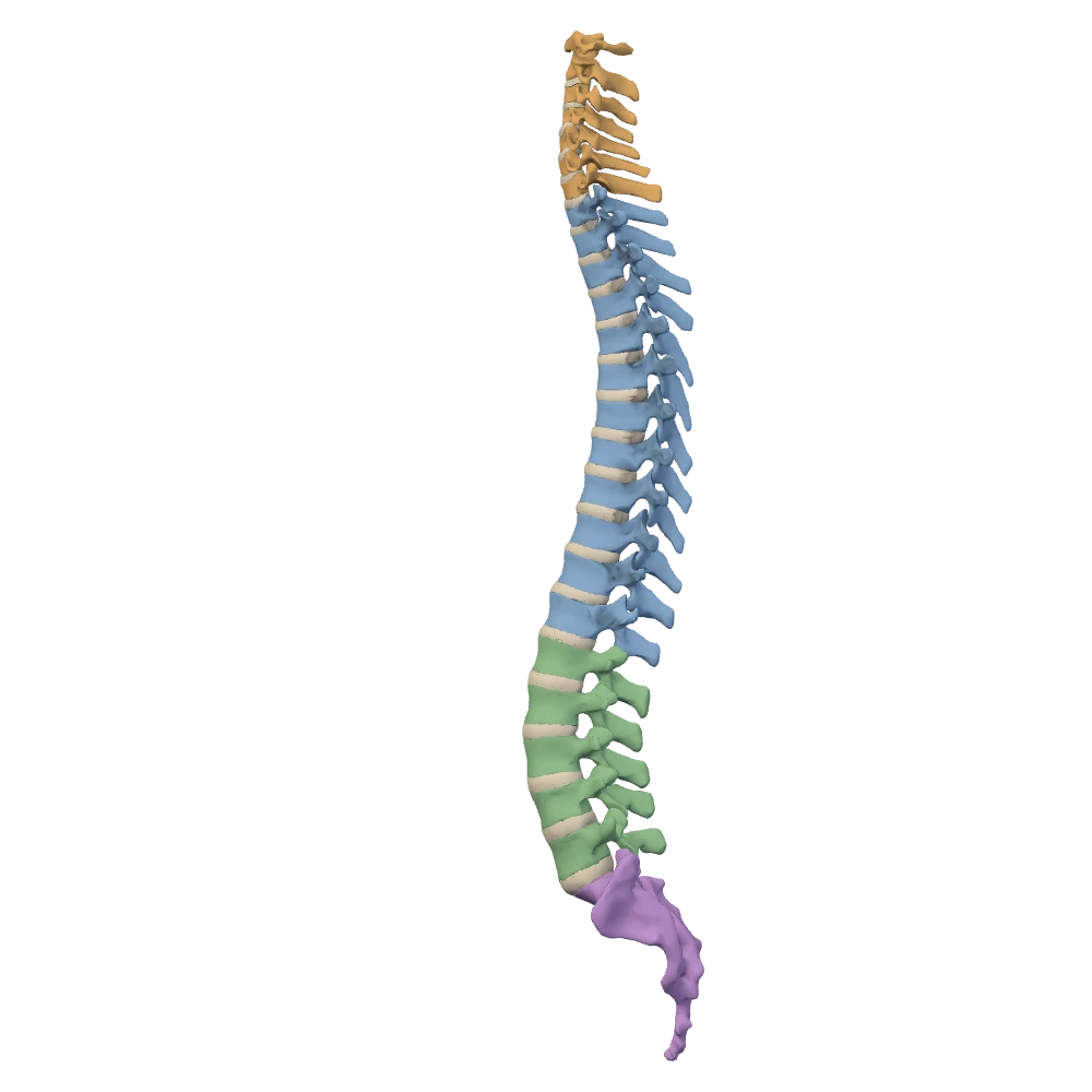
Column alone, left lateral view. Ribs, cartilages and sternum hidden. The patient's left side: the front of the body is toward your left.
Primary and secondary curves
Primary curves
- Thoracic and sacrococcygeal
- Kept from the single curve of the fetal column
- Concave toward the front
Secondary curves
- Cervical and lumbar
- Develop after birth, as the child holds up the head, sits, stands and walks
- Concave toward the back, opposite to the fetal curve
The curves are part of how the column carries weight. Under a heavier load they deepen, and they spring back when the load is removed. The two secondary curves are also where the column moves most freely: the cervical and lumbar regions allow more movement than the thoracic region, which is held by the ribs.
One plan for every vertebra
Almost every vertebra is built from the same parts. In front is the body, the block that carries weight. Behind it is the vertebral arch: a pedicle on each side, anchored to the back of the body, and a lamina on each side that completes the roof. Body and arch enclose the vertebral foramen, and the foramina of all the vertebrae line up as the vertebral canal, which holds the spinal cord and its coverings.
- One spinous process projects backward in the midline. The row of them is what you see and feel down the middle of the back.
- Two transverse processes project sideways from the junction of pedicle and lamina.
- Four articular processes, two superior and two inferior, join each vertebra to its neighbours at the facet joints.
The pedicles are notched above and below. When two vertebrae are stacked, the notches of neighbouring pedicles form an opening at each side, the intervertebral foramen, through which a spinal nerve leaves the canal. Section 3 comes back to this opening, because it is where a bulging disc does its damage.
Finding your way on this specimen
Left and right always belong to the patient. In the anterior view you face the chest, so the patient's left is on your right. In the posterior view the two agree. A lateral view shows one side only, and the sternum tells you which way the body faces: in the left lateral view the sternum is toward your left. Each view states this under the specimen; read it before you name a side.
Where to look for what
| View | Best for |
|---|---|
| Anterior | Sternum, costal cartilages, the front of the ribs, cervical and lumbar bodies, the front of the sacrum |
| Posterior | Spinous processes, the back of the ribs, the back of the sacrum |
| Left or right lateral | The slope of the ribs and the curves of the column |
| Column alone, left lateral | The four curves, the bodies and the discs, with the cage hidden |
| Superior / inferior | The ring of the first ribs; the cage as a ring around the column |
Anterior
- Best for
- Sternum, costal cartilages, the front of the ribs, cervical and lumbar bodies, the front of the sacrum
Posterior
- Best for
- Spinous processes, the back of the ribs, the back of the sacrum
Left or right lateral
- Best for
- The slope of the ribs and the curves of the column
Column alone, left lateral
- Best for
- The four curves, the bodies and the discs, with the cage hidden
Superior / inferior
- Best for
- The ring of the first ribs; the cage as a ring around the column
Knowledge check · 1
Answer, read why, and try again whenever you like. Answers are kept in both study modes.
- Q1Vertebral countOpenStax 7.3Netter p.179–180
How many separate vertebrae does the adult column have above the sacrum?
- Q2Primary and secondary curvesOpenStax 7.3Netter p.179–180
Which pair lists the two secondary curves of the column?
- Q3Parts of a vertebraOpenStax 7.3
Which parts make up the vertebral arch?
- Q4Vertebral and intervertebral foraminaOpenStax 7.3Netter p.187–188
A spinal nerve leaves the vertebral canal through which opening?
- Q5Abnormal curvaturesOpenStax 7.3Netter p.187–188
A woman in the last weeks of pregnancy stands with her lower back strongly arched forward.
What is an exaggeration of this curve called?
- Q6Left and right in the posterior viewBodyParts3D
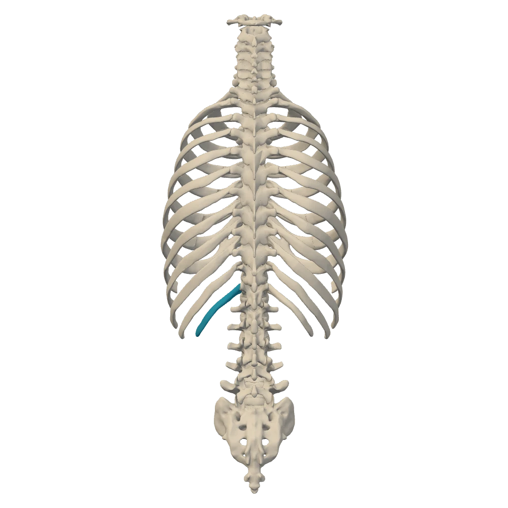
Posterior view. From behind: the patient's left is on your left.
The picture shows the column and cage from behind, with one rib highlighted. On which side of the patient is it?
- Q7Recognising the thoracic regionOpenStax 7.3OpenStax 7.4
On any articulated skeleton, which feature tells you that a vertebra is thoracic?