Discs, facet joints and ligaments: how the pieces are held together
Two neighbouring vertebrae meet at three joints: one disc between the bodies and a pair of small synovial joints between the arches. Ligaments run the length of the column over both.
The intervertebral disc
An intervertebral disc is a pad of fibrocartilage that joins the bodies of two neighbouring vertebrae. The joint is a symphysis, a secondary cartilaginous joint. Each disc has two parts. The anulus fibrosus is a tough fibrous ring that anchors one body to the next. Inside it is the nucleus pulposus, a softer gel with a high water content, which resists compression and changes shape as one vertebra tilts on the other.
Discs are found from the axis to the sacrum. The highest is C2–C3 and the lowest is L5–S1; there is none above or below the atlas, and none between the fused sacral segments. That makes 23, and all 23 are separate pieces on the specimen. Select a few in the Column alone view and compare them: the discs are thin in the neck and thickest in the lumbar region, where the load is greatest.
Facet joints and the intervertebral foramen
Behind the disc, the arches of two neighbouring vertebrae meet at a pair of zygapophysial (facet) joints. Each is a small plane synovial joint between the inferior articular process of the vertebra above and the superior articular process of the vertebra below, wrapped in a thin capsule. The surfaces glide on each other, and their direction in each region guides the movement that region allows.
Between the disc in front and the facet joint behind lies the intervertebral foramen. Its roof and floor are the notched pedicles of the two vertebrae. A spinal nerve leaves the vertebral canal through it, which is why trouble in either neighbour, a bulging disc in front or an arthritic facet joint behind, can press on the nerve.
One level, three joints
- Disc between the two bodies (symphysis): bears weight
- Left and right facet joints between the arches (plane synovial): guide movement
- Intervertebral foramen between them at each side: the spinal nerve's exit
The long ligaments (taught in words)
The ligaments of the column are not part of this model, so this subtopic has no specimen to point at. Learn them by where they lie relative to the parts you can see.
Ligaments of the vertebral column, from front to backNetter p.189–190OpenStax 7.3
| Ligament | Where it runs | What it limits |
|---|---|---|
| Anterior longitudinal | Down the front of the bodies and discs, the whole length of the column | Extension. The stronger of the two longitudinal ligaments |
| Posterior longitudinal | Down the back of the bodies and discs, inside the vertebral canal, in front of the spinal cord | Flexion |
| Ligamentum flavum | Between the laminae of neighbouring vertebrae, behind the spinal cord; elastic | Flexion; helps restore the upright posture |
| Interspinous | Between neighbouring spinous processes | Weak |
| Supraspinous | Along the tips of the spinous processes; in the neck it broadens into the nuchal ligament | Flexion |
Anterior longitudinal
- Where it runs
- Down the front of the bodies and discs, the whole length of the column
- What it limits
- Extension. The stronger of the two longitudinal ligaments
Posterior longitudinal
- Where it runs
- Down the back of the bodies and discs, inside the vertebral canal, in front of the spinal cord
- What it limits
- Flexion
Ligamentum flavum
- Where it runs
- Between the laminae of neighbouring vertebrae, behind the spinal cord; elastic
- What it limits
- Flexion; helps restore the upright posture
Interspinous
- Where it runs
- Between neighbouring spinous processes
- What it limits
- Weak
Supraspinous
- Where it runs
- Along the tips of the spinous processes; in the neck it broadens into the nuchal ligament
- What it limits
- Flexion
When a disc herniates
If the back of the anulus fibrosus weakens, with age or injury, pressure on the disc can push the nucleus pulposus backward through it. This is a herniated (“slipped”) disc. The bulge lies beside the intervertebral foramen and can compress a spinal nerve, giving pain, and sometimes weakness or numbness, in the part of the body that nerve supplies.
Where discs herniate most oftenNetter p.181–182, 185–186OpenStax 7.3
| Region | Usual levels | Typical result |
|---|---|---|
| Lumbar (the most common) | L4–L5 and L5–S1 | Pain down the back of the thigh and leg (sciatica). L4–L5 may compress the L5 root; L5–S1 may compress the S1 root |
| Cervical | C5–C6 and C6–C7 | Pain in the neck, shoulder and upper limb |
Lumbar (the most common)
- Typical result
- Pain down the back of the thigh and leg (sciatica). L4–L5 may compress the L5 root; L5–S1 may compress the S1 root
Cervical
- Typical result
- Pain in the neck, shoulder and upper limb
Worked case · A box lifted badly
A 41-year-old man bends forward to lift a heavy box and feels a sudden pain in his lower back. Over the next day the pain spreads down the back of one leg to the foot.
Which two discs would you look at first, and why does pain appear in the leg when the injury is in the back?
OpenStax 7.3Netter p.185–186Knowledge check · 3
Answer, read why, and try again whenever you like. Answers are kept in both study modes.
- Q1Parts of a discOpenStax 7.3Netter p.187–188
Which part of an intervertebral disc is the gel-like centre that resists compression?
- Q2Extent of the discsNetter p.179–180, 187–188
Between which two vertebrae is there no intervertebral disc?
- Q3Facet jointsNetter p.187–188, 225–226
What kind of joint is a zygapophysial (facet) joint?
- Q4Longitudinal ligamentsNetter p.189–190OpenStax 7.3
Which ligament runs down the front of the vertebral bodies and limits extension of the column?
- Q5Lumbar disc herniationNetter p.185–186
MRI of a patient with pain down the back of the leg shows a herniation of the disc between the fifth lumbar vertebra and the sacrum.
Which nerve root is this herniation most likely to compress?
- Q6Nuchal ligamentOpenStax 7.3
In the neck, the supraspinous ligament broadens into which structure?
- Q7Naming a discBodyParts3D
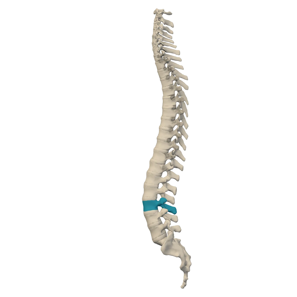
Column alone, left lateral view. Ribs, cartilages and sternum hidden. The patient's left side: the front of the body is toward your left.
The picture shows the column alone from the patient's left, with one vertebra highlighted. Which disc lies directly below it?