Physical injury
Mechanical, thermal, electrical and radiation injuries each leave a recognizable pattern. Read the wound to read the force, and know why a large burn kills by fluid loss before it kills by infection.
Mechanical injury: reading the wound
L6 p.15–16The type of wound tells you the force. The lecture's four skin wounds plus puncture and gunshot cover the forensic basics.
Lecture image
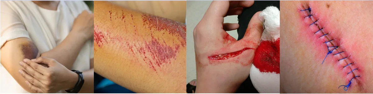
Wound typesL6 p.15–16Ref. · Robbins; forensic wound terminology
| Wound | Mechanism | Edges / clue |
|---|---|---|
| Abrasion | Scraping that removes superficial epidermis | Graze; no deep structures |
| Contusion (bruise) | Blunt force ruptures vessels; blood leaks into tissue | Skin intact; color changes with age |
| Laceration | Blunt force tears skin | Irregular, jagged edges with bridging vessels and nerves across the wound |
| Incision | A sharp edge (knife) | Clean, straight edges; no bridging |
| Puncture / stab | A narrow pointed object | Small surface wound; depth exceeds width; internal damage underestimated |
| Gunshot | Projectile | Entry smaller with an abrasion rim; exit larger and irregular; range read from soot and stippling |
Abrasion
- Mechanism
- Scraping that removes superficial epidermis
- Edges / clue
- Graze; no deep structures
Contusion (bruise)
- Mechanism
- Blunt force ruptures vessels; blood leaks into tissue
- Edges / clue
- Skin intact; color changes with age
Laceration
- Mechanism
- Blunt force tears skin
- Edges / clue
- Irregular, jagged edges with bridging vessels and nerves across the wound
Incision
- Mechanism
- A sharp edge (knife)
- Edges / clue
- Clean, straight edges; no bridging
Puncture / stab
- Mechanism
- A narrow pointed object
- Edges / clue
- Small surface wound; depth exceeds width; internal damage underestimated
Gunshot
- Mechanism
- Projectile
- Edges / clue
- Entry smaller with an abrasion rim; exit larger and irregular; range read from soot and stippling
Lecture image
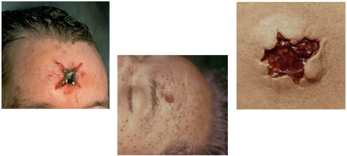
Range is read from what surrounds the entry wound: soot and seared skin at contact range, a stippled zone of unburned powder (tattooing) at intermediate range, and a clean small wound with an abrasion rim at distant range. A contact wound over bone can be torn into a star shape by expanding gas.
Abusive head trauma
L6 p.17Violent shaking of an infant tears the bridging veins that cross from the cortex to the dural sinuses, producing a subdural hemorrhage. The same acceleration–deceleration forces cause retinal hemorrhages, and squeezing the chest fractures the posterior ribs. The triad of subdural hemorrhage, retinal hemorrhages and rib fractures should prompt evaluation for abuse.
Lecture image
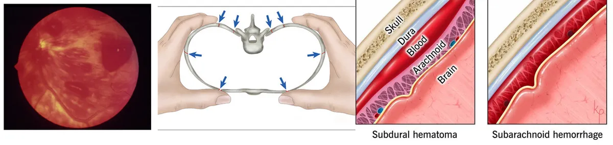
Burns: depth, area and the fluid shift
L6 p.18–20A burn is graded by depth and by the area of body surface involved. Depth decides healing and scarring; area decides how much fluid is lost and whether the patient goes into shock.
Lecture image
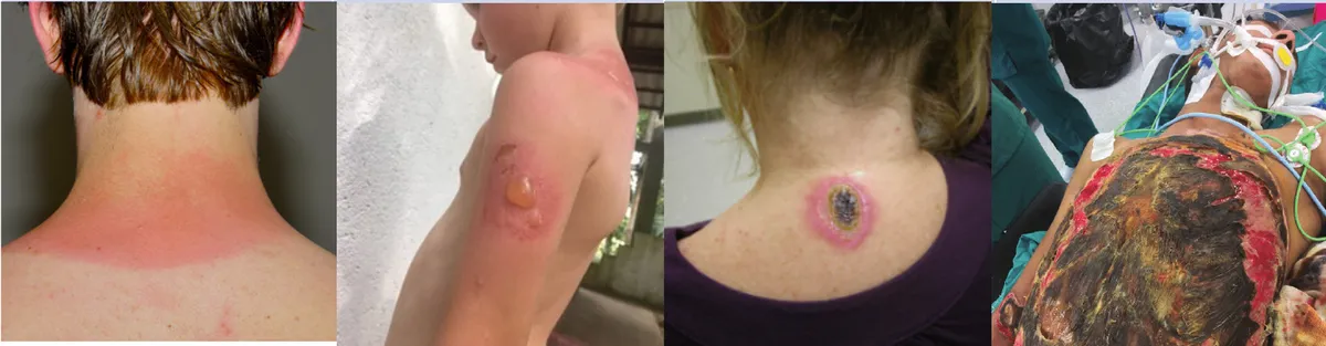
Burn depthL6 p.18
| Degree | Reaches | Features |
|---|---|---|
| First | Epidermis only | Red, painful; heals without scar (sunburn) |
| Second | Into dermis (superficial or deep) | Blisters, very painful; nerves preserved |
| Third | Through epidermis and dermis | Adnexa and nerves destroyed, so painless; needs grafting |
| Fourth | Into muscle and bone | Charring; limb-threatening |
First
- Reaches
- Epidermis only
- Features
- Red, painful; heals without scar (sunburn)
Second
- Reaches
- Into dermis (superficial or deep)
- Features
- Blisters, very painful; nerves preserved
Third
- Reaches
- Through epidermis and dermis
- Features
- Adnexa and nerves destroyed, so painless; needs grafting
Fourth
- Reaches
- Into muscle and bone
- Features
- Charring; limb-threatening
The rule of nines estimates the burned area in an adult: head 9%, each arm 9%, each leg 18%, front of trunk 18%, back of trunk 18%, perineum 1%. The larger the area, the more plasma escapes through heat-injured capillaries.
Why a large burn causes shock within a day
- Large burn
- Heat-injured capillaries become leaky; mediators raise permeability
- Plasma and protein escape into burned tissue (edema) and evaporate from the surface
- Intravascular volume falls
- Hypovolemic shock (the leading early cause of death)
Heat and cold
L6 p.20–21Heat illness is a spectrum. Heat cramps are painful muscle spasms from salt loss during exertion. Heat exhaustion is volume depletion with a core temperature below 40 °C, sweating preserved and no major CNS change. Heat stroke is a core temperature above 40 °C with central nervous system dysfunction, and it comes in two forms.
Two kinds of heat stroke
Classic (non-exertional)
- Elderly, chronically ill, underventilated apartments in a heat wave
- Hot, dry skin (anhidrosis)
- Altered mental status, coma, seizures
Exertional
- Young, healthy athletes and military recruits
- May still sweat
- Rhabdomyolysis, acute kidney injury, DIC, lactic acidosis, hypocalcemia
Lecture image
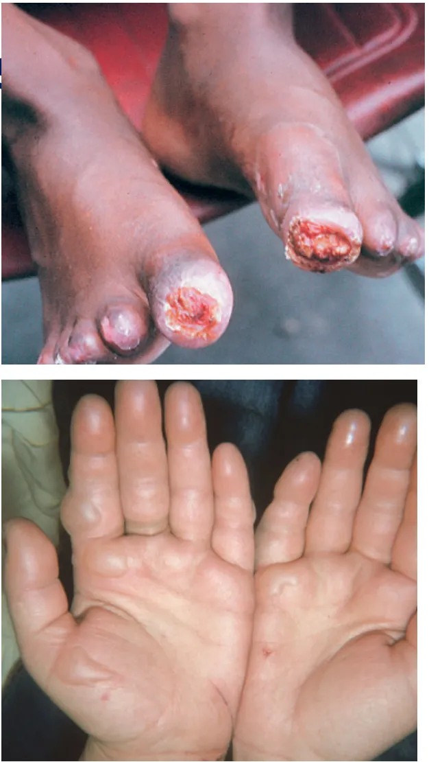
- Frostbite: tissue below 0 °C. Ice crystals form in the extracellular space, drawing water out of cells; microvascular collapse, sludging and stasis then deprive tissue of oxygen and cause necrosis. A prefreeze phase of anesthesia precedes the freeze–thaw phase.
- Trench foot / immersion foot: a nonfreezing injury from prolonged wet cold, without ice crystal formation.
Electrical, drowning and altitude
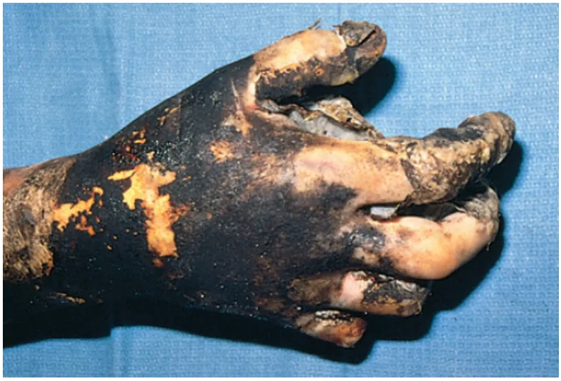
L6 p.22–24Electrical injury follows Ohm's law (I = V/R): current, not voltage alone, injures. Wet skin lowers resistance and raises current. Alternating current at a given voltage is about three times more dangerous than direct current because it causes tetanic muscle contraction that locks the victim to the source; DC tends to throw the victim off. The most common cause of death is cardiorespiratory arrest (ventricular fibrillation or respiratory muscle tetany); deep tissue is burned along the current path.
Lecture image
- Drowning: suffocation from immersion. In wet drowning, laryngospasm relaxes and water is aspirated; surfactant is washed out, causing atelectasis and pulmonary edema. In dry drowning, intense laryngospasm asphyxiates without aspiration. Alcohol, seizures and trauma are major risk factors.
- High-altitude (acute mountain) sickness: above ~8000 ft (2440 m), low barometric pressure lowers inspired PO₂. Headache is the most common symptom, with fatigue, dizziness, anorexia, nausea and insomnia. Severe forms are high-altitude pulmonary edema (HAPE, from hypoxic pulmonary vasoconstriction) and cerebral edema (HACE).
Radiation and UV
L6 p.25–27Ionizing radiation has enough energy to eject electrons and ionize atoms; it injures DNA mainly by generating hydroxyl radicals from water. The most radiosensitive tissues are those that divide fastest: lymphoid tissue, bone marrow, gastrointestinal mucosa and germ cells.
Teaching schematic
< 0.5 Gy
Subclinical
No systemic effect; stem-cell mutations may predispose to cancer
0.5–2 Gy
Radiation sickness
Lethargy, nausea; transient drop in lymphocytes and neutrophils
2–6 Gy
Hematopoietic
Marrow hypoplasia; anemia 2 weeks later
3–10 Gy
Gastrointestinal
Death of gut epithelium, hair loss, diarrhea within hours
> 10 Gy
Cerebral
Hemorrhagic brain necrosis; death within hours; convulsions
Thresholds are approximate and sources differ; higher doses add the lower-dose effects.
Radiation effects by tissueL6 p.25
| Tissue | Acute effect | Chronic effect |
|---|---|---|
| Skin | Erythema, edema, blistering | Radiodermatitis, cancer |
| Bone marrow | Lymphopenia first, then thrombocytopenia, marrow hypoplasia | Acute leukemia |
| GI mucosa | Diarrhea, mucosal necrosis | Fibrosis, strictures |
| Gonads | Germ cell death | Atrophy, fibrosis, infertility |
| Lung | Acute pneumonitis | Interstitial fibrosis |
Skin
- Acute effect
- Erythema, edema, blistering
- Chronic effect
- Radiodermatitis, cancer
Bone marrow
- Acute effect
- Lymphopenia first, then thrombocytopenia, marrow hypoplasia
- Chronic effect
- Acute leukemia
GI mucosa
- Acute effect
- Diarrhea, mucosal necrosis
- Chronic effect
- Fibrosis, strictures
Gonads
- Acute effect
- Germ cell death
- Chronic effect
- Atrophy, fibrosis, infertility
Lung
- Acute effect
- Acute pneumonitis
- Chronic effect
- Interstitial fibrosis
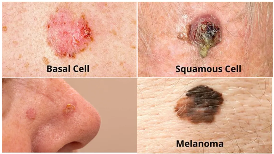
Nonionizing (ultraviolet) radiation does not ionize but damages DNA by forming pyrimidine (thymine) dimers that distort the helix. UVB is the main carcinogen. The dimers are normally removed by nucleotide excision repair; unrepaired damage, together with inactivation of p53 and activation of RAS, produces actinic keratosis, basal cell and squamous cell carcinoma, and melanoma.
Lecture image
Knowledge check · 6.3
Answer, read why, and try again whenever you like. Answers are kept in both study modes.
- Q1Burn shock (fluid loss)Midterm sim. p.8L6 p.18–19
A 50-year-old firefighter has third-degree burns over 70% of his body. He dies 24 hours later.
What is the most likely cause of death?
- Q2Abusive head trauma triadCh. VI cases p.9L6 p.17
A 2-month-old is brought in lethargic with a bulging fontanelle. Fundoscopy shows bilateral retinal hemorrhages, and MRI shows a crescent of blood between the dura and arachnoid.
Which additional finding on a chest x-ray would support the diagnosis?
- Q3Laceration vs incisionL6 p.15
A forensic pathologist examines a skin wound that is irregular and jagged with thin strands of tissue bridging across the defect.
Which wound is this?
- Q4UV, pyrimidine dimers and NERL6 p.27Ref. · Robbins (UV carcinogenesis, XP)
A 68-year-old fair-skinned farmer who worked outdoors for decades develops a scaly, ulcerated plaque on his lower lip. A relative has an inherited condition that caused dozens of skin cancers in childhood.
Which molecular event links the sun exposure to his tumor, and which defect explains his relative's early cancers?