Shock
Shock is failure of tissue perfusion. Find where the circulation failed first (volume, pump, outflow or vessel tone) and the pressures upstream and downstream follow.
What shock is, and how it looks
L4 p.42–43, 47Shock is reduced perfusion of tissues that impairs their oxygenation. Blood pressure is the product of cardiac output and systemic vascular resistance (MAP ≈ CO × SVR), and cardiac output depends on venous return (preload) and pump function. Each type of shock begins with a different one of these failing: hypovolemic (too little volume), cardiogenic (the pump), obstructive (flow blocked through the heart or lungs) and distributive, of which septic shock is the main example (vessel tone and capillary integrity lost; also anaphylactic and neurogenic shock).
Stages
| Stage | What happens | What you see |
|---|---|---|
| Compensated (nonprogressive) | Baroreceptors trigger catecholamines, ADH and RAAS: tachycardia, arteriolar and venous constriction, renal salt and water retention; blood is diverted to brain and heart | Tachycardia, cold pale skin (in non-septic shock), falling urine output, BP may still be normal |
| Progressive | Tissues switch to anaerobic glycolysis → lactic acidosis; acidosis blunts the vasomotor response, arterioles dilate, blood pools in capillaries; endothelium is injured (DIC) | Hypotension, confusion, oliguria, rising lactate, Kussmaul breathing |
| Irreversible | Lysosomal enzymes leak, myocardium is depressed, the gut wall lets bacteria into the blood, kidneys develop acute tubular necrosis | Refractory hypotension, multiorgan failure, death |
Compensated (nonprogressive)
- What happens
- Baroreceptors trigger catecholamines, ADH and RAAS: tachycardia, arteriolar and venous constriction, renal salt and water retention; blood is diverted to brain and heart
- What you see
- Tachycardia, cold pale skin (in non-septic shock), falling urine output, BP may still be normal
Progressive
- What happens
- Tissues switch to anaerobic glycolysis → lactic acidosis; acidosis blunts the vasomotor response, arterioles dilate, blood pools in capillaries; endothelium is injured (DIC)
- What you see
- Hypotension, confusion, oliguria, rising lactate, Kussmaul breathing
Irreversible
- What happens
- Lysosomal enzymes leak, myocardium is depressed, the gut wall lets bacteria into the blood, kidneys develop acute tubular necrosis
- What you see
- Refractory hypotension, multiorgan failure, death
Cold shock versus warm shock
Cold shock (hypovolemic, cardiogenic, obstructive; late septic)
- Catecholamines constrict skin vessels: cold, clammy, pale skin, slow capillary refill
- Rapid, weak (thready) pulse: stroke volume is small
- SVR ↑, CO ↓
Warm shock (early septic / distributive)
- Arterioles dilated: warm, flushed skin, brisk refill
- Strong, bounding pulse, wide pulse pressure; CO ↑ in compensation
- SVR ↓, CO ↑
Some signs are shared by every type. Oliguria (urine < 0.5 mL/kg/h) reflects low renal blood flow and GFR plus ADH and aldosterone; if prolonged it becomes acute tubular necrosis. Lactic acidosis, a high anion gap metabolic acidosis, reflects tissue oxygen debt; a lactate above 2 mmol/L is abnormal and above 4 mmol/L signals severe hypoperfusion. Altered mental status reflects brain hypoperfusion.
Hemodynamic profiles
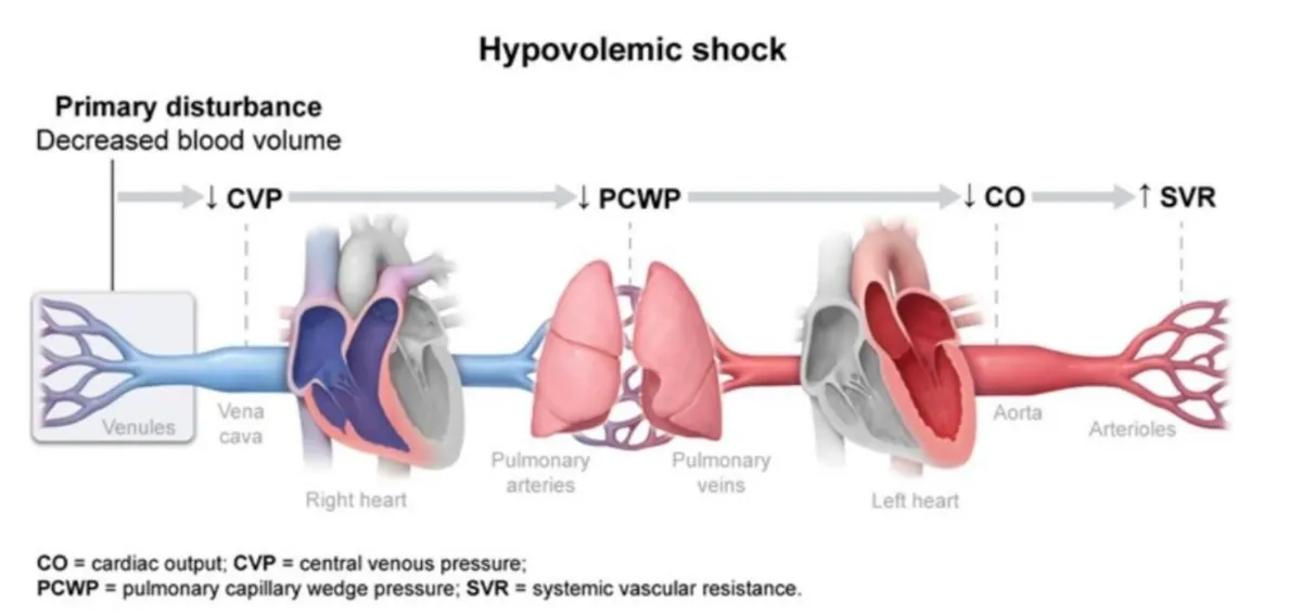
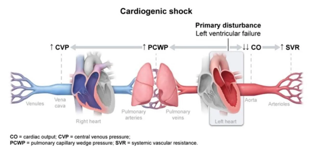
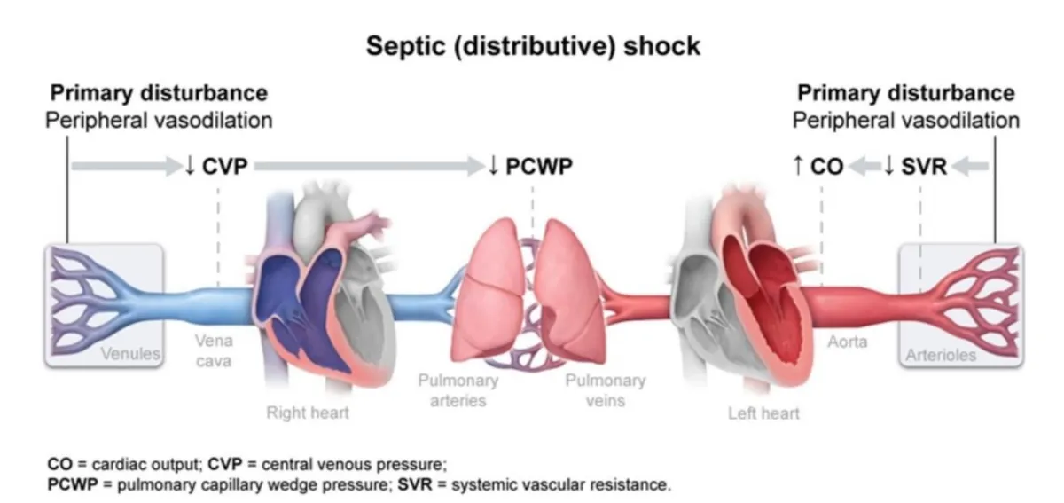
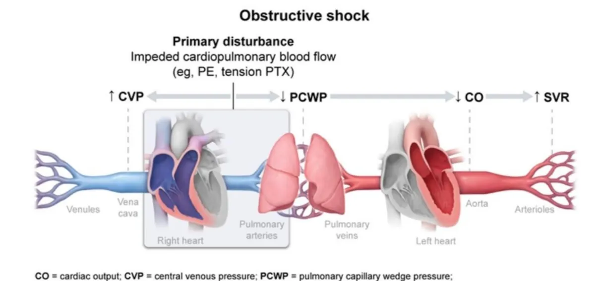
L4 p.43–50Read each profile by walking the circuit from the primary disturbance: venules and vena cava (CVP, right-sided filling), lungs and left atrium (PCWP, left-sided filling), left ventricle and aorta (CO), arterioles (SVR), and back through the tissues (SvO₂, mixed venous oxygen saturation: how much oxygen the tissues left behind). The lecture's diagrams do exactly this; the lecture calls SVR “PVR” and SvO₂ “MVO₂”.
Profiles of the five typesL4 p.43–50
| Type | Primary disturbance | CO | SVR | CVP | PCWP | SvO₂ |
|---|---|---|---|---|---|---|
| Hypovolemic | ↓ Blood volume | ↓ | ↑ | ↓ | ↓ | ↓ |
| Cardiogenic | Left ventricular failure | ↓↓ | ↑ | ↑ | ↑ | ↓ |
| Obstructive (PE, tension pneumothorax) | Flow blocked before the left heart | ↓ | ↑ | ↑ | ↓ | ↓ |
| Septic, early (distributive) | Arteriolar and venous dilation, leaky capillaries | ↑ | ↓ | ↓ | ↓ | ↑ or normal |
| Neurogenic (distributive) | Loss of sympathetic tone | ↓ or normal | ↓ | ↓ | ↓ | Normal or ↓ |
Hypovolemic
- Primary disturbance
- ↓ Blood volume
- CO
- ↓
- SVR
- ↑
- CVP
- ↓
- PCWP
- ↓
- SvO₂
- ↓
Cardiogenic
- Primary disturbance
- Left ventricular failure
- CO
- ↓↓
- SVR
- ↑
- CVP
- ↑
- PCWP
- ↑
- SvO₂
- ↓
Obstructive (PE, tension pneumothorax)
- Primary disturbance
- Flow blocked before the left heart
- CO
- ↓
- SVR
- ↑
- CVP
- ↑
- PCWP
- ↓
- SvO₂
- ↓
Septic, early (distributive)
- Primary disturbance
- Arteriolar and venous dilation, leaky capillaries
- CO
- ↑
- SVR
- ↓
- CVP
- ↓
- PCWP
- ↓
- SvO₂
- ↑ or normal
Neurogenic (distributive)
- Primary disturbance
- Loss of sympathetic tone
- CO
- ↓ or normal
- SVR
- ↓
- CVP
- ↓
- PCWP
- ↓
- SvO₂
- Normal or ↓
Lecture slide
Lecture slide
Lecture slide
Lecture slide
Activity · Shock Profiles
Predict the pressures, then name the shock
A 50-year-old firefighter has third-degree burns over 70% of his body. Six hours later he is cold and clammy, pulse 138/min, BP 74/46 mm Hg, urine output 5 mL/h, hematocrit 56%.
Type of shock
Choose an arrow for each pressure and a type of shock, then check.
Core activity · Almost Twins
Almost Twins
Look-alike A
Hypovolemic shock
- Cold, clammy skin; rapid weak pulse
- CO ↓, SVR ↑, SvO₂ ↓
- Flat neck veins, clear lungs: CVP ↓, PCWP ↓
- Responds to volume
Look-alike A
Hypovolemic shock
- Cold, clammy skin; rapid weak pulse
- CO ↓, SVR ↑, SvO₂ ↓
- Flat neck veins, clear lungs: CVP ↓, PCWP ↓
- Responds to volume
Look-alike B
Cardiogenic shock
- Cold, clammy skin; rapid weak pulse
- CO ↓↓, SVR ↑, SvO₂ ↓
- Distended neck veins, crackles: CVP ↑, PCWP ↑
- Fluid worsens pulmonary edema
Step 1 · What they share
What do they share?
Select every feature that both show.
Hypovolemic shock and the hemoglobin that does not fall
L4 p.43–44Hypovolemic shock follows loss of blood or sodium-containing fluid: external or internal hemorrhage (ruptured spleen or liver, ruptured abdominal aortic aneurysm), vomiting and diarrhea, massive sweating, and burns, where capillaries in burned and unburned tissue leak plasma into the interstitium and fluid evaporates from the wound. Cardiac output and left ventricular end-diastolic pressure fall; catecholamines, ADH and angiotensin II constrict arterioles, so SVR rises; SvO₂ falls because tissues extract more from the slow flow. The patient is cold and clammy with a rapid weak pulse, low urine output and a high anion gap lactic acidosis.
In acute hemorrhage the hemoglobin and hematocrit do not fall at first, because whole blood is lost: red cells and plasma leave in the same proportion. Over hours to days, interstitial fluid moves into the plasma (and intravenous fluids add more), diluting the red cells that remain and uncovering the deficit. The marrow answers with a reticulocytosis that becomes evident over several days and peaks about a week later. In burns the opposite happens: plasma is lost without red cells, so the hematocrit rises.
Worked calculation · Why hemoglobin lags behind bleeding
- Blood volume before
- 5.0 L
- Hemoglobin before
- 15 g/dL (150 g/L)
- Acute loss
- 1.0 L of whole blood
Formula
Hb = total hemoglobin mass / blood volume
L4 p.43Core activity · One Fact Changes
One Fact Changes
1 · The original case
A 40-year-old man is stabbed in the abdomen. Pulse 128/min, BP 82/54 mm Hg, cold clammy skin, flat neck veins. Hemoglobin 14.2 g/dL, drawn 20 minutes after the injury.
What type of shock is this?
Which fact decided it?
Cardiogenic shock and the markers of infarction
L4 p.45–46Cardiogenic shock is pump failure, most often from a large acute myocardial infarction; other causes are myocarditis, acute valvular failure (for example a cusp destroyed by infective endocarditis or papillary muscle rupture) and cardiomyopathy. The damaged ventricle ejects less, so CO falls; blood accumulates in the left ventricle, raising LVEDP and PCWP (pulmonary edema) and eventually CVP. Catecholamines, ADH and angiotensin II raise SVR, which adds afterload to the failing ventricle. Clinically: chest pain, then cold clammy skin, hypotension, low urine output and crackles.
Dying myocytes leak their contents into the blood (the membrane damage of irreversible injury, Chapter 1). Cardiac troponins I and T are the most sensitive and specific markers: they rise about 3–4 hours after onset, peak around 24–48 hours and stay elevated for 7–10 days (troponin I) to 10–14 days (troponin T). CK-MB rises within about 4–6 hours, peaks near 24 hours and returns to normal by 48–72 hours, which makes it useful for detecting a reinfarction a few days after the first event, when troponin is still high.
Teaching schematic
Schematic: relative levels, not units. Troponin: up 3–4 h, peak 24–48 h, 7–14 days. CK-MB: up 4–6 h, peak ≈ 24 h, normal by 48–72 h.
Septic shock: mechanisms, DIC and ARDS
L4 p.47–48Septic shock follows bloodstream infection and is the most common cause of death in intensive care units. The lecture's figures: Gram-positive organisms in about 65% of cases (coagulase-negative staphylococci, Staphylococcus aureus; lipoteichoic acid), Gram-negative in about 25% (most often E. coli; endotoxin, lipopolysaccharide), and fungi in about 9% (Candida). Sepsis is now defined as life-threatening organ dysfunction caused by a dysregulated host response to infection; septic shock adds vasopressor-requiring hypotension with a lactate above 2 mmol/L despite fluid.
From microbe to shock
- Microbial products (LPS, lipoteichoic acid) bind Toll-like receptors on macrophages, neutrophils, endothelium
- TNF, IL-1, IL-6 and complement (C3a, C5a) released
- Endothelium activated: inducible NO synthase → NO → arteriolar and venous dilation
- Capillaries leak → plasma lost to interstitium → preload ↓
- SVR ↓, CVP ↓, PCWP ↓; CO ↑ (tachycardia, low afterload): warm shock
- Tissue factor ↑, thrombomodulin and protein C ↓ → DIC
- Myocardial depression, ARDS, acute kidney injury → late cold shock
Early septic shock is warm: the skin is warm and flushed, the pulse is strong and bounding, and cardiac output rises to compensate for the low resistance. Blood races through dilated beds, and cells poisoned by cytokines extract oxygen poorly, so lactate rises even though venous blood stays relatively oxygen-rich. Hematologic findings in the lecture: anemia (bleeding and venipuncture), thrombocytopenia (platelets trapped and consumed in thrombi) and neutropenia (neutrophils marginated by activated adhesion molecules), although neutrophilia is more typical early.
ARDS is the same endothelial and epithelial injury in the lungs: activated neutrophils and cytokines damage alveolar capillaries, a protein-rich exudate floods the alveoli and hyaline membranes form (diffuse alveolar damage). It is a permeability edema with a normal PCWP, which separates it from the pressure edema of left heart failure (4.5).
Core activity · The Missing Arrow
The Missing Arrow
A 70-year-old woman's E. coli pyelonephritis seeds her bloodstream. Complete the chain to warm shock.
- Gram-negative bacteria in the blood
- Arterioles and veins dilate; capillaries leak
- SVR falls; venous return falls
- endothelial tissue factor ↑, thrombomodulin and protein C ↓
- DIC: microthrombi and bleeding
Cards
Tap or drag a card. Placing a used card moves it.
Obstructive and neurogenic shock
L4 p.49–50In obstructive shock blood cannot get through the heart or lungs: massive pulmonary embolism, tension pneumothorax (rising intrathoracic pressure kinks and compresses the great veins), cardiac tamponade and constrictive pericarditis (the heart cannot fill). Pressure rises upstream of the block, so the neck veins distend (CVP ↑); the left heart is underfilled, so CO falls and SVR rises. A saddle embolus is the version you meet in 4.6: sudden collapse with raised JVP after immobility.
Neurogenic shock follows high spinal cord injury (above about T6) or spinal anesthesia. Sympathetic outflow to vessels and heart is lost: arterioles and veins dilate (SVR ↓, venous pooling, CVP ↓) and the heart rate is slow because vagal tone is unopposed. The skin is warm and dry. Bradycardia with hypotension in a trauma patient points here rather than to hemorrhage, which causes tachycardia.
Knowledge check · 4.7
Answer, read why, and try again whenever you like. Answers are kept in both study modes.
- Q1Hypovolemic shock after burnsMidterm sim. p.8L4 p.43
A 50-year-old firefighter emerges from a burning house with third-degree burns over 70% of his body. He dies 24 hours later.
What was the most likely cause of death?
- Q2Early septic shock profileMidterm sim. p.8–9L4 p.47–48
A 92-year-old woman is brought unconscious from a nursing home. BP 70/30 mm Hg, temperature 38 °C, tachypneic. WBC 22,000/µL with 92% neutrophils; urine contains numerous Gram-negative organisms.
Which hemodynamic profile is most likely (cardiac output · PCWP · SVR · CVP)?
- Q3Hemoglobin in acute hemorrhageL4 p.43
A 28-year-old man arrives 30 minutes after a motorcycle crash with a ruptured spleen. Pulse 132/min, BP 78/50 mm Hg, cold clammy skin. Hemoglobin is 14.6 g/dL.
Why is his hemoglobin normal?
- Q4Cardiac markers and reinfarctionL4 p.45
A 61-year-old man had an anterior myocardial infarction 4 days ago. Today he has new chest pain and new ST elevation.
Which marker best confirms a reinfarction?
- Q5Warm septic shock and SvO₂L4 p.47
A 55-year-old woman with pyelonephritis has warm, flushed skin and a bounding pulse; BP 84/38 mm Hg; lactate 4.5 mmol/L. Her mixed venous oxygen saturation is 82% (normal ≈ 70–75%).
Why is her venous blood so well oxygenated while lactate is high?
- Q6Neurogenic shockL4 p.42
A 19-year-old diver strikes his head in shallow water and cannot move his legs. BP 76/40 mm Hg, pulse 48/min, skin warm and dry. There is no sign of bleeding.
Which type of shock is this?