ADH, RAAS and potassium
ADH decides how much water the kidney keeps; aldosterone decides how much sodium. Potassium lives inside cells, and anything that moves it across the membrane shows on the ECG.
Two control systems: ADH for water, RAAS for sodium
L4 p.17, 30, 43ADH (vasopressin) is made in the supraoptic and paraventricular nuclei of the hypothalamus and released from the posterior pituitary. Its main trigger is a rise in plasma osmolality, sensed by hypothalamic osmoreceptors, which respond to changes of 1–2%. A large fall in blood volume or pressure (more than about 5–10%) also releases ADH and overrides osmolality, which is why hypovolemic patients retain water even when their sodium is already low. Nausea, pain and many drugs release it too.
How the kidney handles water. About two-thirds of filtered water is reabsorbed with sodium in the proximal tubule whatever the body needs. The thick ascending limb then pumps NaCl out without water: this dilutes the tubular fluid (to roughly 100 mOsm/kg) and, through countercurrent multiplication, builds a medullary interstitium of up to about 1200 mOsm/kg. Whether that dilute fluid leaves as dilute urine or is concentrated depends only on whether the collecting duct is permeable to water, which is what ADH decides. Urine osmolality can therefore range from about 50 to 1200 mOsm/kg: a low value means ADH is absent or ignored, a high one means it is acting.
ADH binds V2 receptors on collecting-duct principal cells. Through cAMP it inserts aquaporin-2 channels into the luminal membrane, and water flows out of the tubule into the concentrated medullary interstitium. Urine becomes concentrated; the body keeps free water, which lowers plasma osmolality and sodium. On V1 receptors in arterioles it causes vasoconstriction.
The renin–angiotensin–aldosterone system responds to low renal perfusion, low NaCl at the macula densa and sympathetic (β1) stimulation. Renin cleaves angiotensinogen to angiotensin I; ACE converts it to angiotensin II, which constricts arterioles, stimulates thirst and ADH, increases proximal sodium reabsorption and releases aldosterone. Aldosterone acts on principal cells (more ENaC channels and Na⁺/K⁺-ATPase): sodium is reabsorbed and K⁺ and H⁺ are secreted in exchange. Water follows the sodium, so aldosterone expands the ECF without changing sodium concentration.
Teaching schematic
ADH (water)
- Plasma osmolality ↑ (or large volume ↓)
- Hypothalamus → posterior pituitary releases ADH
- V2 receptors → aquaporin-2 in collecting duct
- Free water retained; urine concentrated
- Serum Na⁺ and osmolality fall
RAAS (sodium)
- Renal perfusion ↓, macula densa NaCl ↓, β1
- Renin → angiotensin I → (ACE) angiotensin II
- Vasoconstriction, thirst, ADH, aldosterone
- Collecting duct: Na⁺ reabsorbed, K⁺ and H⁺ secreted
- ECF volume restored; [Na⁺] unchanged
Diabetes insipidus and the water deprivation test
L4 p.17–18, 20In diabetes insipidus the kidney cannot keep free water. In central DI the pituitary does not release ADH (head trauma, pituitary or hypothalamic surgery, tumors such as craniopharyngioma, infiltrative disease, idiopathic). In nephrogenic DI ADH is present but the collecting duct does not respond: lithium, hypercalcemia, hypokalemia (vacuolar nephropathy), demeclocycline, or inherited V2-receptor or aquaporin-2 mutations. Either way the patient passes large volumes of dilute urine (polyuria), plasma osmolality and sodium rise (pure water loss, TBNa⁺/↓↓TBW), and thirst drives polydipsia. With free access to water the sodium may stay high-normal; without it, hypernatremia develops quickly.
The water deprivation test asks two questions in order. First, withhold water: does the urine concentrate by itself? If it does, the ADH axis works, and the patient was simply drinking too much (primary polydipsia). If urine stays dilute (< 300 mOsm/kg) while plasma osmolality rises, the patient has DI. Second, give desmopressin (synthetic ADH): if the urine now concentrates, the kidney could respond and ADH was missing (central); if it stays dilute, the kidney cannot respond (nephrogenic).
Teaching schematic
- Normal or primary polydipsia: concentrates with deprivation
- Central DI: concentrates only after desmopressin
- Nephrogenic DI: stays dilute
Dashed line: 300 mOsm/kg. Illustrative values.
Worked calculation · Reading the desmopressin step
- Patient 1, urine osmolality
- 150 → 450 mOsm/kg after desmopressin
- Patient 2, urine osmolality
- 170 → 185 mOsm/kg after desmopressin
Formula
% rise = (after − before) / before × 100
L4 p.18Core activity · Almost Twins
Almost Twins
Look-alike A
Central diabetes insipidus
- Polyuria, polydipsia, dilute urine
- Plasma osmolality and Na⁺ high-normal or high
- Urine concentrates after desmopressin
- Head trauma, pituitary surgery, craniopharyngioma
Look-alike A
Central diabetes insipidus
- Polyuria, polydipsia, dilute urine
- Plasma osmolality and Na⁺ high-normal or high
- Urine concentrates after desmopressin
- Head trauma, pituitary surgery, craniopharyngioma
Look-alike B
Nephrogenic diabetes insipidus
- Polyuria, polydipsia, dilute urine
- Plasma osmolality and Na⁺ high-normal or high
- Urine stays dilute after desmopressin
- Lithium, hypercalcemia, hypokalemia, V2-receptor mutation
Step 1 · What they share
What do these two patients share?
Select every feature that both show.
Core activity · One Fact Changes
One Fact Changes
1 · The original case
A 58-year-old man is confused. Serum Na⁺ 121 mEq/L, plasma osmolality 252 mOsm/kg; no edema, normal neck veins, moist mucosa. Urine osmolality 480 mOsm/kg, urine Na⁺ 45 mEq/L.
What is the most likely mechanism?
Which fact decided it?
SIADH versus primary polydipsia
L4 p.10, 19In SIADH, ADH is released although plasma osmolality is low and volume is not reduced. The lecture names the commonest cause it emphasizes: ectopic ADH from small cell carcinoma of the lung. Others are CNS disease (meningitis, hemorrhage, trauma), lung disease (pneumonia, tuberculosis), drugs (SSRIs, carbamazepine, cyclophosphamide, vincristine) and postoperative pain and nausea.
SIADH step by step
- ADH secreted regardless of osmolality
- Aquaporin-2 stays in collecting-duct membranes
- Free water retained; urine concentrated (UOsm > 100, often > POsm)
- Hypotonic ECF → water enters cells (2/3 of it)
- Slight ECF expansion → renin ↓, natriuretic peptide ↑ → urine Na⁺ > 20 mEq/L
- Hyponatremia without edema; brain swelling → confusion, seizures, coma
Primary (psychogenic) polydipsia produces hyponatremia the other way: the patient drinks more than the kidney can excrete, while ADH is correctly switched off. The urine is therefore maximally dilute, urine osmolality < 100 mOsm/kg. The single number that separates the two is the urine osmolality: above 100 means ADH is acting (SIADH, or ADH released by low volume); below 100 means it is not.
Two euvolemic hyponatremiasL4 p.10, 19
| SIADH | Primary polydipsia | |
|---|---|---|
| ADH | High (inappropriate) | Suppressed (appropriate) |
| Urine osmolality | > 100 mOsm/kg, often > plasma | < 100 mOsm/kg (maximally dilute) |
| Urine Na⁺ | > 20 mEq/L (off diuretics) | Low or variable (diluted) |
| Urine volume | Normal or low | Very high |
| Fix | Fluid restriction; treat the cause; V2 antagonists or hypertonic saline if severe | Stop drinking excess water |
ADH
- SIADH
- High (inappropriate)
- Primary polydipsia
- Suppressed (appropriate)
Urine osmolality
- SIADH
- > 100 mOsm/kg, often > plasma
- Primary polydipsia
- < 100 mOsm/kg (maximally dilute)
Urine Na⁺
- SIADH
- > 20 mEq/L (off diuretics)
- Primary polydipsia
- Low or variable (diluted)
Urine volume
- SIADH
- Normal or low
- Primary polydipsia
- Very high
Fix
- SIADH
- Fluid restriction; treat the cause; V2 antagonists or hypertonic saline if severe
- Primary polydipsia
- Stop drinking excess water
Core activity · The Missing Arrow
The Missing Arrow
A 64-year-old smoker with small cell carcinoma of the lung has a seizure. Serum Na⁺ is 116 mEq/L. Complete the chain from the tumor to the seizure.
Two gaps need a mechanism (how one step causes the next); two need an event (what has happened).
- Small cell carcinoma secretes ADH whatever the plasma osmolality
- Free water reabsorbed; urine concentrated (UOsm > 100 mOsm/kg)
- retained water dilutes the ECF
- Brain cells swell inside the skull (cerebral edema)
- raised intracranial pressure and neuronal dysfunction
Cards
Tap or drag a card. Placing a used card moves it.
Core activity · Almost Twins
Almost Twins
Look-alike A
SIADH
- Hyponatremia, low plasma osmolality
- Euvolemic, no edema
- Urine osmolality > 100 mOsm/kg, often above plasma; urine Na⁺ > 20
- Improves with fluid restriction
Look-alike A
SIADH
- Hyponatremia, low plasma osmolality
- Euvolemic, no edema
- Urine osmolality > 100 mOsm/kg, often above plasma; urine Na⁺ > 20
- Improves with fluid restriction
Look-alike B
Primary polydipsia
- Hyponatremia, low plasma osmolality
- Euvolemic, no edema
- Urine osmolality < 100 mOsm/kg (maximally dilute), very large urine volume
- Resolves when drinking stops
Step 1 · What they share
What do they share?
Select every feature that both show.
Correcting sodium too fast: two opposite injuries
L4 p.12Brain cells defend their volume. Over about 48 hours of hyponatremia they export potassium, chloride and organic osmolytes (glutamine, taurine, myo-inositol), so they shrink back toward normal despite the dilute ECF. Re-importing those osmolytes takes days. If the serum sodium is then raised quickly, the ECF becomes hypertonic relative to the adapted cells, water leaves them, and oligodendrocytes in the densely packed white matter of the central pons are injured: osmotic demyelination syndrome (central pontine myelinolysis). Signs appear 2–6 days after the correction: dysarthria, dysphagia, diplopia, spastic quadriparesis, falling consciousness, and sometimes a locked-in state.
Lecture image
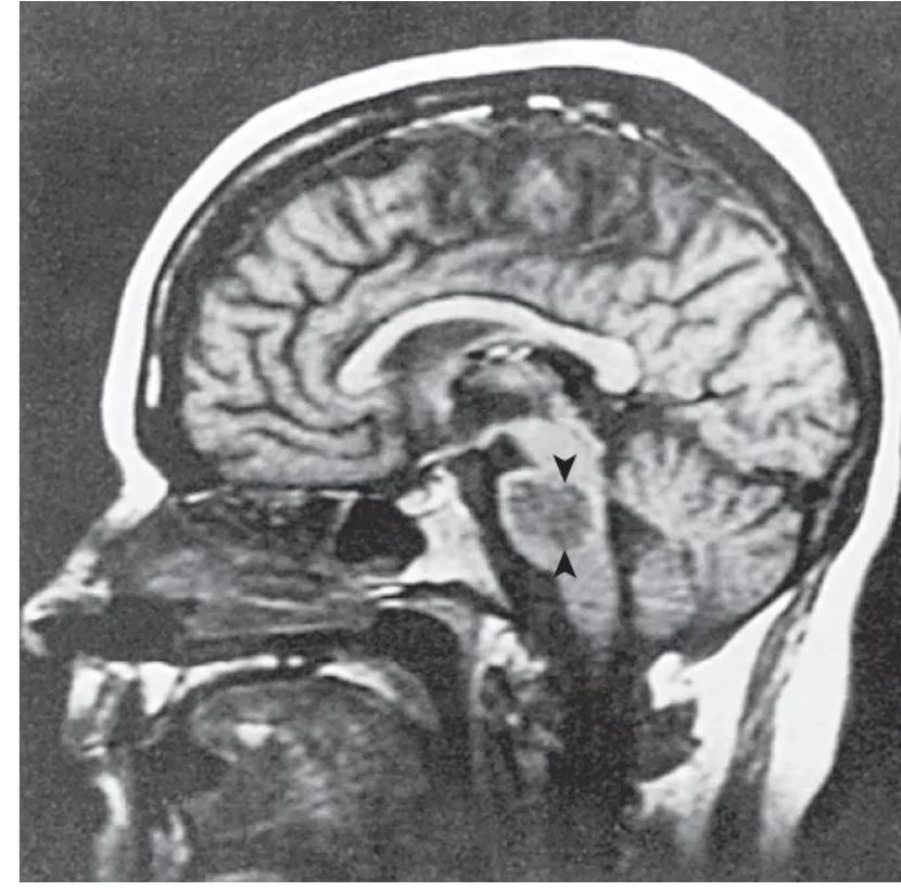
In chronic hypernatremia the reverse adaptation happens: brain cells accumulate osmolytes (sometimes called idiogenic osmoles) to hold on to water. Lower the sodium too fast with free water and water rushes into those cells: cerebral edema, seizures and herniation, most often in children.
The lecture's two rhymes
“From low to high, your pons will die”
- Chronic hyponatremia corrected too fast
- Water leaves adapted brain cells
- Osmotic demyelination (pons and elsewhere)
- Correct slowly: the lecture says over the first 24 hours; a common limit is 8–10 mEq/L per 24 h
“From high to low, your brains will blow”
- Chronic hypernatremia corrected too fast
- Water enters adapted brain cells
- Cerebral edema, herniation
- Lower by no more than about 10–12 mEq/L per day
Teaching schematic
Chronic hyponatremia corrected too fast
Adapted: osmolytes exported, volume near normal
Na⁺ raised fast: water leaves → oligodendrocytes injured
→ Osmotic demyelination (pons), 2–6 days later
Chronic hypernatremia corrected too fast
Adapted: osmolytes accumulated, volume near normal
Na⁺ lowered fast: water rushes in
→ Cerebral edema, seizures, herniation
Potassium: where it lives and what moves it
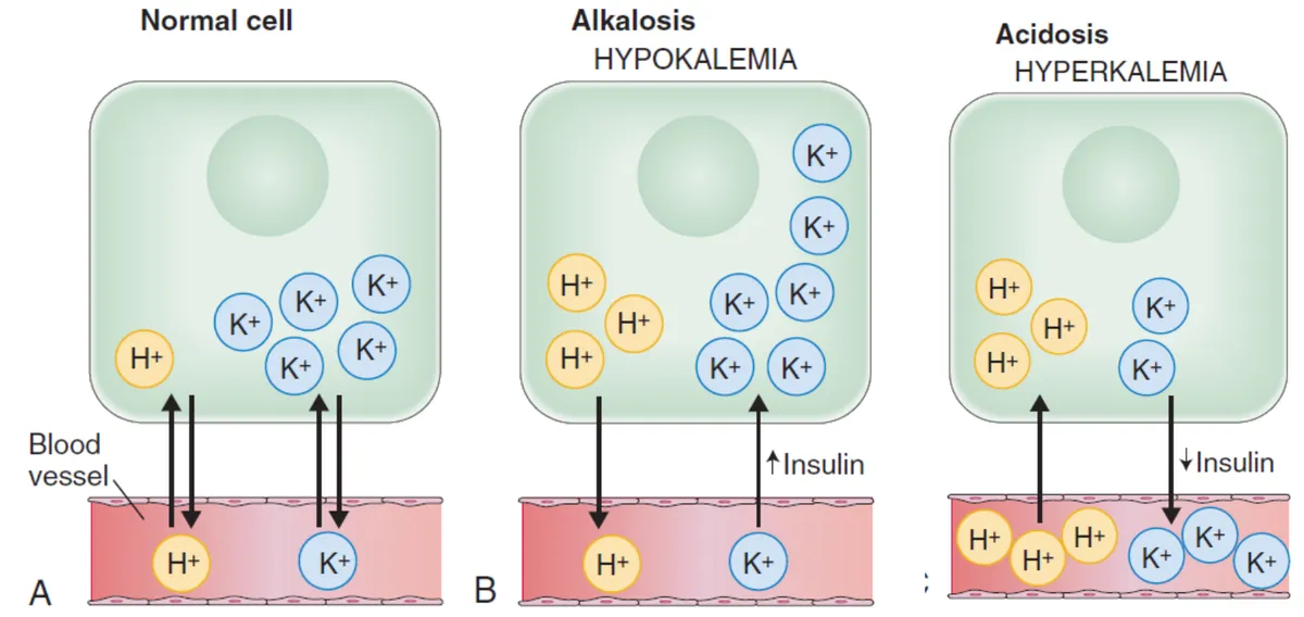
L4 p.20–22About 98% of body potassium is inside cells (about 140 mEq/L, against 3.5–5.0 mEq/L in plasma), held there by the Na⁺/K⁺-ATPase. That steep gradient sets the resting membrane potential of nerve and muscle. Because so little potassium is outside, a small shift of intracellular K⁺ across the membrane changes the serum level a lot, before any potassium has been gained or lost.
Transcellular shiftsL4 p.20–22
| Moves K⁺ into cells (serum K⁺ ↓) | Moves K⁺ out of cells (serum K⁺ ↑) |
|---|---|
| Insulin (stimulates the Na⁺/K⁺-ATPase) | Insulin deficiency (untreated DKA) |
| β2-agonists (albuterol, epinephrine, stress) | β-blockers; digoxin toxicity (pump inhibited) |
| Alkalosis (H⁺ leaves cells, K⁺ enters) | Acidosis, especially with inorganic acids (H⁺ enters cells, K⁺ leaves) |
| Refeeding; rapid cell production | Hyperosmolality (water drags K⁺ out); cell lysis (rhabdomyolysis, tumor lysis, hemolysis) |
Insulin (stimulates the Na⁺/K⁺-ATPase)
- Moves K⁺ out of cells (serum K⁺ ↑)
- Insulin deficiency (untreated DKA)
β2-agonists (albuterol, epinephrine, stress)
- Moves K⁺ out of cells (serum K⁺ ↑)
- β-blockers; digoxin toxicity (pump inhibited)
Alkalosis (H⁺ leaves cells, K⁺ enters)
- Moves K⁺ out of cells (serum K⁺ ↑)
- Acidosis, especially with inorganic acids (H⁺ enters cells, K⁺ leaves)
Refeeding; rapid cell production
- Moves K⁺ out of cells (serum K⁺ ↑)
- Hyperosmolality (water drags K⁺ out); cell lysis (rhabdomyolysis, tumor lysis, hemolysis)
Lecture image
Hypokalemia
L4 p.20Hypokalemia is serum K⁺ < 3.5 mEq/L. Causes: low intake (rarely alone), GI loss (diarrhea, laxatives), renal loss (loop and thiazide diuretics, mineralocorticoid excess, vomiting through secondary aldosteronism and alkalosis, hypomagnesemia, some renal tubular acidoses) and shift into cells (insulin, β2-agonists, alkalosis).
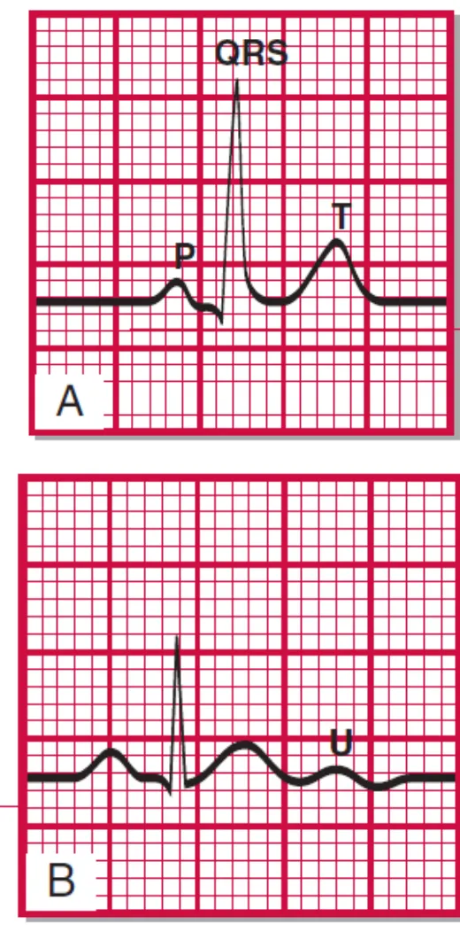
Low extracellular K⁺ makes the resting membrane potential of skeletal and smooth muscle more negative (hyperpolarized), so it is harder to fire: muscle weakness, fatigue, cramps, constipation or ileus. In the heart, repolarization is delayed: the T wave flattens, the ST segment sags and a U wave appears after the T wave; arrhythmias follow, especially with digoxin. In the kidney, severe hypokalemia vacuolates collecting-duct cells and makes them resistant to ADH (polyuria, a nephrogenic DI). Rhabdomyolysis can occur during exertion.
Lecture image
Hyperkalemia
L4 p.22Hyperkalemia is serum K⁺ > 5 mEq/L. Causes: decreased renal excretion (kidney failure; low aldosterone from Addison disease, ACE inhibitors, angiotensin receptor blockers, spironolactone), shift out of cells (acidosis, insulin deficiency, β-blockers, digoxin toxicity), tissue breakdown (rhabdomyolysis, tumor lysis, hemolysis, crush injury) and increased intake when excretion is impaired. Rule out pseudohyperkalemia first: a hemolyzed sample, fist clenching or very high platelet or white cell counts release K⁺ in the tube.
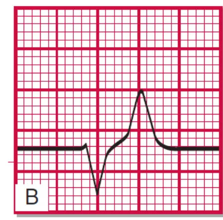
High extracellular K⁺ makes the resting potential less negative (partial depolarization). That sounds excitatory, but sustained depolarization inactivates the fast sodium channels, so cells become less excitable: muscle weakness and depressed or absent tendon reflexes. In the heart, potassium conductance rises and repolarization speeds up, giving tall, narrow, peaked T waves. As K⁺ rises further, P waves flatten, PR and QRS widen, the complex merges into a sine wave, and the heart stops in diastole (severe hyperkalemia, about 7–8 mEq/L).
Lecture image
Teaching schematic
Hypokalemia
Flat T wave, ST depression, U wave after the T
Normal
P, QRS, T
Hyperkalemia
Tall, narrow, peaked T; P flattening
Severe hyperkalemia
Wide QRS merging into a sine wave → arrest
Schematic strips, not recordings.
Knowledge check · 4.3
Answer, read why, and try again whenever you like. Answers are kept in both study modes.
- Q1Water deprivation testL4 p.17–18
A 45-year-old woman who has taken lithium for bipolar disorder for 12 years passes 6 L of urine a day. Serum Na⁺ is 147 mEq/L. After 8 hours without water her urine osmolality is 170 mOsm/kg and plasma osmolality 302 mOsm/kg. One hour after desmopressin, urine osmolality is 185 mOsm/kg.
What is the diagnosis?
- Q2SIADH from small cell carcinomaL4 p.19
A 66-year-old man with a 50 pack-year smoking history becomes confused. Serum Na⁺ is 122 mEq/L, plasma osmolality 254 mOsm/kg, urine osmolality 520 mOsm/kg and urine Na⁺ 52 mEq/L. He has no edema and normal neck veins. CT shows a central hilar mass.
What links the mass to the laboratory findings?
- Q3Hyperkalemia ECG and mechanismL4 p.22
This rhythm strip comes from a 70-year-old man with chronic kidney disease who recently started spironolactone in addition to an ACE inhibitor. He feels weak.
Schematic rhythm strip, lead II.
What disturbance explains the strip, and what is happening at the cell membrane?
- Q4Transcellular potassium shiftL4 p.21
A 22-year-old woman in diabetic ketoacidosis has a serum K⁺ of 5.6 mEq/L on arrival. Two hours after an insulin infusion is started, her K⁺ is 3.1 mEq/L, although she has passed little urine.
Why did the potassium fall?
- Q5Osmotic demyelination syndromeL4 p.12
A 58-year-old woman with alcohol use disorder and a serum Na⁺ of 108 mEq/L for several days receives intravenous saline; her sodium is 128 mEq/L 24 hours later. Three days afterwards she cannot speak clearly, has difficulty swallowing and develops weakness of all four limbs.
What has happened?