Thrombosis, embolism and DIC
A thrombus forms where the vessel wall, the flow or the blood itself is abnormal. Where it forms decides what it is made of, and where its fragments travel decides which organ infarcts.
Virchow triad
L4 p.34Thrombosis is the formation of a blood clot (thrombus) inside a vessel in a living person. Three kinds of abnormality favor it, and most thrombi need more than one.
Teaching schematic
Endothelial injury
Turbulence at bifurcations, plaque rupture, homocysteine, oxidized LDL, smoke, cytokines
Stasis or turbulence
Bed rest, long sitting, dilated left atrium (mitral stenosis, AF), aneurysm
Hypercoagulability
DIC, factor V Leiden and other factor defects, antiphospholipid syndrome, thrombocytosis, pregnancy, estrogen, cancer
Each corner, what it does and the lecture's examplesL4 p.34
| Corner | Why it promotes clotting | Examples |
|---|---|---|
| Endothelial injury | Exposes collagen and von Willebrand factor (platelet adhesion) and tissue factor; the injured cell stops making prostacyclin, NO and thrombomodulin | Turbulent flow at arterial bifurcations, ruptured atherosclerotic plaque, homocysteine, oxidized LDL, cigarette smoke, cytokines |
| Abnormal flow: stasis or turbulence | Platelets reach the wall, activated clotting factors are not washed away, inhibitors do not arrive, endothelium becomes activated | Prolonged bed rest or sitting, dilated left atrium in mitral stenosis (and atrial fibrillation), aneurysms |
| Hypercoagulability | More procoagulant activity or less natural anticoagulant | DIC, inherited or acquired factor defects (factor V Leiden, prothrombin mutation, antithrombin, protein C or S deficiency), antiphospholipid syndrome, thrombocytosis; also pregnancy, estrogens, cancer, surgery |
Endothelial injury
- Why it promotes clotting
- Exposes collagen and von Willebrand factor (platelet adhesion) and tissue factor; the injured cell stops making prostacyclin, NO and thrombomodulin
- Examples
- Turbulent flow at arterial bifurcations, ruptured atherosclerotic plaque, homocysteine, oxidized LDL, cigarette smoke, cytokines
Abnormal flow: stasis or turbulence
- Why it promotes clotting
- Platelets reach the wall, activated clotting factors are not washed away, inhibitors do not arrive, endothelium becomes activated
- Examples
- Prolonged bed rest or sitting, dilated left atrium in mitral stenosis (and atrial fibrillation), aneurysms
Hypercoagulability
- Why it promotes clotting
- More procoagulant activity or less natural anticoagulant
- Examples
- DIC, inherited or acquired factor defects (factor V Leiden, prothrombin mutation, antithrombin, protein C or S deficiency), antiphospholipid syndrome, thrombocytosis; also pregnancy, estrogens, cancer, surgery
Arterial versus venous thrombi; lines of Zahn
L4 p.35Flow speed decides composition. In arteries and the heart, fast flow sweeps red cells past, and the thrombus grows from platelets that stick to injured endothelium, held together by fibrin: a pale, gray-white thrombus, usually on a ruptured atherosclerotic plaque in coronary, cerebral or femoral arteries. In veins, slow flow lets the whole blood clot: a soft, dark red thrombus made mostly of red cells trapped in fibrin, with fewer platelets and white cells. Venous thrombi grow in the direction of flow, toward the heart, which is why their loose tails embolize.
The lecture's comparisonL4 p.35
| Arterial thrombus | Venous thrombus | |
|---|---|---|
| Main cause | Endothelial injury, turbulence at bifurcations and over disrupted plaques | Stasis (most common) + hypercoagulability |
| Sites | Coronary, cerebral, femoral arteries over plaques; mural thrombi in heart chambers and aneurysms | Deep veins of the legs (popliteal, femoral, peroneal), pelvic veins; also SVC, hepatic vein |
| Composition | Platelets + fibrin: gray-white | Red cells in fibrin, some platelets and leukocytes: red |
| Consequence | Occlusion → infarct of the territory; fragments → systemic emboli | Local swelling and pain; fragments → pulmonary emboli |
Main cause
- Arterial thrombus
- Endothelial injury, turbulence at bifurcations and over disrupted plaques
- Venous thrombus
- Stasis (most common) + hypercoagulability
Sites
- Arterial thrombus
- Coronary, cerebral, femoral arteries over plaques; mural thrombi in heart chambers and aneurysms
- Venous thrombus
- Deep veins of the legs (popliteal, femoral, peroneal), pelvic veins; also SVC, hepatic vein
Composition
- Arterial thrombus
- Platelets + fibrin: gray-white
- Venous thrombus
- Red cells in fibrin, some platelets and leukocytes: red
Consequence
- Arterial thrombus
- Occlusion → infarct of the territory; fragments → systemic emboli
- Venous thrombus
- Local swelling and pain; fragments → pulmonary emboli
A thrombus that formed in flowing blood is laminated: pale layers of platelets and fibrin alternate with darker layers of red cells. These lines of Zahn prove it formed during life. A postmortem clot forms in still blood after death: it is gelatinous, not attached to the wall, and settles into a dark red dependent part (“currant jelly”) under a yellow, plasma-rich upper part (“chicken fat”), with no laminations.
Teaching schematic
Core activity · Almost Twins
Almost Twins
Look-alike A
Antemortem thrombus
- Fibrin and red cells
- Lines of Zahn; attached to the wall
- Firm, dry, friable
Look-alike A
Antemortem thrombus
- Fibrin and red cells
- Lines of Zahn; attached to the wall
- Firm, dry, friable
Look-alike B
Postmortem clot
- Fibrin and red cells
- No laminations; lies free in the lumen
- Gelatinous: dark red “currant jelly” below, yellow “chicken fat” above
Step 1 · What they share
What do they share?
Select every feature that both show.
What happens to a thrombus
L4 p.36–37Teaching schematic
Propagation
Grows until it occludes
Embolization
Fragment travels downstream
Dissolution
Fibrinolysis of a recent clot
Organization + recanalization
Ingrowth of cells; new channels
- Propagation: more platelets and fibrin are added and the thrombus grows until it occludes the vessel.
- Embolization: all or part breaks off and travels downstream.
- Dissolution: plasmin (fibrinolysis) removes it, mainly when it is recent; the basis of thrombolytic therapy, which works only in the first hours.
- Organization and recanalization: endothelial cells, smooth muscle cells and fibroblasts grow into an older thrombus; new channels form through it and partly restore flow, while the rest becomes a fibrous scar in the wall.
A thrombus can also become infected (septic thrombophlebitis, mycotic aneurysm). Which fate occurs depends on size, age and site; most deep vein thrombi either dissolve or organize, and the dangerous ones are those that embolize first.
From deep vein thrombosis to pulmonary embolism
L4 p.36–37, 49Embolism is a detached mass (clot, fat, gas, amniotic fluid) carried by the blood to a distant site. More than 95% of pulmonary emboli come from deep veins of the legs above the knee (popliteal, femoral, iliac) and the pelvis. The thrombus breaks off, passes through the right heart and lodges where the pulmonary artery tree becomes too narrow. What follows depends on the size of the embolus and on the patient's circulation.
Outcomes of pulmonary embolismL4 p.37, 49
| Embolus | Effect | Clues |
|---|---|---|
| Small, single (most) | Clinically silent; the lung's dual supply keeps tissue alive; organizes | None, or brief dyspnea |
| Medium or small, with poor bronchial flow (e.g. left heart failure) | Pulmonary infarction: wedge-shaped, pleura-based, hemorrhagic (red) | Pleuritic chest pain, hemoptysis, pleural rub or effusion |
| Large or saddle embolus at the bifurcation of the pulmonary trunk | Obstructs > 60% of the pulmonary circulation: acute right heart failure (acute cor pulmonale), obstructive shock, sudden death | Collapse, hypotension, raised JVP, hypoxemia |
| Many small emboli over time | Pulmonary hypertension and chronic right heart strain | Progressive dyspnea |
Small, single (most)
- Effect
- Clinically silent; the lung's dual supply keeps tissue alive; organizes
- Clues
- None, or brief dyspnea
Medium or small, with poor bronchial flow (e.g. left heart failure)
- Effect
- Pulmonary infarction: wedge-shaped, pleura-based, hemorrhagic (red)
- Clues
- Pleuritic chest pain, hemoptysis, pleural rub or effusion
Large or saddle embolus at the bifurcation of the pulmonary trunk
- Effect
- Obstructs > 60% of the pulmonary circulation: acute right heart failure (acute cor pulmonale), obstructive shock, sudden death
- Clues
- Collapse, hypotension, raised JVP, hypoxemia
Many small emboli over time
- Effect
- Pulmonary hypertension and chronic right heart strain
- Clues
- Progressive dyspnea
Lecture image
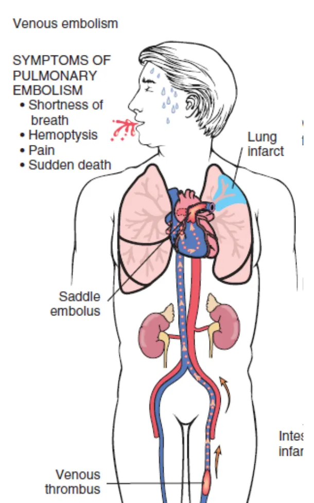
Gas exchange changes in a recognizable way. Blood is diverted from the blocked arteries to the rest of the lung, creating low V/Q units and hypoxemia; hypoxemia, pain and receptor stimulation drive hyperventilation, so the classic gas is a low PaO₂ with a low PaCO₂ and respiratory alkalosis (Sim. Q32, worked in 4.4). The dead space itself wastes ventilation but is not what lowers PaO₂.
Activity · 3D teaching model
Thrombus to pulmonary embolism: trace the route
The 2D diagram shows the same route, steps and questions as the 3D model.
Stage 1 of 5
A mass in the femoral vein
A 62-year-old man is on bed rest five days after a hip replacement. His right calf and thigh are swollen. The cutaway shows a dark red mass fixed to the wall of the right femoral vein, in the pocket behind a valve cusp.
The thrombus is attached to the wall of the right femoral vein; nothing has detached yet.
The mass is attached to the vein wall where it formed. What is it, and which part of the Virchow triad matters most here?
Core activity · The Missing Arrow
The Missing Arrow
A 58-year-old woman sits for a 10-hour flight. Two days later she collapses. Complete the chain.
One gap is the site where the embolus stops; it is a mechanism, not an event.
- Hours of immobility: calf veins barely empty
- a loose fragment breaks off
- Embolus travels through the inferior vena cava and right heart
- cardiac output falls; catecholamines raise SVR
- Obstructive shock: hypotension, distended neck veins, hypoxemia
Cards
Tap or drag a card. Placing a used card moves it.
Systemic and paradoxical emboli
L4 p.37–38A systemic (arterial) embolus travels in arteries. Most come from thrombi in the left heart: a mural thrombus on an infarcted left ventricular wall, or a thrombus in a dilated left atrium in mitral stenosis or atrial fibrillation. Others come from atrial myxoma fragments, valve vegetations (sterile or infective), and ulcerated plaques or aneurysms of the aorta. They lodge in the lower limbs most often, then the brain, small bowel, spleen, kidneys and upper limbs, and the territory beyond them infarcts.
A paradoxical embolus starts in a vein but ends in an artery. It crosses from the right to the left side of the heart through a septal defect: most often a patent foramen ovale, or an atrial or ventricular septal defect. It needs right-sided pressure to exceed left, which happens transiently with coughing or straining and persistently after earlier pulmonary emboli. The clue is a systemic infarct (often a stroke) in a patient with a leg DVT.
Fat, amniotic fluid and gas emboli
L4 p.39–41Fat embolism follows fractures of long bones (femur) or the pelvis, and less often liposuction or fatty liver. Marrow fat globules enter torn marrow sinusoids and venules and lodge first in pulmonary capillaries; some pass through pulmonary arteriovenous shunts into the systemic circulation. The fat embolism syndrome appears 1–3 days after the injury in a minority of patients: dyspnea and tachypnea, confusion, delirium or coma, and petechiae over the chest, axillae and upper limbs, often with thrombocytopenia and anemia. The globules block vessels mechanically, and free fatty acids released from them injure endothelium.
Lecture image
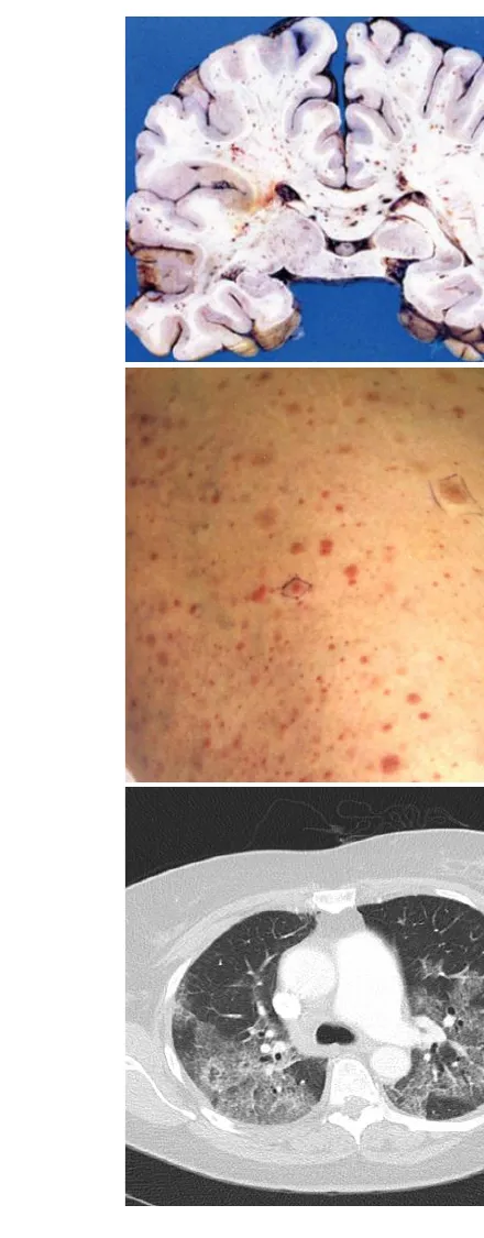
Amniotic fluid embolism occurs during labor or just after delivery through tears in placental membranes or uterine veins. Amniotic fluid in the maternal circulation causes sudden cardiorespiratory collapse (possibly an anaphylactoid reaction to fetal material) and DIC, because amniotic fluid is rich in procoagulants (tissue factor). Abrupt dyspnea, cyanosis, hypotension and bleeding follow; more than half die within an hour. At autopsy, fetal squamous cells, lanugo hair, vernix fat and mucin are found in the maternal pulmonary vessels.
Decompression sickness is a gas embolism. Breathing air at depth dissolves more nitrogen in blood and tissue; a rapid ascent lets it come out of solution as bubbles in tissues and vessels. Bubbles cause severe pain in joints, muscles and bones (“the bends”), pulmonary edema, hemorrhage and alveolar collapse (“the chokes”), and spinal cord ischemia mimicking cord trauma (back pain, loss of sphincter tone). Treatment is recompression in a hyperbaric chamber. Persistent bubbles in bone cause ischemic necrosis of the femoral and humeral heads (caisson disease).
Disseminated intravascular coagulation
L4 p.34, 40, 47In DIC, a trigger exposes the blood to tissue factor or damages endothelium everywhere at once. Thrombin forms throughout the microcirculation, and two opposite problems follow. Microthrombi block small vessels (ischemic injury of kidneys, lungs, brain, adrenals) and shear red cells into fragments (schistocytes: microangiopathic hemolytic anemia). Meanwhile platelets and clotting factors are consumed and fibrinolysis is activated, so the patient bleeds: oozing from venipuncture sites, mucosal and skin hemorrhages.
Teaching schematic
Microthrombi
Ischemia of kidney, lung, brain, adrenals; schistocytes (microangiopathic hemolysis)
Consumption + fibrinolysis
Platelets ↓, PT/aPTT ↑, fibrinogen ↓, D-dimer ↑ → oozing and hemorrhage
Triggers and laboratory patternL4 p.34, 40, 47
| Triggers | Laboratory findings |
|---|---|
| Sepsis (especially Gram-negative); amniotic fluid embolism, placental abruption; trauma, burns; acute promyelocytic leukemia, mucin-producing adenocarcinomas; snake venoms | Platelets ↓; PT and aPTT prolonged; fibrinogen ↓; D-dimer and fibrin degradation products ↑; schistocytes on the smear |
Sepsis (especially Gram-negative); amniotic fluid embolism, placental abruption; trauma, burns; acute promyelocytic leukemia, mucin-producing adenocarcinomas; snake venoms
- Laboratory findings
- Platelets ↓; PT and aPTT prolonged; fibrinogen ↓; D-dimer and fibrin degradation products ↑; schistocytes on the smear
Case clues at a glance
L4 p.34–41What the stem tells youL4 p.34–41
| Clue in the case | Think |
|---|---|
| Long flight, recent surgery, immobilization, swollen calf, then sudden dyspnea and tachycardia | DVT → pulmonary embolism |
| Pleuritic pain and hemoptysis days after a DVT, in a patient with heart failure | Hemorrhagic pulmonary infarct |
| Sudden collapse with raised JVP after leg surgery | Saddle embolus, obstructive shock |
| Atrial fibrillation or mitral stenosis, then a cold, pale, pulseless leg or a stroke | Left atrial thrombus → systemic embolus |
| Large anterior MI two weeks ago, now flank pain and hematuria or a stroke | LV mural thrombus → systemic embolus |
| Stroke in a young patient with a DVT | Paradoxical embolus through a patent foramen ovale |
| Femur fracture two days ago; confusion, dyspnea, axillary petechiae | Fat embolism syndrome |
| Collapse and uncontrolled bleeding during labor | Amniotic fluid embolism with DIC |
| Joint pain and back pain after a rapid ascent from a dive | Decompression sickness |
| Layered thrombus attached to the wall at autopsy | Lines of Zahn: formed before death |
Long flight, recent surgery, immobilization, swollen calf, then sudden dyspnea and tachycardia
- Think
- DVT → pulmonary embolism
Pleuritic pain and hemoptysis days after a DVT, in a patient with heart failure
- Think
- Hemorrhagic pulmonary infarct
Sudden collapse with raised JVP after leg surgery
- Think
- Saddle embolus, obstructive shock
Atrial fibrillation or mitral stenosis, then a cold, pale, pulseless leg or a stroke
- Think
- Left atrial thrombus → systemic embolus
Large anterior MI two weeks ago, now flank pain and hematuria or a stroke
- Think
- LV mural thrombus → systemic embolus
Stroke in a young patient with a DVT
- Think
- Paradoxical embolus through a patent foramen ovale
Femur fracture two days ago; confusion, dyspnea, axillary petechiae
- Think
- Fat embolism syndrome
Collapse and uncontrolled bleeding during labor
- Think
- Amniotic fluid embolism with DIC
Joint pain and back pain after a rapid ascent from a dive
- Think
- Decompression sickness
Layered thrombus attached to the wall at autopsy
- Think
- Lines of Zahn: formed before death
Bridge to Chapter 1
- Virchow triad
- Thrombus (arterial or venous)
- Embolus travels
- Vessel occluded
- Territory infarcts
- Pattern set by the organ (Chapter 1, 1.6)
Knowledge check · 4.6
Answer, read why, and try again whenever you like. Answers are kept in both study modes.
- Q1Venous thrombosis: Virchow triad and compositionL4 p.34–35
Five days after a hip replacement, a 74-year-old woman who has stayed in bed develops a swollen, tender left calf. Ultrasound shows a thrombus in the popliteal and femoral veins.
Which component of Virchow triad is the main driver here, and what is the thrombus mostly made of?
- Q2Lines of Zahn versus postmortem clotL4 p.35
At autopsy, a clot in the inferior vena cava is soft and gelatinous and comes away from the wall easily. Its lower part is dark red and its upper part yellow and translucent. No layering is seen on the cut surface.
What is it?
- Q3Fat embolismL4 p.39
A 22-year-old man with a closed femoral shaft fracture from a motorcycle crash becomes confused and short of breath 48 hours after admission. Petechiae are scattered over his chest and axillae; platelets are 92,000/µL.
What is the most likely cause?
- Q4Paradoxical embolismL4 p.37
A 34-year-old woman develops sudden weakness of her right arm and face during a long-haul flight. On arrival her left calf is swollen and tender. Echocardiography with a bubble study shows contrast crossing from the right to the left atrium.
What explains her stroke?
- Q5DIC laboratory patternL4 p.40
Minutes after delivery, a 31-year-old woman becomes cyanotic and hypotensive, and blood oozes from her IV sites and the uterus.
Which laboratory pattern do you expect?
- Q6Hemorrhagic pulmonary infarctL4 p.37L1 p.33
A 68-year-old man with chronic left heart failure develops pleuritic chest pain and hemoptysis a week after hip surgery. Imaging shows a wedge-shaped opacity with its base on the pleura.
Why is this pulmonary infarct hemorrhagic?