Fluid shifts: hyponatremia, hypernatremia and hyperglycemia
Every gain or loss of salt and water can be read with two separate questions: which way does water move between cells and ECF, and does the ECF grow or shrink?
Two questions for every disorder
L4 p.5, 8–11, 13–16The lecture draws each disorder as a pair of boxes (a Darrow–Yannet diagram): width is volume, height is osmolality, ICF on the left and ECF on the right. The dashed outline is the starting state and the solid outline is the new one. Read any of them with two questions, asked separately:
- Does ECF tonicity change? If what was gained or lost has the same tonicity as plasma, nothing pulls water across cell membranes and the ICF is untouched. If the ECF becomes hypotonic, water moves into cells; if hypertonic, water moves out of cells.
- Did ECF volume go up or down? That follows the change in total body sodium (and, for a moment, glucose or mannitol) plus any water shift from step 1.
Teaching schematic
Isotonic loss
ECF ↓ · ICF —
Cholera, hemorrhage
Isotonic gain
ECF ↑ · ICF —
Excess 0.9% saline
Na⁺ loss > water
ECF ↓ · ICF ↑ · osm ↓
Diuretics, Addison
Pure water gain
ECF ↑ · ICF ↑ · osm ↓
SIADH, polydipsia
Na⁺ and more water gained
ECF ↑↑ · ICF ↑ · osm ↓
RHF, cirrhosis, nephrotic
Water loss > Na⁺
ECF ↓ · ICF ↓ · osm ↑
Sweating, osmotic diuresis
Pure water loss
ECF ↓ · ICF ↓ · osm ↑
DI, fever
Na⁺ gain > water
ECF ↑ · ICF ↓ · osm ↑
NaHCO₃, hypertonic saline
Hyperglycemia (early)
ECF ↑ · ICF ↓ · osm ↑
DKA, HHS before diuresis
Width = volume, height = osmolality. Dashed = before; solid = after. Teaching schematics, sizes exaggerated.
Isotonic loss and gain
L4 p.5, 8An isotonic loss removes sodium and water in plasma proportions: secretory diarrhea (cholera) or bleeding. The remaining ECF has the same tonicity, so there is no gradient and no shift; the ECF shrinks and the ICF is unchanged. Serum Na⁺ stays normal (hypovolemic normonatremia), and the patient shows the volume-depletion signs of 4.1.
An isotonic gain, typically too much 0.9% saline, does the reverse: the ECF expands, the ICF is unchanged, serum Na⁺ is normal. The extra ECF accumulates in the interstitium as pitting edema and effusions, most readily in older people and those with renal dysfunction, who excrete sodium slowly.
Three hyponatremias
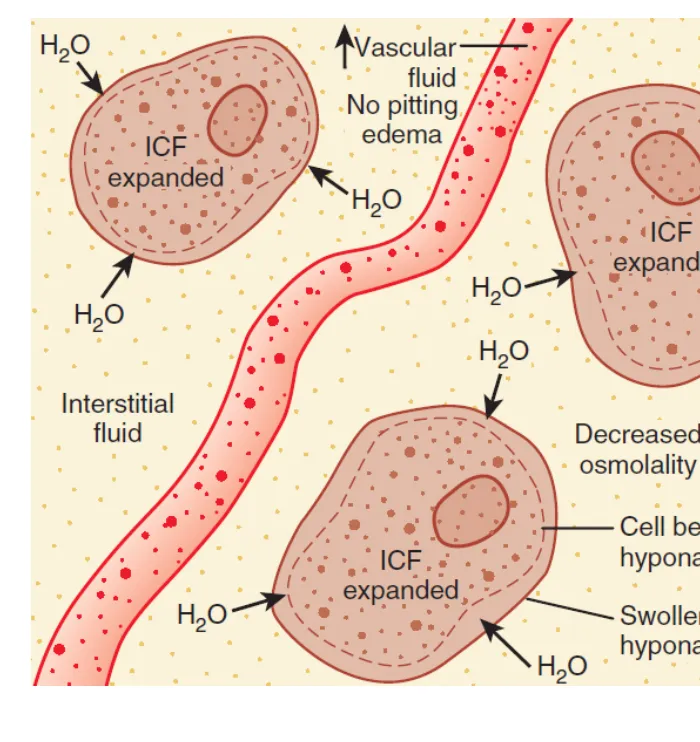
L4 p.9–11In all three, the ECF is hypotonic, so water moves into cells and the ICF expands. They differ in total body sodium, and therefore in ECF volume.
Low sodium, three volumesL4 p.9–11
| Type (lecture name) | Serum Na⁺ = TBNa⁺/TBW | ECF | ICF | Examples | Clue |
|---|---|---|---|---|---|
| Hypovolemic (Na⁺ lost in excess of water) | ↓↓TBNa⁺ / ↓TBW | ↓ | ↑ | Thiazide or loop diuretics, Addison disease; GI losses replaced with water | Volume-depletion signs; urine Na⁺ > 20 mEq/L if the kidney is losing salt |
| Euvolemic (gain of pure water) | TBNa⁺ / ↑↑TBW | ↑ (slight, no edema) | ↑ | SIADH, compulsive water drinking | No edema, no volume depletion |
| Hypervolemic (Na⁺ and even more water gained) | ↑TBNa⁺ / ↑↑TBW | ↑↑ | ↑ | Right heart failure, cirrhosis, nephrotic syndrome | Pitting edema, effusions |
Hypovolemic (Na⁺ lost in excess of water)
- Serum Na⁺ = TBNa⁺/TBW
- ↓↓TBNa⁺ / ↓TBW
- ECF
- ↓
- ICF
- ↑
- Examples
- Thiazide or loop diuretics, Addison disease; GI losses replaced with water
- Clue
- Volume-depletion signs; urine Na⁺ > 20 mEq/L if the kidney is losing salt
Euvolemic (gain of pure water)
- Serum Na⁺ = TBNa⁺/TBW
- TBNa⁺ / ↑↑TBW
- ECF
- ↑ (slight, no edema)
- ICF
- ↑
- Examples
- SIADH, compulsive water drinking
- Clue
- No edema, no volume depletion
Hypervolemic (Na⁺ and even more water gained)
- Serum Na⁺ = TBNa⁺/TBW
- ↑TBNa⁺ / ↑↑TBW
- ECF
- ↑↑
- ICF
- ↑
- Examples
- Right heart failure, cirrhosis, nephrotic syndrome
- Clue
- Pitting edema, effusions
Why does the hypovolemic patient's ICF expand while the ECF shrinks? He loses salty fluid and replaces it with water (thirst and ADH are both stimulated by volume loss). Sodium has been taken out of the ECF; the water left behind is now hypotonic, so some of it moves into cells. This is the only common hyponatremia in which the two compartments move in opposite directions.
Why does the SIADH patient look euvolemic? Pure water spreads through all body water, so two-thirds of it goes into cells. The small ECF expansion also suppresses renin and raises natriuretic peptides, so the kidney excretes sodium (urine Na⁺ > 20 mEq/L), which keeps the ECF from growing enough to produce edema.
Lecture image
Three hypernatremias
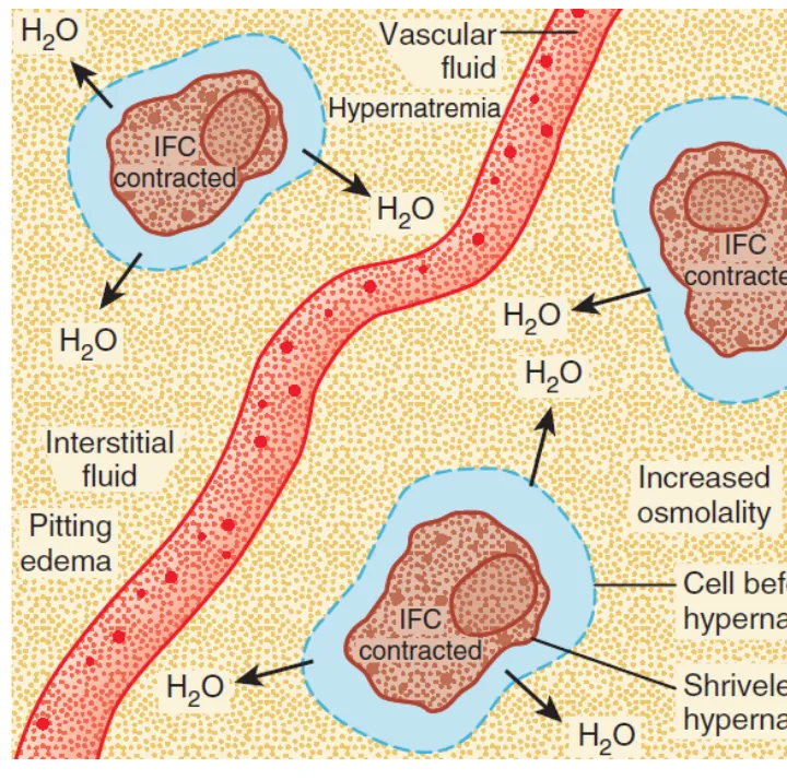
L4 p.13–15In all three the ECF is hypertonic, so water moves out of cells and the ICF contracts. Thirst is the main defense: hypernatremia is rare in anyone awake with access to water, and common in infants, the very old and the unconscious.
High sodium, three volumesL4 p.13–15
| Type (lecture name) | Serum Na⁺ = TBNa⁺/TBW | ECF | ICF | Examples |
|---|---|---|---|---|
| Hypovolemic (water lost in excess of Na⁺) | ↓TBNa⁺ / ↓↓TBW | ↓ | ↓ | Sweating, osmotic diuresis (glucosuria, mannitol), osmotic diarrhea (lactulose, magnesium, polyethylene glycol), vomiting |
| Euvolemic (pure water loss) | TBNa⁺ / ↓↓TBW | ↓ (small) | ↓ | Diabetes insipidus, insensible loss (fever) |
| Hypervolemic (Na⁺ gained in excess of water) | ↑↑TBNa⁺ / ↑TBW | ↑ | ↓ | NaHCO₃ infusion, hypertonic saline, sodium-containing antibiotics, salt ingestion |
Hypovolemic (water lost in excess of Na⁺)
- Serum Na⁺ = TBNa⁺/TBW
- ↓TBNa⁺ / ↓↓TBW
- ECF
- ↓
- ICF
- ↓
- Examples
- Sweating, osmotic diuresis (glucosuria, mannitol), osmotic diarrhea (lactulose, magnesium, polyethylene glycol), vomiting
Euvolemic (pure water loss)
- Serum Na⁺ = TBNa⁺/TBW
- TBNa⁺ / ↓↓TBW
- ECF
- ↓ (small)
- ICF
- ↓
- Examples
- Diabetes insipidus, insensible loss (fever)
Hypervolemic (Na⁺ gained in excess of water)
- Serum Na⁺ = TBNa⁺/TBW
- ↑↑TBNa⁺ / ↑TBW
- ECF
- ↑
- ICF
- ↓
- Examples
- NaHCO₃ infusion, hypertonic saline, sodium-containing antibiotics, salt ingestion
Pure water loss shrinks the ECF too, but only by a third of the water lost (the rest comes out of cells), so volume signs are mild and the patient is called euvolemic. A hypertonic sodium gain is the mirror image of the hypovolemic hyponatremia: the compartments move in opposite directions, ECF up and ICF down.
Lecture image
Urine sodium and concentration: the kidney's testimony
L4 p.9, 14–15, 17–19A spot urine answers two questions about how the kidney is responding. Urine osmolality shows whether ADH is acting (concentrated urine) or switched off (dilute urine). Urine sodium shows whether the kidney is conserving sodium (< 20 mEq/L, as it does when it senses a poorly filled circulation) or excreting it (> 20 mEq/L, because of a diuretic, missing aldosterone, a sodium load or SIADH). Read them against the patient's volume.
Hyponatremia: what the urine addsL4 p.9, 19
| Urine finding | Meaning | Fits |
|---|---|---|
| Osmolality < 100 mOsm/kg | ADH appropriately off; water is being excreted as fast as the kidney can | Primary polydipsia; very low solute intake |
| Osmolality > 100 mOsm/kg | ADH is acting | SIADH; hypovolemia; heart failure, cirrhosis |
| Na⁺ < 20 mEq/L + volume depletion | Kidney conserving sodium; the loss was elsewhere | Vomiting or diarrhea replaced with water |
| Na⁺ < 20 mEq/L + edema | Kidney senses a poorly filled arterial tree and retains sodium | Heart failure, cirrhosis, nephrotic syndrome |
| Na⁺ > 20 mEq/L + volume depletion | Kidney itself losing sodium | Thiazide or loop diuretics, Addison disease |
| Na⁺ > 20 mEq/L + euvolemia | Water retention with natriuresis | SIADH |
Osmolality < 100 mOsm/kg
- Meaning
- ADH appropriately off; water is being excreted as fast as the kidney can
- Fits
- Primary polydipsia; very low solute intake
Osmolality > 100 mOsm/kg
- Meaning
- ADH is acting
- Fits
- SIADH; hypovolemia; heart failure, cirrhosis
Na⁺ < 20 mEq/L + volume depletion
- Meaning
- Kidney conserving sodium; the loss was elsewhere
- Fits
- Vomiting or diarrhea replaced with water
Na⁺ < 20 mEq/L + edema
- Meaning
- Kidney senses a poorly filled arterial tree and retains sodium
- Fits
- Heart failure, cirrhosis, nephrotic syndrome
Na⁺ > 20 mEq/L + volume depletion
- Meaning
- Kidney itself losing sodium
- Fits
- Thiazide or loop diuretics, Addison disease
Na⁺ > 20 mEq/L + euvolemia
- Meaning
- Water retention with natriuresis
- Fits
- SIADH
Water-loss hypernatremia versus sodium-gain hypernatremia
Water loss (TBNa⁺/↓↓TBW)
- Fever, sweating, no access to water; diabetes insipidus
- ECF small: dry mucosa, tachycardia (mild if pure water)
- Urine Na⁺ low when volume is low
- Urine maximally concentrated if ADH and kidney work (extrarenal loss); inappropriately dilute in DI
- Replace water
Sodium gain (↑↑TBNa⁺/↑TBW)
- NaHCO₃ during resuscitation, hypertonic saline, Na⁺-containing antibiotics, salt ingestion
- ECF large: edema, raised JVP, sometimes pulmonary edema
- Urine Na⁺ high (the kidney is excreting the load)
- Urine concentrated
- Stop the sodium; water plus a loop diuretic
Osmotic diuresis (glucosuria, mannitol) sits between the two: large volumes of urine that is neither dilute nor highly concentrated (roughly plasma-like osmolality, carrying a big solute load), with water lost in excess of sodium.
Hyperglycemia: DKA, HHS and two kinds of low sodium
L4 p.16, 27Without insulin, glucose cannot enter muscle and fat cells, so it acts as an effective osmole in the ECF. Water moves out of cells, the ICF contracts and, at first, the ECF expands and its sodium is diluted. This real dilution is translocational (hypertonic) hyponatremia: the sodium is low but measured osmolality is high. Soon the filtered glucose exceeds tubular reabsorption, and osmotic diuresis removes water (and sodium and potassium) in the urine, so both compartments end up contracted.
Worked calculation · Translocational hyponatremia
- Measured Na⁺
- 128 mEq/L
- Glucose
- 700 mg/dL
- BUN
- 28 mg/dL
Formula
Corrected Na⁺ = measured Na⁺ + 1.6 × (glucose − 100)/100; Posm = 2 × Na⁺ + glucose/18 + BUN/2.8
L4 p.16Translocational hyponatremia versus pseudohyponatremia
Translocational (real)
- Cause: an effective osmole in the ECF (glucose, mannitol)
- Water really moved out of cells and diluted sodium
- Measured osmolality high
- Cells shrunken; corrects as glucose falls
Pseudohyponatremia (lab artifact)
- Cause: severe hypertriglyceridemia or paraprotein (multiple myeloma) takes up plasma volume
- Indirect methods report sodium per liter of plasma, not per liter of plasma water
- Measured osmolality normal
- No water shift; a direct ion-selective electrode gives the true, normal sodium
DKA and HHS: the same osmotic diuresis, different acidL4 p.16, 27
| Diabetic ketoacidosis (DKA) | Hyperglycemic hyperosmolar state (HHS) | |
|---|---|---|
| Typical patient | Type 1 diabetes, often young; omitted insulin or infection | Type 2 diabetes, older; infection, poor water intake |
| Insulin | Absent: lipolysis and ketogenesis run unchecked | Enough to suppress ketogenesis, not hyperglycemia |
| Glucose | Usually 250–600 mg/dL | Often > 600 mg/dL, sometimes > 1000 |
| Acid–base | High anion gap metabolic acidosis (acetoacetate, β-hydroxybutyrate), Kussmaul breathing | Little or no acidosis |
| Osmolality and brain | Raised | Very high (> 320 mOsm/kg); confusion and coma from cell shrinking |
| Volume | Depleted by osmotic diuresis | Severely depleted (longer, slower onset) |
| Potassium | Serum K⁺ often normal or high, total body K⁺ depleted (shift out of cells, urinary loss) | Same pattern |
Typical patient
- Diabetic ketoacidosis (DKA)
- Type 1 diabetes, often young; omitted insulin or infection
- Hyperglycemic hyperosmolar state (HHS)
- Type 2 diabetes, older; infection, poor water intake
Insulin
- Diabetic ketoacidosis (DKA)
- Absent: lipolysis and ketogenesis run unchecked
- Hyperglycemic hyperosmolar state (HHS)
- Enough to suppress ketogenesis, not hyperglycemia
Glucose
- Diabetic ketoacidosis (DKA)
- Usually 250–600 mg/dL
- Hyperglycemic hyperosmolar state (HHS)
- Often > 600 mg/dL, sometimes > 1000
Acid–base
- Diabetic ketoacidosis (DKA)
- High anion gap metabolic acidosis (acetoacetate, β-hydroxybutyrate), Kussmaul breathing
- Hyperglycemic hyperosmolar state (HHS)
- Little or no acidosis
Osmolality and brain
- Diabetic ketoacidosis (DKA)
- Raised
- Hyperglycemic hyperosmolar state (HHS)
- Very high (> 320 mOsm/kg); confusion and coma from cell shrinking
Volume
- Diabetic ketoacidosis (DKA)
- Depleted by osmotic diuresis
- Hyperglycemic hyperosmolar state (HHS)
- Severely depleted (longer, slower onset)
Potassium
- Diabetic ketoacidosis (DKA)
- Serum K⁺ often normal or high, total body K⁺ depleted (shift out of cells, urinary loss)
- Hyperglycemic hyperosmolar state (HHS)
- Same pattern
Fluid Compartment Predictor
L4 p.5, 8–16Choose an event. Before anything moves, predict for each compartment whether volume and osmolality rise, fall or stay the same, and which way water crosses the cell membrane. Then reveal and watch the boxes change.
Centerpiece · Fluid Compartment Predictor
Where does the water go?
Event
A 70-kg man receives 2 L of 0.9% NaCl intravenously over an hour.
+ isotonic NaCl → ECF
Make all five predictions, then check. The boxes will change to the real result.
Core activity · Almost Twins
Almost Twins
Look-alike A
Translocational hyponatremia
- Reported Na⁺ 128 mEq/L
- Measured osmolality high (≈ 305 mOsm/kg)
- Glucose pulled water out of cells and diluted sodium
- Sodium rises as glucose falls
Look-alike A
Translocational hyponatremia
- Reported Na⁺ 128 mEq/L
- Measured osmolality high (≈ 305 mOsm/kg)
- Glucose pulled water out of cells and diluted sodium
- Sodium rises as glucose falls
Look-alike B
Pseudohyponatremia
- Reported Na⁺ 128 mEq/L
- Measured osmolality normal (≈ 288 mOsm/kg)
- Lipid takes up plasma volume; the method reports Na⁺ per liter of plasma, not of plasma water
- Direct ion-selective electrode reads a normal Na⁺
Step 1 · What they share
What do they share?
Select every feature that both show.
Knowledge check · 4.2
Answer, read why, and try again whenever you like. Answers are kept in both study modes.
- Q1Hypovolemic hyponatremiaL4 p.9
A 72-year-old woman started a thiazide for hypertension three weeks ago. She is lightheaded, has dry mucous membranes and a postural drop in blood pressure. Serum Na⁺ is 126 mEq/L; urine Na⁺ is 48 mEq/L.
Which change in the compartments fits her?
- Q2Reading a Darrow–Yannet diagramL4 p.15
In the Darrow–Yannet diagram shown, the solid ICF box is narrower than the dashed outline, the solid ECF box is wider, and both solid boxes are taller.
Dashed = before; solid = after. Width = volume, height = osmolality.
Which event produced it?
- Q3Translocational hyponatremiaL4 p.16
A 19-year-old with type 1 diabetes is admitted with vomiting and deep, rapid breathing. Glucose is 800 mg/dL, serum Na⁺ 128 mEq/L and measured plasma osmolality 312 mOsm/kg.
What best explains the low sodium?
- Q4PseudohyponatremiaL4 p.3
A 61-year-old man with multiple myeloma has a total serum protein of 12 g/dL. The chemistry analyzer reports Na⁺ 127 mEq/L; measured plasma osmolality is 288 mOsm/kg and he has no neurologic symptoms.
What is the best interpretation?
- Q5Pure water lossL4 p.14, 17
After pituitary surgery, a 40-year-old man passes 8 L of dilute urine a day. He is kept nil by mouth by mistake overnight.
Where does most of the water he loses overnight come from?