Autoimmune disease
Self-reactive lymphocytes are made every day. Autoimmune disease follows when the checks that delete, silence or suppress them fail in a genetically susceptible person and something in the environment sets them off. The injury then uses the same hypersensitivity mechanisms as any other immune attack.
Tolerance and how it fails
L3 p.34Because receptors are made at random (3.1), the body continually produces lymphocytes that recognize self. Tolerance is the set of checks that keeps them harmless. Central tolerance acts while lymphocytes are developing; peripheral tolerance catches those that escape.
Teaching schematic
Central tolerance (developing lymphocytes)
- Thymus: negative selection with AIREAIRE mutation → APS-1
- Thymus: some self-reactive cells become Tregs
- Marrow: B-cell receptor editing or deletionSelf-reactive B cells survive (estrogen)
Peripheral tolerance (mature lymphocytes)
- Anergy: antigen without B7–CD28; CTLA-4, PD-1Checkpoint-blocking drugs → colitis, thyroiditis
- Tregs (FOXP3, CD25): IL-10, TGF-βFOXP3 mutation → IPEX; CD25 variants → MS, T1DM
- Deletion through Fas–FasLFas mutation → ALPS
Susceptibility × trigger. HLA (B27, DR4, DR3), sex hormones, complement defects; then infection (molecular mimicry, costimulation), tissue injury releasing hidden antigens, UV light.
The checks, and what happens when each failsL3 p.34
| Check | Where | How it works | If it fails |
|---|---|---|---|
| Negative selection with AIRE | Thymic medulla (central) | Tissue antigens are displayed; strongly self-reactive thymocytes die or become Tregs | APS-1 (autoimmune polyendocrine syndrome): hypoparathyroidism, adrenal failure, chronic mucocutaneous candidiasis |
| Receptor editing, deletion | Bone marrow (central, B cells) | A self-reactive immature B cell rearranges a new light chain or dies | Self-reactive B cells survive (estrogen may reduce their deletion, one reason SLE favors women) |
| Anergy | Periphery | Antigen seen without costimulation (no B7–CD28), or with inhibitory CTLA-4 and PD-1, switches the T cell off | Checkpoint-inhibitor cancer drugs cause autoimmune colitis, thyroiditis, hepatitis |
| Suppression by Tregs (FOXP3, CD25) | Periphery | Tregs consume IL-2 and secrete IL-10 and TGF-β | IPEX (FOXP3 mutation, X-linked): neonatal diabetes, enteropathy, eczema in boys; CD25 polymorphisms → MS, type 1 diabetes |
| Deletion (apoptosis) | Periphery | Repeatedly stimulated T cells die through Fas–Fas ligand | Autoimmune lymphoproliferative syndrome (Fas defect): lymphadenopathy, autoimmune cytopenias |
Negative selection with AIRE
- Where
- Thymic medulla (central)
- How it works
- Tissue antigens are displayed; strongly self-reactive thymocytes die or become Tregs
- If it fails
- APS-1 (autoimmune polyendocrine syndrome): hypoparathyroidism, adrenal failure, chronic mucocutaneous candidiasis
Receptor editing, deletion
- Where
- Bone marrow (central, B cells)
- How it works
- A self-reactive immature B cell rearranges a new light chain or dies
- If it fails
- Self-reactive B cells survive (estrogen may reduce their deletion, one reason SLE favors women)
Anergy
- Where
- Periphery
- How it works
- Antigen seen without costimulation (no B7–CD28), or with inhibitory CTLA-4 and PD-1, switches the T cell off
- If it fails
- Checkpoint-inhibitor cancer drugs cause autoimmune colitis, thyroiditis, hepatitis
Suppression by Tregs (FOXP3, CD25)
- Where
- Periphery
- How it works
- Tregs consume IL-2 and secrete IL-10 and TGF-β
- If it fails
- IPEX (FOXP3 mutation, X-linked): neonatal diabetes, enteropathy, eczema in boys; CD25 polymorphisms → MS, type 1 diabetes
Deletion (apoptosis)
- Where
- Periphery
- How it works
- Repeatedly stimulated T cells die through Fas–Fas ligand
- If it fails
- Autoimmune lymphoproliferative syndrome (Fas defect): lymphadenopathy, autoimmune cytopenias
Susceptibility genes set the stage. HLA alleles shape which self-peptides are presented: HLA-B27 (class I) with ankylosing spondylitis, psoriatic arthritis, IBD-associated and reactive arthritis; HLA-DR4 (class II) with rheumatoid arthritis; DR2 and DR3 with SLE. Triggers then break tolerance: infections that look like self (molecular mimicry, as streptococcal M protein and cardiac myosin in rheumatic fever) or that switch on costimulators in tissues; tissue injury that releases hidden antigens (sympathetic ophthalmia, 3.1); ultraviolet light, which makes skin cells die and expose nuclear antigens in SLE; and female sex hormones.
Core activity · The Missing Arrow
The Missing Arrow
A girl inherits two nonfunctional copies of AIRE. Complete the chain to her autoimmune disease.
One gap needs the mechanism in the thymus; another the event that follows in the tissues.
- Medullary thymic epithelial cells lack AIRE
- in the periphery they meet their antigens with costimulation
- organ failure appears through childhood
- APS-1: chronic mucocutaneous candidiasis, hypoparathyroidism, adrenal insufficiency
Cards
Tap or drag a card. Placing a used card moves it.
Systemic lupus erythematosus: mechanism and manifestations
L3 p.36–38SLE is a chronic multisystem autoimmune disease of women of childbearing age (20–45 years; about 9 women per man). Its central defect is a failure to tolerate nuclear antigens. Several things combine:
- Too much nuclear antigen in view. Dying cells (after UV exposure, for instance) are cleared poorly, and their DNA and nucleosomes linger. Inherited deficiency of the early complement components C1q, C2 and C4, which normally help clear apoptotic debris and immune complexes, raises the risk strongly.
- Loss of tolerance. HLA-DR2/DR3 and other genes allow self-reactive B and T cells to be activated; nucleic acids in immune complexes stimulate TLRs inside B cells and plasmacytoid dendritic cells, which produce type I interferon and keep the response going.
- Antibodies to many nuclear and cell-surface antigens. DNA–anti-DNA complexes deposit in glomeruli, skin, joints, serosa and small vessels and fix complement (type III). Antibodies against red cells, platelets and leukocytes cause cytopenias (type II). Antiphospholipid antibodies cause thrombosis.
Manifestations and the mechanism behind eachL3 p.37
| System | Findings | Mechanism |
|---|---|---|
| General | Fever, fatigue, weight loss | Cytokines, interferon |
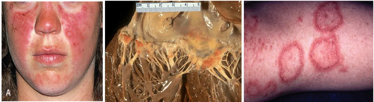
| Skin and mucosa | Malar (butterfly) rash sparing the nasolabial folds; discoid lesions in sun-exposed skin; photosensitivity; oral ulcers; alopecia | Immune complexes at the dermal–epidermal junction; UV-induced keratinocyte death |
| Joints | Arthralgia/arthritis of ≥2 joints, often PIP and MCP, symmetric, nonerosive and nondeforming | Immune complexes in synovium |
| Blood | Autoimmune hemolytic anemia (Coombs positive), thrombocytopenia, leukopenia, lymphopenia; lymphadenopathy, splenomegaly | Type II antibodies |
| Kidney | Nephritic or nephrotic syndrome (below); chronic renal failure is a common cause of death | Immune complexes in glomeruli |
| Heart and lungs | Fibrinous pericarditis, pleuritis, Libman–Sacks (sterile verrucous) endocarditis, myocarditis, interstitial fibrosis, Raynaud phenomenon | Immune complexes, vasculitis |
| Nervous system | Headache, psychosis, seizures, chorea, strokes | Vasculitis, antibodies, antiphospholipid thrombosis |
| Pregnancy | Recurrent miscarriage; congenital heart block in the baby | Antiphospholipid antibodies; maternal IgG anti-Ro/La crossing the placenta and damaging the fetal conduction system |
| Bone | Avascular necrosis of the femoral head, osteoporosis | Corticosteroid treatment, vasculitis |
General
- Findings
- Fever, fatigue, weight loss
- Mechanism
- Cytokines, interferon
Skin and mucosa
- Findings
- Malar (butterfly) rash sparing the nasolabial folds; discoid lesions in sun-exposed skin; photosensitivity; oral ulcers; alopecia
- Mechanism
- Immune complexes at the dermal–epidermal junction; UV-induced keratinocyte death
Joints
- Findings
- Arthralgia/arthritis of ≥2 joints, often PIP and MCP, symmetric, nonerosive and nondeforming
- Mechanism
- Immune complexes in synovium
Blood
- Findings
- Autoimmune hemolytic anemia (Coombs positive), thrombocytopenia, leukopenia, lymphopenia; lymphadenopathy, splenomegaly
- Mechanism
- Type II antibodies
Kidney
- Findings
- Nephritic or nephrotic syndrome (below); chronic renal failure is a common cause of death
- Mechanism
- Immune complexes in glomeruli
Heart and lungs
- Findings
- Fibrinous pericarditis, pleuritis, Libman–Sacks (sterile verrucous) endocarditis, myocarditis, interstitial fibrosis, Raynaud phenomenon
- Mechanism
- Immune complexes, vasculitis
Nervous system
- Findings
- Headache, psychosis, seizures, chorea, strokes
- Mechanism
- Vasculitis, antibodies, antiphospholipid thrombosis
Pregnancy
- Findings
- Recurrent miscarriage; congenital heart block in the baby
- Mechanism
- Antiphospholipid antibodies; maternal IgG anti-Ro/La crossing the placenta and damaging the fetal conduction system
Bone
- Findings
- Avascular necrosis of the femoral head, osteoporosis
- Mechanism
- Corticosteroid treatment, vasculitis
Lecture image
Lupus nephritis: nephritic or nephrotic?
Diffuse proliferative (class IV)
- Most common and most severe form
- Complexes in the subendothelial space and mesangium, close to the blood, so neutrophils and complement reach them
- Proliferation, thick 'wire-loop' capillaries
- Nephritic: hematuria, red-cell casts, proteinuria, hypertension, falling GFR
Membranous (class V)
- Complexes in the subepithelial space, outside the basement membrane, away from inflammatory cells
- Little proliferation; the podocyte barrier is damaged
- Nephrotic: massive proteinuria, hypoalbuminemia, edema, hyperlipidemia
- Little hematuria
Autoantibodies and laboratory tests
L3 p.35, 39Every autoantibody test trades sensitivity (how often it is positive in the disease, good for ruling out) against specificity (how rarely it is positive in anything else, good for ruling in). The screening test for SLE is sensitive but not specific; the confirmatory antibodies are specific but miss many patients.
Antibodies to knowL3 p.35, 39
| Test | What it means | Use |
|---|---|---|
| ANA (immunofluorescence) | Antibodies to any nuclear antigen; positive in ~98% of SLE but also in drug-induced lupus, systemic sclerosis, Sjögren syndrome, infections and healthy older people | Screening: a negative ANA makes SLE very unlikely |
| Anti-dsDNA | Highly specific for SLE; levels rise with active nephritis | Confirm SLE; follow renal activity |
| Anti-Smith (anti-Sm) | The most specific SLE antibody, but present in only about a quarter of patients | Confirm SLE |
| Anti-Ro (SS-A), anti-La (SS-B) | Cross the placenta → neonatal lupus and congenital heart block; also Sjögren syndrome, subacute cutaneous lupus | Screen lupus pregnancies |
| Antihistone | In over 90% of drug-induced lupus (hydralazine, procainamide, isoniazid) | Drug-induced lupus spares the kidney and brain and fades when the drug stops |
| Antiphospholipid | Lupus anticoagulant (prolonged PTT), anticardiolipin (false-positive VDRL/RPR), anti-β₂-glycoprotein I | Explain clots and pregnancy loss (next topic) |
| Complement C3, C4 | Low in active disease because immune complexes consume them | Follow activity |
| ESR and CRP | ESR raised in active SLE; CRP often normal unless serositis or infection | A high CRP in a lupus flare suggests infection |
| Anti-U1 RNP | High titer is the marker of mixed connective tissue disease; also found in SLE | — |
| Organ-specific | Anti–parietal cell (proton pump) and anti–intrinsic factor antibodies in pernicious anemia | — |
ANA (immunofluorescence)
- What it means
- Antibodies to any nuclear antigen; positive in ~98% of SLE but also in drug-induced lupus, systemic sclerosis, Sjögren syndrome, infections and healthy older people
- Use
- Screening: a negative ANA makes SLE very unlikely
Anti-dsDNA
- What it means
- Highly specific for SLE; levels rise with active nephritis
- Use
- Confirm SLE; follow renal activity
Anti-Smith (anti-Sm)
- What it means
- The most specific SLE antibody, but present in only about a quarter of patients
- Use
- Confirm SLE
Anti-Ro (SS-A), anti-La (SS-B)
- What it means
- Cross the placenta → neonatal lupus and congenital heart block; also Sjögren syndrome, subacute cutaneous lupus
- Use
- Screen lupus pregnancies
Antihistone
- What it means
- In over 90% of drug-induced lupus (hydralazine, procainamide, isoniazid)
- Use
- Drug-induced lupus spares the kidney and brain and fades when the drug stops
Antiphospholipid
- What it means
- Lupus anticoagulant (prolonged PTT), anticardiolipin (false-positive VDRL/RPR), anti-β₂-glycoprotein I
- Use
- Explain clots and pregnancy loss (next topic)
Complement C3, C4
- What it means
- Low in active disease because immune complexes consume them
- Use
- Follow activity
ESR and CRP
- What it means
- ESR raised in active SLE; CRP often normal unless serositis or infection
- Use
- A high CRP in a lupus flare suggests infection
Anti-U1 RNP
- What it means
- High titer is the marker of mixed connective tissue disease; also found in SLE
- Use
- —
Organ-specific
- What it means
- Anti–parietal cell (proton pump) and anti–intrinsic factor antibodies in pernicious anemia
- Use
- —
Lecture image
Antiphospholipid syndrome: the clotting paradox
L3 p.37, 39Antiphospholipid antibodies are aimed at proteins that bind phospholipid, chiefly β₂-glycoprotein I and prothrombin. They occur in about a third of SLE patients (secondary syndrome) or alone (primary). The syndrome is diagnosed when the antibodies persist and the patient has thrombosis (venous: deep-vein thrombosis, pulmonary embolism; or arterial: stroke in a young person) or pregnancy morbidity (recurrent miscarriage, fetal loss, placental insufficiency).
Teaching schematic
In the test tube
- Antiphospholipid antibodies bind the phospholipid in the clotting reagent
- Factor complexes cannot assemble on it
- PTT prolonged; mixing with normal plasma does not correct it
- Anticardiolipin reacts with VDRL/RPR antigen → false-positive syphilis screen
Looks like a bleeding tendency: hence the misleading name 'lupus anticoagulant'.
In the patient
- Antibodies bind β₂-glycoprotein I on endothelium, platelets, monocytes, trophoblast
- Cells activated; tissue factor expressed; complement activated
- Natural anticoagulants (protein C) impaired
- Venous and arterial thrombosis; placental thrombosis → pregnancy loss
Treat with anticoagulation. Continues in Chapter 4 (thrombosis).
- The paradox. The lupus anticoagulant prolongs phospholipid-dependent clotting tests such as the PTT, and mixing with normal plasma does not correct it (an inhibitor, not a factor deficiency). Yet the patient is prothrombotic: in vivo the antibodies activate endothelial cells and platelets, induce tissue factor on monocytes, activate complement and interfere with natural anticoagulants. The name is a laboratory artifact.
- False-positive syphilis test. The VDRL and RPR detect antibodies against cardiolipin, a phospholipid. Anticardiolipin antibodies react with the test antigen, so the screening test is positive while specific treponemal tests are negative. A young woman with a 'positive VDRL', no sexual history and oral ulcers should make you think of SLE (Case III Q10).
- Pregnancy loss comes from thrombosis and complement-driven inflammation in the placenta.
- Treatment is anticoagulation (heparin with aspirin in pregnancy). The thrombotic side of the story continues in Chapter 4: thrombosis and embolism.
Systemic sclerosis: from damaged vessels to fibrosis
L3 p.40–43Systemic sclerosis (scleroderma) is excess deposition of normal collagen in skin and organs on a background of small-vessel damage. Women are affected about four times as often as men, usually between 35 and 64. The sequence explains the clinical picture:
Vascular injury to fibrosis
- Autoantibodies and T cells injure endothelium (digital vessels first)
- NO and prostacyclin fall; vessels constrict → Raynaud phenomenon
- Damaged endothelium releases PDGF and TGF-β
- CD4 Th2 cells add IL-13 and TGF-β
- Fibroblasts lay down collagen; perivascular fibrosis narrows lumens
- Ischemia and fibrosis of skin, gut, lungs, kidneys
Diffuse and limited (CREST) forms
Diffuse cutaneous
- Skin thickening spreads proximally to the arms and trunk, rapidly
- Early visceral disease: interstitial lung fibrosis, renal crisis, heart
- Anti-Scl-70 (DNA topoisomerase I): highly specific
- Worse prognosis
Limited cutaneous (CREST)
- Skin of fingers, hands, forearms and face; Raynaud for years first
- Calcinosis cutis, Raynaud, Esophageal dysmotility, Sclerodactyly, Telangiectasia
- Anticentromere antibody
- Late pulmonary arterial hypertension
Lecture image
Organ by organL3 p.41
| Organ | What happens | Clinical result |
|---|---|---|
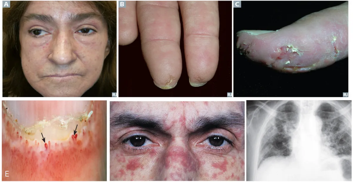
| Skin | Swollen fingers, then firm thickened skin extending proximally; taut face without wrinkles, small mouth; telangiectasias; abnormal nailfold capillaries | Sclerodactyly, digital ulcers, 'stone facies' |
| Esophagus | Smooth muscle of the lower two-thirds atrophies and is replaced by collagen; the lower esophageal sphincter becomes incompetent | Dysphagia, reflux, strictures, Barrett esophagus |
| Stomach and intestine | Dysmotility, villous atrophy, wide-mouthed diverticula | Postprandial bloating, malabsorption, constipation |
| Lungs | Interstitial fibrosis; pulmonary arterial hypertension | Restrictive disease, dyspnea, dry cough; leading cause of death |
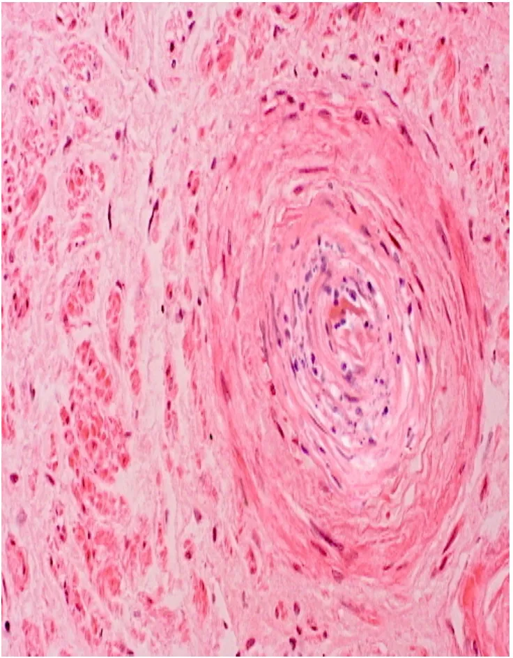
| Kidneys | Intimal thickening of interlobular arteries ('onion-skin'), fibrinoid necrosis of arterioles, thrombosis | Scleroderma renal crisis: malignant hypertension, acute kidney failure (treated with ACE inhibitors) |
Skin
- What happens
- Swollen fingers, then firm thickened skin extending proximally; taut face without wrinkles, small mouth; telangiectasias; abnormal nailfold capillaries
- Clinical result
- Sclerodactyly, digital ulcers, 'stone facies'
Esophagus
- What happens
- Smooth muscle of the lower two-thirds atrophies and is replaced by collagen; the lower esophageal sphincter becomes incompetent
- Clinical result
- Dysphagia, reflux, strictures, Barrett esophagus
Stomach and intestine
- What happens
- Dysmotility, villous atrophy, wide-mouthed diverticula
- Clinical result
- Postprandial bloating, malabsorption, constipation
Lungs
- What happens
- Interstitial fibrosis; pulmonary arterial hypertension
- Clinical result
- Restrictive disease, dyspnea, dry cough; leading cause of death
Kidneys
- What happens
- Intimal thickening of interlobular arteries ('onion-skin'), fibrinoid necrosis of arterioles, thrombosis
- Clinical result
- Scleroderma renal crisis: malignant hypertension, acute kidney failure (treated with ACE inhibitors)
Lecture image
Knowledge check · 3.5
Answer, read why, and try again whenever you like. Answers are kept in both study modes.
- Q1SLE: anti-dsDNAL3 p.35, 37, 39Ch. III cases p.7
A 21-year-old woman has had malaise, joint pain, weight loss and intermittent fever for 3 months. Temperature is 38 °C. She has a malar rash, erythematous plaques with telangiectatic vessels, oral ulcers and nonblanching purpuric papules on her legs. Blood urea nitrogen and creatinine are raised.
Antibodies to which antigen would be expected in her serum?
- Q2SLE: anti-Smith; false-positive VDRLL3 p.35, 39Ch. III cases p.11
A 17-year-old girl has a painful, swollen left elbow and fever; days earlier her right knee was swollen. She has several oral ulcers. ANA is weakly positive, she is anemic, and a VDRL test is positive although she has never been sexually active.
Which antibody is most specific for her condition?
- Q3Systemic sclerosis: anti-Scl-70L3 p.40–43Ch. III cases p.5
A 42-year-old woman has severe headaches and difficulty swallowing. Over 6 months she has noticed small red lesions around her mouth and thickening of her skin. She has a taut, expressionless 'stone' facies.
Which antigen is the most specific target of autoantibody in this disease?
- Q4Scleroderma esophagusL3 p.41Ch. III cases p.10
A 45-year-old woman with scleroderma diagnosed 5 years ago has increasing difficulty swallowing.
Which abnormality of esophageal muscle most likely causes her symptoms?
- Q5Antiphospholipid paradoxL3 p.37, 39Ref.
A 31-year-old woman has had two miscarriages and now a deep-vein thrombosis. Her PTT is prolonged and does not correct when her plasma is mixed with normal plasma. A screening VDRL is positive, but a treponemal test is negative.
Which explanation fits all of these findings?
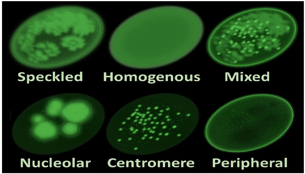
- Q6Image: centromere ANA patternL3 p.35, 40–43
A 58-year-old woman has had Raynaud phenomenon for 12 years and now has tight skin on her fingers, small calcified nodules on her fingertips and telangiectasias on her face. Her ANA shows many discrete, evenly sized dots scattered across each nucleus.
Lecture image
Which panel matches her ANA, and which antibody does it indicate?
- Q7Tolerance: FOXP3 and IPEXL3 p.34
A 3-month-old boy has insulin-requiring diabetes, chronic watery diarrhea with failure to thrive, and widespread eczema. His maternal uncle died in infancy with similar problems.
Which gene is most likely mutated, and which tolerance mechanism has failed?