Antigen presentation, T-cell help and antibodies
T cells see only peptide displayed on MHC. Where a protein came from decides which MHC shows it and which T cell answers; costimulation decides whether the answer is attack or tolerance; T-cell help decides which antibody class a B cell makes.
MHC class I and class II: two routes to the surface
L3 p.12–14A T cell cannot see a whole protein. It sees a short peptide held in the groove of an MHC molecule (in humans, an HLA molecule). The two classes sample two different places in the cell, and that is the whole logic of who gets killed and who gets helped.
Teaching schematic
MHC class I: what the cell is making
- Cytosolic protein (viral, tumor, self) tagged with ubiquitin
- Proteasome cuts it into peptides
- TAP pumps peptides into the ER
- Loaded on class I heavy chain + β₂-microglobulin
- Surface: read by CD8 cytotoxic T cells
On every nucleated cell and platelets, so any infected cell can be killed.
MHC class II: what the cell has taken in
- Class II made in the ER; invariant chain blocks the groove
- Travels to endosomes where engulfed protein is digested
- Invariant chain trimmed to CLIP
- HLA-DM swaps CLIP for a foreign peptide
- Surface: read by CD4 helper T cells
On antigen-presenting cells: dendritic cells, macrophages, B cells.
- MHC class I (HLA-A, -B, -C) is on all nucleated cells and platelets. Cytosolic proteins, including viral and mutant tumor proteins, are tagged with ubiquitin and cut by the proteasome. TAP (transporter associated with antigen processing) pumps the peptides into the endoplasmic reticulum, where they load onto the class I heavy chain paired with β₂-microglobulin. CD8 binds the class I molecule, so CD8 cytotoxic T cells read it: an infected cell announces itself and is killed.
- MHC class II (HLA-DP, -DQ, -DR) is on antigen-presenting cells: dendritic cells (including Langerhans cells of the skin), macrophages and B cells. It is assembled in the ER with the invariant chain blocking its groove, so it cannot pick up the cell's own ER peptides. In endosomes, where engulfed proteins are being digested, the invariant chain is trimmed to a fragment called CLIP, and HLA-DM exchanges CLIP for a foreign peptide. CD4 binds class II, so CD4 helper T cells read it and respond with cytokines.
HLA genes on chromosome 6
L3 p.12The MHC is a cluster of genes on the short arm of chromosome 6. Each person inherits one set of linked HLA genes (a haplotype) from each parent and expresses both, so HLA genes are codominant. Because the genes are so polymorphic, two unrelated people almost never match completely, and only identical twins share the whole set.
- Class I region: HLA-A, -B, -C.
- Class II region: HLA-DP, -DQ, -DR.
- Class III region: not antigen-presenting molecules but inflammatory proteins, including complement components C2, C4 and factor B, TNF and heat-shock proteins. Inherited C2 and C4 deficiencies therefore travel with HLA haplotypes, which partly explains their link with SLE (3.5, 3.7).
- β₂-microglobulin is encoded outside the MHC (chromosome 15). In long-term dialysis it is not cleared and can deposit as Aβ₂M amyloid (3.7).
Where HLA molecules are foundL3 p.12
| Class | Tissue distribution | Consequence |
|---|---|---|
| I (A, B, C) | Every nucleated cell and platelets; not mature red cells | Any infected or transplanted cell can be recognized by CD8 cells; platelets can be refractory to HLA-mismatched transfusion |
| II (DP, DQ, DR) | Dendritic and Langerhans cells, macrophages, B cells, thymic epithelium; induced on endothelium and other cells by IFN-γ | Only these cells start CD4 responses; donor dendritic cells in a graft drive early rejection |
I (A, B, C)
- Tissue distribution
- Every nucleated cell and platelets; not mature red cells
- Consequence
- Any infected or transplanted cell can be recognized by CD8 cells; platelets can be refractory to HLA-mismatched transfusion
II (DP, DQ, DR)
- Tissue distribution
- Dendritic and Langerhans cells, macrophages, B cells, thymic epithelium; induced on endothelium and other cells by IFN-γ
- Consequence
- Only these cells start CD4 responses; donor dendritic cells in a graft drive early rejection
Two signals: activation, anergy and class switching
L3 p.8, 11Recognizing peptide on MHC is signal 1. A naive T cell also needs signal 2, costimulation: B7 molecules (CD80/CD86) on the antigen-presenting cell binding CD28 on the T cell. APCs raise B7 only after their pattern receptors have sensed microbes or damage (3.1). So a T cell is activated when its antigen arrives together with a danger signal. Signal 1 without signal 2 makes the T cell anergic, unresponsive for good: one of the peripheral tolerance mechanisms in 3.5.
Teaching schematic
T-cell activation
- 1 + 2 → IL-2, proliferation, effector cells.
- 1 alone → anergy (peripheral tolerance). CTLA-4 binds B7 to switch the response off.
B-cell help and class switching
- BCR binds antigen; B cell presents its peptide on MHC II
- Helper T cell recognizes it: CD40L ↔ CD40
- Cytokine chooses the class: IFN-γ → IgG, IL-4 → IgE, TGF-β → IgA
- Germinal center → plasma cells and memory B cells
No CD40L → only IgM (hyper-IgM syndrome, 3.6).
B cells need help too. A B cell binds antigen with its receptor, internalizes it and presents its peptides on class II to a helper T cell that recognizes the same antigen. The T cell then supplies CD40 ligand (CD40L, CD154), which binds CD40 on the B cell, plus cytokines. Together these start germinal centers, affinity maturation and class (isotype) switching: the B cell keeps its antigen specificity but changes the heavy-chain constant region, and so the antibody's job.
Which cytokine chooses the classL3 p.8
| Cytokine (with CD40L) | Switch to | Typical setting |
|---|---|---|
| IFN-γ (Th1) | IgG (opsonizing, complement-fixing subclasses) | Bacteria and viruses; type II and III injury |
| IL-4, IL-13 (Th2) | IgE | Helminths; allergy (type I) |
| TGF-β (mucosa) | IgA | Gut and airway secretions, milk |
IFN-γ (Th1)
- Switch to
- IgG (opsonizing, complement-fixing subclasses)
- Typical setting
- Bacteria and viruses; type II and III injury
IL-4, IL-13 (Th2)
- Switch to
- IgE
- Typical setting
- Helminths; allergy (type I)
TGF-β (mucosa)
- Switch to
- IgA
- Typical setting
- Gut and airway secretions, milk
In the germinal center, the helped B cell proliferates, mutates its variable genes and is selected for higher affinity. It then leaves as one of two cell types: a plasma cell, a factory with abundant rough ER and a clock-face nucleus that secretes thousands of antibody molecules per second (many home to the bone marrow and live for years), or a memory B cell that waits for the next exposure. Plasma cells no longer carry surface immunoglobulin or class II, so they no longer present antigen. A malignant clone of them makes a monoclonal protein, and its free light chains are the source of AL amyloid (3.7).
Helper subsets: Th1, Th2, Th17 and Treg
L3 p.9–10A naive CD4 T cell becomes one of several kinds of helper, depending on the cytokines around it when it is activated. Each subset recruits a different effector, and each over-reaction gives a different disease. If you know which subset a disease uses, you know which cells you will see in the tissue.
Teaching schematic
Th1
- Induced by
- IL-12, IFN-γ
- Master gene
- T-bet
- Makes
- IFN-γ, IL-2
- Job
- Activate macrophages; help CD8; IgG
- Disease
- Type IV, granulomas · IL-12R defect → mycobacteria
Th2
- Induced by
- IL-4
- Master gene
- GATA3
- Makes
- IL-4, IL-5, IL-13
- Job
- IgE, eosinophils, mucus; helminths
- Disease
- Type I allergy, asthma
Th17
- Induced by
- TGF-β + IL-6 (IL-1, IL-23)
- Master gene
- RORγt
- Makes
- IL-17, IL-22
- Job
- Neutrophils, epithelial defense; bacteria, Candida
- Disease
- Psoriasis, IBD, RA · STAT3 (Job) → abscesses
Treg
- Induced by
- TGF-β + IL-2
- Master gene
- FOXP3 (CD25⁺)
- Makes
- IL-10, TGF-β, IL-35
- Job
- Suppress other T cells
- Disease
- FOXP3 → IPEX; CD25 → MS, T1DM
- Th1: induced by IL-12 from macrophages and dendritic cells (and IFN-γ). Makes IFN-γ and IL-2, which activate macrophages to kill intracellular microbes and help CD8 cells. Drives delayed hypersensitivity and granulomas (type IV). The lecture notes Th1 memory cells as a reservoir of HIV in latency.
- Th2: induced by IL-4. Makes IL-4 (switch to IgE), IL-5 (eosinophils) and IL-13 (IgE, mucus). Fights helminths; drives allergy and asthma (type I).
- Th17: induced by TGF-β with IL-6 (and IL-1, IL-23). Makes IL-17 and IL-22, which bring in neutrophils and stimulate epithelial defenses against extracellular bacteria and fungi. Overactivity drives psoriasis, IBD and rheumatoid arthritis; loss (STAT3 mutation, Job syndrome) gives staphylococcal abscesses and candidiasis (3.6).
- Treg: induced by TGF-β with IL-2; express FOXP3 and the IL-2 receptor α chain CD25; secrete IL-10, TGF-β and IL-35 and keep other T cells in check. FOXP3 mutation causes IPEX (3.5).
- CD8 cytotoxic T cells are activated by dendritic cells (which can show engulfed viral or tumor antigen on class I, cross-presentation) with IL-2 from CD4 helpers. Once armed, a CD8 cell kills any cell that shows the same peptide on class I: perforin opens pores, granzymes enter and activate caspases, and Fas ligand triggers the extrinsic pathway. The target dies by apoptosis, so virus is not spilled. The same killing destroys donor graft cells, β cells in type 1 diabetes and virus-infected hepatocytes in hepatitis B. See cytotoxic T-cell killing in Chapter 1.
Immunoglobulin classes and what each one does
L3 p.8, 21Every antibody has two identical light chains and two identical heavy chains. The variable ends bind antigen; the heavy-chain constant region decides what happens next: whether the antibody activates complement, binds phagocytes or mast cells, crosses the placenta or is secreted onto mucosa. Naive B cells start with surface IgM and IgD; class switching later swaps in another constant region.
Lecture slide
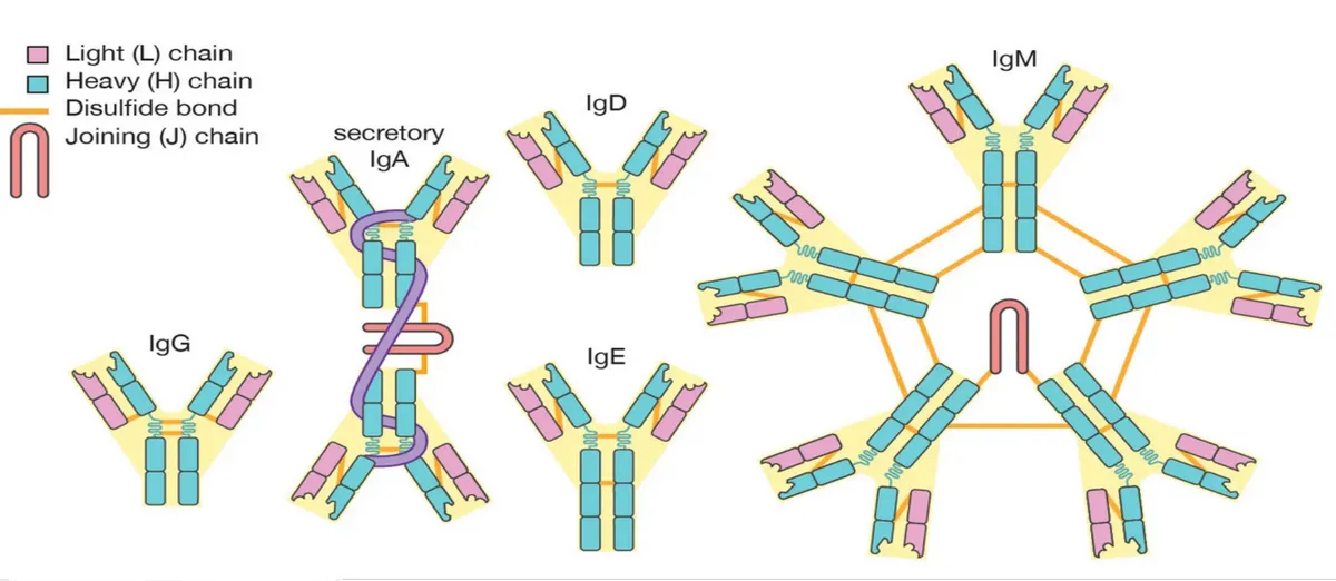
Function follows the constant regionL3 p.8, 20–21
| Class | Form | Key functions | Clinical hooks |
|---|---|---|---|
| IgM | Pentamer (surface monomer on naive B cells) | First antibody in a primary response; best complement activator; agglutinates | ABO isohemagglutinins (anti-A, anti-B) → intravascular hemolysis; cannot cross the placenta |
| IgG | Monomer; most abundant in serum | Secondary response; opsonizes (Fc receptors), fixes complement, neutralizes toxins, ADCC by NK cells; crosses the placenta | Newborn protection; HDN (anti-D); most type II and III injury |
| IgA | Dimer + secretory component in secretions | Mucosal neutralization; most abundant antibody produced daily; in milk | Selective IgA deficiency; IgA vasculitis and IgA nephropathy |
| IgE | Monomer bound to FcεRI on mast cells and basophils | Helminth defense with eosinophils; allergy | Type I hypersensitivity; skin-prick test |
| IgD | Monomer on naive B cells | Antigen receptor with IgM | — |
IgM
- Form
- Pentamer (surface monomer on naive B cells)
- Key functions
- First antibody in a primary response; best complement activator; agglutinates
- Clinical hooks
- ABO isohemagglutinins (anti-A, anti-B) → intravascular hemolysis; cannot cross the placenta
IgG
- Form
- Monomer; most abundant in serum
- Key functions
- Secondary response; opsonizes (Fc receptors), fixes complement, neutralizes toxins, ADCC by NK cells; crosses the placenta
- Clinical hooks
- Newborn protection; HDN (anti-D); most type II and III injury
IgA
- Form
- Dimer + secretory component in secretions
- Key functions
- Mucosal neutralization; most abundant antibody produced daily; in milk
- Clinical hooks
- Selective IgA deficiency; IgA vasculitis and IgA nephropathy
IgE
- Form
- Monomer bound to FcεRI on mast cells and basophils
- Key functions
- Helminth defense with eosinophils; allergy
- Clinical hooks
- Type I hypersensitivity; skin-prick test
IgD
- Form
- Monomer on naive B cells
- Key functions
- Antigen receptor with IgM
- Clinical hooks
- —
Bridge: the Th1–macrophage axis and the granuloma
L3 p.26, 45L2 p.42–43When a macrophage cannot destroy what it has eaten, typically Mycobacterium tuberculosis, it presents mycobacterial peptides on class II and secretes IL-12. CD4 cells become Th1 cells and release IFN-γ, which activates the macrophages; TNF holds the structure together. Activated macrophages become epithelioid cells and fuse into multinucleated giant cells, and a cuff of lymphocytes forms around them: a granuloma, the persistent form of type IV hypersensitivity. In tuberculosis its center undergoes caseous necrosis.
- Macrophage cannot kill mycobacteria
- IL-12 → naive CD4 becomes Th1
- IFN-γ activates macrophages
- Epithelioid cells, giant cells (TNF maintains)
- Granuloma walls off the organism
Knowledge check · 3.2
Answer, read why, and try again whenever you like. Answers are kept in both study modes.
- Q1Class II presentation in delayed hypersensitivityL3 p.12, 26Ch. III cases p.8
During a physical examination, purified protein derivative from Mycobacterium tuberculosis is injected into the skin of a 22-year-old man. Three days later the site is raised and indurated.
Which glycoprotein was directly involved in presenting antigen during the initiation phase of this delayed hypersensitivity reaction?
- Q2Class I pathway (TAP)L3 p.12, 14
A herpesvirus makes a protein that blocks TAP in the cells it infects.
What is the most likely consequence for the infected cells?
- Q3Class switching signalsL3 p.8, 11, 16
A B cell specific for a dust-mite protein presents it to a Th2 cell. Which combination of signals makes the B cell switch to producing IgE?
- Q4Immunoglobulin classesL3 p.21
Lecture slide
The largest antibody in the figure, a pentamer joined by a J chain, is the class that: