Hypersensitivity reactions
Hypersensitivity is a normal immune mechanism doing harm. Read every case in the same order: what triggered it, where the antigen sits, which effector did the damage, what injury resulted, and which clue in the case proves it.
One method for four types
L3 p.15The four Gell–Coombs types are remembered as ABCD: Anaphylactic and Atopic (type I), antiBody-mediated (type II), immune Complex (type III) and Delayed, cell-mediated (type IV). Types I–III are all made by antibody; type IV is made by T cells. The names are less useful than the questions behind them, so the chapter reads every case the same way.
Teaching schematic
Type I · Immediate
- Target
- Free antigen meeting IgE on mast cells
- Effector
- Histamine, tryptase, lipid mediators
- Injury
- Leak, bronchospasm, itch
- Clue
- Minutes; wheal; tryptase
Type II · Antibody
- Target
- Antigen fixed on a cell or matrix
- Effector
- IgG/IgM + complement, phagocytes, NK; or a receptor
- Injury
- Lysis, removal, inflammation, receptor on/off
- Clue
- Coombs+; linear IF
Type III · Immune complex
- Target
- Complexes formed in blood
- Effector
- Deposition, complement, neutrophils
- Injury
- Vasculitis, GN, arthritis
- Clue
- Granular IF; low C3/C4
Type IV · Delayed
- Target
- Peptide on MHC (no antibody)
- Effector
- Th1 → macrophages; CD8 killing
- Injury
- Induration, granuloma, cell death
- Clue
- 48–72 h; lymphocytes
Trigger → target → effector → injury → case clueL3 p.15–26
| Type | Target | Effector | Injury | Case clue |
|---|---|---|---|---|
| I | Free (soluble) antigen meeting IgE already on mast cells | Mast-cell and basophil mediators; later eosinophils | Leak, smooth-muscle spasm, mucus, itch | Minutes after re-exposure; wheal and flare; raised tryptase or specific IgE |
| II | Antigen fixed on a cell surface or in matrix | IgG/IgM with complement, phagocytes or NK cells; or the antibody alters receptor function | Lysis, removal of coated cells, local inflammation, or a receptor switched on or off | Positive direct Coombs; linear immunofluorescence; antireceptor antibody |
| III | Antigen–antibody complexes formed in blood (or locally) | Deposited complexes fix complement; C5a and Fc receptors bring neutrophils | Vasculitis, glomerulonephritis, arthritis wherever complexes lodge | Granular immunofluorescence; low C3/C4; days to weeks after the antigen |
| IV | Peptide on MHC recognized by sensitized T cells (no antibody) | Th1/Th17 cytokines activating macrophages; CD8 cells killing | Induration, granulomas, vesicular dermatitis, cell death | Peaks 48–72 h; lymphocytes and macrophages; positive patch or tuberculin test |
I
- Target
- Free (soluble) antigen meeting IgE already on mast cells
- Effector
- Mast-cell and basophil mediators; later eosinophils
- Injury
- Leak, smooth-muscle spasm, mucus, itch
- Case clue
- Minutes after re-exposure; wheal and flare; raised tryptase or specific IgE
II
- Target
- Antigen fixed on a cell surface or in matrix
- Effector
- IgG/IgM with complement, phagocytes or NK cells; or the antibody alters receptor function
- Injury
- Lysis, removal of coated cells, local inflammation, or a receptor switched on or off
- Case clue
- Positive direct Coombs; linear immunofluorescence; antireceptor antibody
III
- Target
- Antigen–antibody complexes formed in blood (or locally)
- Effector
- Deposited complexes fix complement; C5a and Fc receptors bring neutrophils
- Injury
- Vasculitis, glomerulonephritis, arthritis wherever complexes lodge
- Case clue
- Granular immunofluorescence; low C3/C4; days to weeks after the antigen
IV
- Target
- Peptide on MHC recognized by sensitized T cells (no antibody)
- Effector
- Th1/Th17 cytokines activating macrophages; CD8 cells killing
- Injury
- Induration, granulomas, vesicular dermatitis, cell death
- Case clue
- Peaks 48–72 h; lymphocytes and macrophages; positive patch or tuberculin test
Hypersensitivity case sorter
L3 p.15–27Ch. III cases p.3, 8Midterm sim. p.6–8Practise the method on whole cases. For each vignette, decide first where the antigen is and what does the damage; only then are you asked for the type. After a correct answer, one clue in the case changes. Decide again: the mechanism moves, and you see exactly which clue moved it.
Centerpiece · Hypersensitivity case sorter
Hypersensitivity case sorter
Solved 0 of 9
- 1. Target + effector
- 2. Type
- 3. One clue changes
- 4. Type again
The case
A 24-year-old woman with a sore throat takes amoxicillin; she had a full course of penicillin two years ago without problems. Within 15 minutes of the first tablet she has generalized hives, wheezing and a blood pressure of 80/50 mm Hg; serum tryptase is raised.
What caused the reaction?
Step 1 · Where is the antigen, and what does the damage?
Choose one target and one effector, then check.
Type I: IgE and mast cells
L3 p.16–19Type I hypersensitivity needs two meetings with the allergen. The first meeting sensitizes; the second elicits the reaction. Atopy, the inherited tendency to make IgE to harmless environmental antigens, runs strongly in families.
- Sensitization. Dendritic cells carry the allergen (pollen, dust mite, food, drug) to lymph nodes. With IL-4 in the environment, naive CD4 cells become Th2 cells that make IL-4, IL-5 and IL-13. IL-4 (with CD40L) switches allergen-specific B cells to IgE; IL-5 raises and activates eosinophils. IgE binds the high-affinity receptor FcεRI on mast cells and basophils and waits there.
- Re-exposure, early phase (minutes). The allergen cross-links two IgE molecules on the mast cell. The cell degranulates, releasing preformed histamine (vasodilation, leak, bronchoconstriction, itch), tryptase (a marker of mast-cell activation) and eosinophil chemotactic factor, and within minutes it makes leukotrienes C4/D4/E4, prostaglandin D2 and platelet-activating factor from membrane lipids.
- Late phase (about 2–24 hours). Mast-cell cytokines (TNF, IL-4, IL-5, IL-13) and leukotrienes recruit eosinophils, neutrophils, basophils and Th2 cells. This cellular infiltrate prolongs swelling and, in the airway, causes the sustained obstruction and hyperreactivity of asthma.
Early and late phase comparedL3 p.16–17
| Early (immediate) phase | Late phase | |
|---|---|---|
| Timing | Within minutes; fades in about an hour | Hours later (about 2–24 h), lasting a day or more |
| Main mediators | Histamine, tryptase, then leukotrienes, PGD₂, PAF | Cytokines and lipid mediators recruiting eosinophils, neutrophils, Th2 cells |
| What you see | Wheal and flare, sneezing, bronchospasm, hypotension | Persistent swelling, airway inflammation, mucus |
| Treatment logic | Epinephrine, antihistamines | Corticosteroids, leukotriene antagonists |
Timing
- Early (immediate) phase
- Within minutes; fades in about an hour
- Late phase
- Hours later (about 2–24 h), lasting a day or more
Main mediators
- Early (immediate) phase
- Histamine, tryptase, then leukotrienes, PGD₂, PAF
- Late phase
- Cytokines and lipid mediators recruiting eosinophils, neutrophils, Th2 cells
What you see
- Early (immediate) phase
- Wheal and flare, sneezing, bronchospasm, hypotension
- Late phase
- Persistent swelling, airway inflammation, mucus
Treatment logic
- Early (immediate) phase
- Epinephrine, antihistamines
- Late phase
- Corticosteroids, leukotriene antagonists
Lecture image
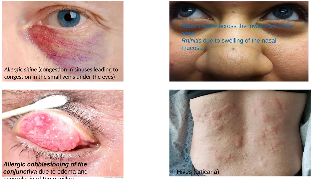
The same mechanism looks different depending on where the allergen enters: the nose (allergic rhinitis), the conjunctiva, the skin (urticaria, angioedema), the bronchi (allergic asthma), the gut (food allergy) or the bloodstream, where mediator release is systemic. Anaphylaxis is the systemic form: vasodilation and leak cause shock, laryngeal edema and bronchospasm threaten the airway, and only intramuscular epinephrine reverses all three quickly.
Lecture image
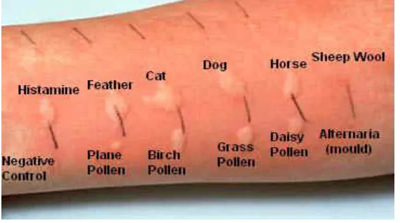
- Skin-prick (scratch) test: the standard first test; reads IgE on skin mast cells. Antihistamines must be stopped beforehand.
- Allergen-specific serum IgE (the older RAST, now immunoassays): measures IgE against a named allergen without exposing the patient. Useful with extensive eczema or when antihistamines cannot be stopped.
- Total serum IgE: raised in atopy, helminth infection and hyper-IgE syndrome; not specific for one allergen.
- Serum tryptase: rises within about an hour of anaphylaxis; confirms mast-cell activation after the event.
Core activity · The Missing Arrow
The Missing Arrow
A child inhales grass pollen in spring for the first time, and again the next spring. Complete the chain to the itchy wheal on her skin-prick test.
One gap asks for the event that sensitizes the mast cell; another asks for the mechanism that sets it off on re-exposure.
- Dendritic cells present pollen peptide to naive CD4 T cells
- B cells switch to IgE against pollen
- IgE circulates and binds the high-affinity Fc receptor
- histamine dilates venules and opens endothelial junctions
- Wheal and flare within 15–20 minutes; late-phase eosinophils hours later
Cards
Tap or drag a card. Placing a used card moves it.
Type II: antibody against a fixed antigen
L3 p.20–22In type II reactions, IgG or IgM binds an antigen that is part of a cell surface or of extracellular matrix: a red-cell antigen, a platelet glycoprotein, collagen in a basement membrane, a hormone receptor. The antigen may be self (autoimmunity), foreign (a transfused red cell), or a drug sitting on a cell as a hapten. What happens next depends on the effector the antibody recruits.
Four ways an antibody on a cell causes diseaseL3 p.20, 22
| Mechanism | How it works | Examples |
|---|---|---|
| Complement lysis | IgM (or IgG) fixes C1q; the membrane attack complex punctures the cell inside the vessel | ABO-incompatible transfusion (IgM anti-A/anti-B); cold autoimmune hemolysis |
| Opsonization and phagocytosis | IgG and C3b coat the cell; splenic and hepatic macrophages with Fc and C3b receptors eat it (extravascular) | Warm autoimmune hemolytic anemia; immune thrombocytopenia (ITP); hemolytic disease of the newborn (maternal IgG anti-D); penicillin bound to red cells |
| Inflammation and ADCC | Antibody on matrix or a cell activates complement (C5a draws neutrophils) or engages Fc receptors on neutrophils, macrophages and NK cells, which release enzymes and oxygen radicals or kill the coated cell | Goodpasture syndrome (type IV collagen of glomerular and alveolar basement membranes); acute rheumatic fever; pemphigus vulgaris; hyperacute rejection; NK killing of IgG-coated cells |
| Receptor dysfunction without injury | Antibody stimulates or blocks a receptor; the cell is not destroyed | Graves (stimulates the TSH receptor → hyperthyroidism); myasthenia gravis (blocks and removes the acetylcholine receptor → weakness); pernicious anemia (anti–intrinsic factor and anti-parietal-cell antibodies → B12 deficiency) |
Complement lysis
- How it works
- IgM (or IgG) fixes C1q; the membrane attack complex punctures the cell inside the vessel
- Examples
- ABO-incompatible transfusion (IgM anti-A/anti-B); cold autoimmune hemolysis
Opsonization and phagocytosis
- How it works
- IgG and C3b coat the cell; splenic and hepatic macrophages with Fc and C3b receptors eat it (extravascular)
- Examples
- Warm autoimmune hemolytic anemia; immune thrombocytopenia (ITP); hemolytic disease of the newborn (maternal IgG anti-D); penicillin bound to red cells
Inflammation and ADCC
- How it works
- Antibody on matrix or a cell activates complement (C5a draws neutrophils) or engages Fc receptors on neutrophils, macrophages and NK cells, which release enzymes and oxygen radicals or kill the coated cell
- Examples
- Goodpasture syndrome (type IV collagen of glomerular and alveolar basement membranes); acute rheumatic fever; pemphigus vulgaris; hyperacute rejection; NK killing of IgG-coated cells
Receptor dysfunction without injury
- How it works
- Antibody stimulates or blocks a receptor; the cell is not destroyed
- Examples
- Graves (stimulates the TSH receptor → hyperthyroidism); myasthenia gravis (blocks and removes the acetylcholine receptor → weakness); pernicious anemia (anti–intrinsic factor and anti-parietal-cell antibodies → B12 deficiency)
The lecture adds two antibody-dependent killing mechanisms that do not need complement: NK cells kill IgG-coated virus-infected or tumor cells through their Fc receptors (antibody-dependent cell-mediated cytotoxicity), and eosinophils bind IgE on a tissue helminth and release major basic protein onto it.
Receptor antibodies: stimulate or block
Graves disease
- IgG against the TSH receptor acts like TSH
- Thyroid cells make excess T3/T4 and the gland grows
- TSH falls (negative feedback) while the gland keeps working
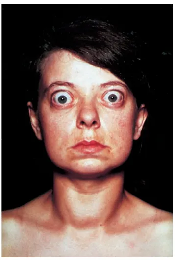
- Diffuse warm goiter, exophthalmos, pretibial myxedema
Myasthenia gravis
- IgG against the acetylcholine receptor blocks it, cross-links it for removal and fixes complement at the end plate
- Fewer receptors → smaller end-plate potentials
- Fatigable weakness: ptosis, diplopia, worse at day's end
- Associated with thymic hyperplasia or thymoma
Case image
Teaching schematic
Direct Coombs (direct antiglobulin test)
Asks: is antibody or C3d already on the patient's red cells? Autoimmune hemolysis, a newborn with HDN, a hemolytic transfusion reaction, drug-induced hemolysis.
Indirect Coombs
Asks: does the patient's serum contain antibody against red-cell antigens? Antenatal anti-D screening, antibody screen and crossmatch before transfusion.
Type III: immune complexes
L3 p.23–25, 39Antigen–antibody complexes form whenever antibody meets soluble antigen, and the mononuclear phagocyte system normally clears them. Complexes become harmful when they are made faster than they are cleared, which usually means a persistent antigen (a chronic infection, a drug, nuclear antigens in SLE) and complexes of small to intermediate size, formed in slight antigen excess. These escape clearance and lodge where blood is filtered under pressure: glomeruli, synovium, small vessels of the skin.
From complex to injury
- Persistent soluble antigen + IgG
- Complexes circulate
- Deposit in glomeruli, joints, small vessels
- Classical complement activated (C3, C4 consumed)
- C5a and Fc receptors recruit neutrophils
- Enzymes and radicals: vasculitis, glomerulonephritis, arthritis
Because the injury is made by complement and neutrophils, three findings follow. Serum C3 and C4 fall while complement is consumed. Immunofluorescence shows IgG and C3 as granular, lumpy deposits wherever complexes settled. And small vessels may show fibrinoid necrosis, the bright pink smudge of plasma proteins and fibrin in a damaged wall (fibrinoid necrosis).
Systemic versus local: serum sickness and the Arthus reaction
Serum sickness (systemic)
- Foreign serum or protein (antivenom, antithymocyte globulin, monoclonal antibodies), drugs (penicillin), chronic antigen (HBsAg)
- Antibody appears after about a week; complexes form as it meets remaining antigen
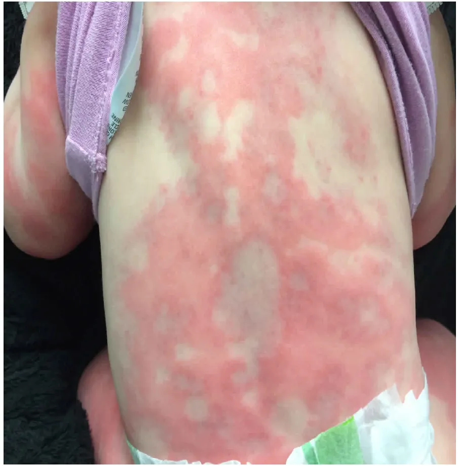
- 1–2 weeks after exposure: fever, urticarial rash, arthralgia, painful lymphadenopathy, splenomegaly, proteinuria; low complement
- Resolves when the antigen is cleared
Arthus reaction (local)
- Antigen injected into the skin of someone who already has a high level of IgG against it (for example an extra tetanus booster)
- Complexes form in the wall of small vessels at the injection site
- Hours (about 4–10 h): painful swelling, hemorrhage, then necrosis and ulceration
- Neutrophils, fibrinoid necrosis and thrombosis in local vessels
Lecture image
Immune-complex diseases in this chapterL3 p.23–24, 36, 39
| Disease | Antigen | Where complexes lodge | Clues |
|---|---|---|---|
| Poststreptococcal glomerulonephritis | Nephritogenic group A streptococcal antigens | Glomerular capillary wall: subepithelial humps on EM | Child, 1–3 weeks after pharyngitis; cola urine, periorbital edema, hypertension; low C3; raised ASO/anti-DNase B; granular IgG + C3 |
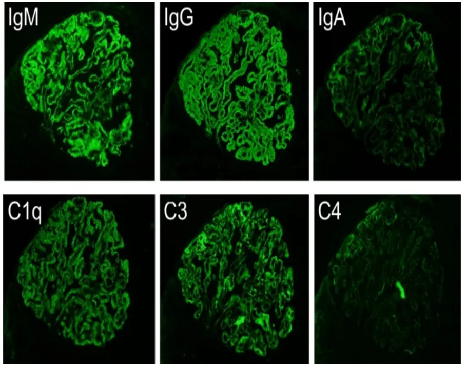
| Lupus nephritis | DNA and nucleosomes (anti-dsDNA) | Glomeruli (mesangial, subendothelial, subepithelial), skin, joints, serosa | Granular 'full-house' IF (IgG, IgA, IgM, C1q, C3); low C3 and C4; anti-dsDNA (3.5) |
| IgA vasculitis (Henoch–Schönlein purpura) | IgA-containing complexes | Small vessels of skin, gut, glomerular mesangium | Child after an upper respiratory infection; palpable purpura on buttocks and legs with normal platelets; arthralgia, colicky abdominal pain, hematuria |
| Polyarteritis nodosa (HBV-associated) | Hepatitis B surface antigen | Medium-sized muscular arteries | Segmental fibrinoid necrosis, aneurysms, renal infarcts, mononeuritis; ANCA-negative; HBsAg positive |
| Serum sickness; Arthus reaction | Foreign protein, drug; injected antigen | Systemic small vessels and glomeruli; local dermal vessels | Timing (1–2 weeks vs hours) |
| Rheumatoid arthritis, reactive arthritis (as listed in the lecture) | Rheumatoid factor (IgM anti-IgG) complexes; bacterial antigens | Synovium | RA also has a large T-cell (type IV) component |
Poststreptococcal glomerulonephritis
- Antigen
- Nephritogenic group A streptococcal antigens
- Where complexes lodge
- Glomerular capillary wall: subepithelial humps on EM
- Clues
- Child, 1–3 weeks after pharyngitis; cola urine, periorbital edema, hypertension; low C3; raised ASO/anti-DNase B; granular IgG + C3
Lupus nephritis
- Antigen
- DNA and nucleosomes (anti-dsDNA)
- Where complexes lodge
- Glomeruli (mesangial, subendothelial, subepithelial), skin, joints, serosa
- Clues
- Granular 'full-house' IF (IgG, IgA, IgM, C1q, C3); low C3 and C4; anti-dsDNA (3.5)
IgA vasculitis (Henoch–Schönlein purpura)
- Antigen
- IgA-containing complexes
- Where complexes lodge
- Small vessels of skin, gut, glomerular mesangium
- Clues
- Child after an upper respiratory infection; palpable purpura on buttocks and legs with normal platelets; arthralgia, colicky abdominal pain, hematuria
Polyarteritis nodosa (HBV-associated)
- Antigen
- Hepatitis B surface antigen
- Where complexes lodge
- Medium-sized muscular arteries
- Clues
- Segmental fibrinoid necrosis, aneurysms, renal infarcts, mononeuritis; ANCA-negative; HBsAg positive
Serum sickness; Arthus reaction
- Antigen
- Foreign protein, drug; injected antigen
- Where complexes lodge
- Systemic small vessels and glomeruli; local dermal vessels
- Clues
- Timing (1–2 weeks vs hours)
Rheumatoid arthritis, reactive arthritis (as listed in the lecture)
- Antigen
- Rheumatoid factor (IgM anti-IgG) complexes; bacterial antigens
- Where complexes lodge
- Synovium
- Clues
- RA also has a large T-cell (type IV) component
Lecture image
Core activity · Almost Twins
Almost Twins
Look-alike A
Anti-GBM (Goodpasture) disease
- Hematuria, red-cell casts, rising creatinine
- Smooth linear IgG along the basement membrane; antibody against type IV collagen fixed in the GBM
- Serum C3 usually normal
- May bleed into the lungs (alveolar basement membrane shares the antigen)
Look-alike A
Anti-GBM (Goodpasture) disease
- Hematuria, red-cell casts, rising creatinine
- Smooth linear IgG along the basement membrane; antibody against type IV collagen fixed in the GBM
- Serum C3 usually normal
- May bleed into the lungs (alveolar basement membrane shares the antigen)
Look-alike B
Poststreptococcal glomerulonephritis
- Hematuria, red-cell casts, edema, hypertension
- Granular IgG and C3 with subepithelial humps: complexes of streptococcal antigen and antibody
- Serum C3 low
- Child, 1–3 weeks after a sore throat; raised ASO
Step 1 · What they share
What do the two diseases share?
Select every feature that both show.
Core activity · The Missing Arrow
The Missing Arrow
A man with chronic hepatitis B makes antibody against the surface antigen that his liver keeps releasing. Complete the chain to the necrotizing arteritis.
- HBsAg circulates for months; IgG anti-HBs is made
- antigen and antibody combine in slight antigen excess
- Fibrinoid necrosis of medium-sized arteries: polyarteritis nodosa
Cards
Tap or drag a card. Placing a used card moves it.
Necrotizing vasculitis: ANCA or immune complex?
L3 p.23–24Midterm sim. p.6–7A biopsy that shows fibrinoid necrosis of a small artery tells you the vessel wall has been destroyed and plasma proteins have leaked into it. It does not tell you how. Two antibody mechanisms produce the same picture, and they are separated by the immunofluorescence, the complement level and the serology.
Case image
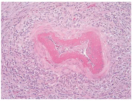
Teaching schematic
ANCA-associated (pauci-immune)
- IgG autoantibodies against neutrophil granule enzymes: myeloperoxidase (p-ANCA) or proteinase 3 (c-ANCA)
- Cytokines (for example during an infection) prime neutrophils, which move MPO or PR3 to their surface; ANCA bind them and engage Fc receptors
- Activated neutrophils degranulate and release radicals at the endothelium: necrotizing small-vessel vasculitis, crescentic glomerulonephritis
- IF: few or no deposits; serum complement usually normal
- p-ANCA: microscopic polyangiitis, eosinophilic granulomatosis with polyangiitis
Immune-complex
- Antibody meets a soluble antigen (HBsAg, streptococcal antigen, DNA) and complexes deposit in the wall
- Complement is consumed; C5a and Fc receptors bring neutrophils
- IF: granular IgG and C3; serum C3 and C4 low
- Classic polyarteritis nodosa with HBV (ANCA-negative), lupus vasculitis, cryoglobulinemia, IgA vasculitis
Core activity · One Fact Changes
One Fact Changes
1 · The original case
A 50-year-old man has fever, weight loss, abdominal pain, bloody urine and red-purple palpable skin lesions. Skin biopsy shows fibrinoid necrosis of small arteries with neutrophils. p-ANCA (anti-myeloperoxidase) is strongly positive; immunofluorescence shows almost no immunoglobulin or complement; serum C3 and C4 are normal.
Which mechanism best explains the vasculitis?
Which fact decided it?
Type IV: T cells, delayed
L3 p.26–27Type IV reactions involve no antibody. Sensitized T cells recognize peptide on MHC in the tissue and cause injury in one of two ways, and both take time because the T cells must be recruited and must make new proteins. Reactions therefore peak at 48–72 hours.
CD4 T-cell inflammation (delayed-type hypersensitivity)
- Antigen on class II → memory Th1 (and Th17) cells
- IFN-γ activates macrophages; TNF and chemokines bring more cells
- Induration; with persistent antigen, granulomas
- Tuberculin (PPD) reaction, tuberculosis, contact dermatitis
CD8 T-cell cytotoxicity
- Antigen on class I of altered cells: virus-infected, tumor, donor graft
- Perforin and granzymes, Fas ligand → apoptosis of the target
- Tissue cells die without antibody
- Viral hepatitis, type 1 diabetes, acute cellular rejection, graft-versus-host disease
Allergic contact dermatitis shows both stages clearly. In the sensitization stage, a small reactive molecule (a hapten: urushiol from poison ivy, nickel, rubber-glove chemicals, formaldehyde, topical sulfonamides) binds skin proteins. Langerhans cells carry the modified protein to regional lymph nodes and present it, creating CD4 Th1 memory cells and CD8 effector memory cells. In the elicitation stage, re-exposure brings the hapten back; Langerhans cells present it to the circulating memory cells, which release cytokines or kill keratinocytes. Itching, redness, edema and vesicles of clear fluid appear exactly where the substance touched, 24–72 hours later.
Lecture image
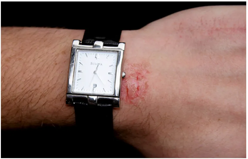
- Tuberculin (PPD) test: protein injected into the dermis; previously sensitized Th1 cells produce a firm induration read at 48–72 hours. The Candida skin test uses the same principle to check whether T-cell immunity works at all.
- Tuberculosis: persistent mycobacterial antigen turns the same response into caseating granulomas (3.2).
- Other delayed examples in the lecture: drug reaction with eosinophilia and systemic symptoms (DRESS) and graft-versus-host disease (3.4).
- Diminished cell-mediated immunity makes the skin test smaller or negative (anergy): older adults, and people with AIDS whose CD4 cells are lost.
Knowledge check · 3.3
Answer, read why, and try again whenever you like. Answers are kept in both study modes.
- Q1Poststreptococcal GN: type IIIL3 p.23Ch. III cases p.3
An 8-year-old boy has periorbital edema and throbbing headaches. His parents report a 'strep throat' 2 weeks ago. Urinalysis shows 3+ hematuria. A renal biopsy shows hypercellular glomeruli, and electron microscopy shows subepithelial 'humps'.
Which best explains the pathogenesis of the glomerulonephritis?
- Q2Tuberculous granuloma: type IVL3 p.26Ch. III cases p.4L2 p.42–43
A 34-year-old woman has had fever, productive cough with blood-tinged sputum and 4 kg of weight loss for 6 weeks. Cervical lymph nodes are enlarged. Chest x-ray shows a 2.5-cm nodule in the right upper lobe; biopsy shows caseating granulomas with multinucleated giant cells.
Which mechanism underlies the pulmonary nodule?
- Q3Contact dermatitis: type IVL3 p.26–27Midterm sim. p.6
A 20-year-old gardener has what he calls 'poison ivy'. His hands and forearms are red and covered with oozing blisters and crusts, two days after clearing weeds.
Which best describes the pathogenesis of these lesions?
- Q4ANCA vs immune-complex vasculitisMidterm sim. p.6–7L3 p.23–24Ref.
A 50-year-old man has fever, weight loss, abdominal pain and bloody urine, with red-purple palpable lesions on his legs. A skin biopsy shows fibrinoid necrosis of small arteries with neutrophils. p-ANCA (anti-myeloperoxidase) is strongly positive. Immunofluorescence of the biopsy shows little or no immunoglobulin or complement, and serum C3 and C4 are normal.
Which immune mechanism best explains the necrotizing vasculitis?
- Q5Graves: stimulating antireceptor antibodyL3 p.20, 22Midterm sim. p.7
A 53-year-old woman (shown) has weight loss, nervousness, sweating, tachycardia and exophthalmos. Her thyroid is diffusely enlarged and warm. TSH is low and T3 and T4 are markedly raised.
Case image
Which mechanism best explains her thyroid disease?
- Q6ABO transfusion reaction: type IIL3 p.20Midterm sim. p.8
A 54-year-old woman needs a transfusion after a car accident. Five hours later she is febrile with severe back pain, and tests show intravascular hemolysis. Type A Rh⁺ blood had been given by mistake to this type B Rh⁺ patient.
Which best explains the intravascular hemolysis?
- Q7Image: granular full-house IFL3 p.39
A 27-year-old woman has hematuria, red-cell casts and proteinuria. Her kidney biopsy immunofluorescence is shown: each panel is the same glomerulus stained for a different protein.
Lecture image
Which serum finding best fits this pattern, and why?