HIV, complement disorders and amyloidosis
Three ways the immune system fails beyond the inherited lymphocyte defects: a virus that destroys the CD4 cells everything else depends on; missing complement components or regulators; and chronic immune activity that leaves misfolded protein in the tissues.
HIV: genes, transmission, entry and drug targets
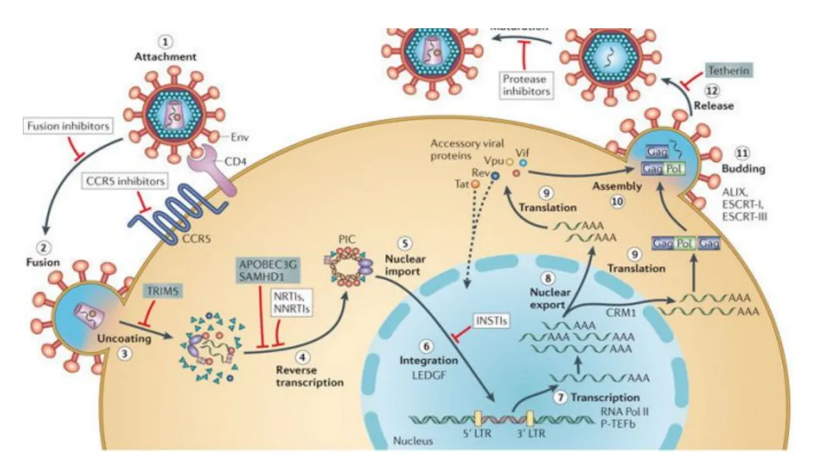
L3 p.48–50HIV is a retrovirus: an enveloped particle carrying two copies of single-stranded RNA plus the enzymes that will copy it into DNA and insert it into the host genome. Three structural genes build it:
| Gene | Proteins | Role | Clinical use |
|---|---|---|---|
| env | gp120 (surface) and gp41 (transmembrane), cut from gp160 | gp120 attaches to CD4 and the coreceptor; gp41 fuses the membranes | Antibodies to gp120/gp41 in screening tests; fusion and attachment inhibitors |
| gag | p24 (capsid), p17 (matrix), nucleocapsid | Core structure | p24 antigen appears early, before antibodies |
| pol | Reverse transcriptase, integrase, protease | Copy RNA to DNA; insert it; cut polyproteins during maturation | Targets of most antiretroviral drugs |
env
- Proteins
- gp120 (surface) and gp41 (transmembrane), cut from gp160
- Role
- gp120 attaches to CD4 and the coreceptor; gp41 fuses the membranes
- Clinical use
- Antibodies to gp120/gp41 in screening tests; fusion and attachment inhibitors
gag
- Proteins
- p24 (capsid), p17 (matrix), nucleocapsid
- Role
- Core structure
- Clinical use
- p24 antigen appears early, before antibodies
pol
- Proteins
- Reverse transcriptase, integrase, protease
- Role
- Copy RNA to DNA; insert it; cut polyproteins during maturation
- Clinical use
- Targets of most antiretroviral drugs
Virus is present in blood, semen, vaginal secretions and breast milk, so it spreads by sexual contact, shared needles and transfusion, and from mother to child during pregnancy, delivery or breastfeeding.
- Attachment. gp120 binds CD4 on helper T cells, macrophages and dendritic cells.
- Coreceptor binding. gp120 then binds a chemokine receptor: CCR5 (on macrophages and memory T cells; the strains that transmit infection) or CXCR4 (on T cells; strains that often emerge late and predict faster decline). People homozygous for a CCR5 deletion are largely resistant; heterozygotes progress more slowly.
- Fusion. gp41 unfolds, inserts into the host membrane and pulls the two membranes together; the core enters the cytoplasm.
- Reverse transcription. RT copies the RNA into double-stranded DNA (error-prone, so resistance mutations arise quickly).
- Integration. Integrase inserts the DNA into the host genome as a provirus, which can stay silent (latency).
- Transcription. When the cell is activated (for example by another infection), host NF-κB switches on the provirus along with the cell's own cytokine genes.
- Assembly, budding and maturation. New cores assemble at the membrane and bud off; viral protease then cuts the Gag and Gag-Pol polyproteins so the particle becomes infectious.
Teaching schematic
1Attachment
gp120 binds CD4
⊣ Attachment inhibitor (binds gp120); anti-CD4 antibody
2Coreceptor
gp120 binds CCR5 or CXCR4
⊣ CCR5 antagonist (maraviroc)
3Fusion
gp41 joins the membranes
⊣ Fusion inhibitor (enfuvirtide)
4Reverse transcription
RNA → dsDNA (RT)
⊣ NRTIs, NNRTIs
5Integration
Integrase inserts provirus
⊣ Integrase inhibitors
6Transcription
Host NF-κB activates the provirus
7Assembly and budding
Gag, Gag-Pol, RNA at membrane
8Maturation
Protease cuts Gag and Gag-Pol
⊣ Protease inhibitors
Lecture slide
Why CD4 cells disappear, and where the virus hides
L3 p.49Losing CD4 helper cells removes the conductor of almost every adaptive response: B cells lose help, CD8 cells lose IL-2, and macrophages lose IFN-γ activation. Several processes kill CD4 cells:
- Direct cytopathic effect: productive infection, with budding and accumulation of viral products, kills the cell (the lecture: HIV is cytotoxic to CD4 T cells).
- Pyroptosis of bystander cells: in lymphoid tissue many resting CD4 cells are infected abortively; incomplete viral DNA is sensed in the cytosol and caspase-1 kills the cell in an inflammatory way (3.1). This explains why far more cells die than are productively infected.
- CD8 killing of infected cells, and chronic immune activation that drives activated cells to die.
- Loss of replacement: infection of thymic and lymph-node structures reduces production. In acute infection a large share of memory CD4 cells in the gut mucosa is lost within weeks.
Reservoirs keep the virus out of reach of drugs and immunity. Macrophages and dendritic cells contain large numbers of particles but resist the cytolytic effect, carry virus to lymph nodes, spleen and brain, and keep producing it. Latently infected resting memory CD4 cells carry silent provirus for years; treatment suppresses replication but does not eliminate them, which is why stopping therapy lets the virus return.
Core activity · The Missing Arrow
The Missing Arrow
Follow HIV from the first contact with a CD4 T cell to the infections of AIDS.
Two gaps ask for a mechanism: how the virus gets in, and how cells die. Don't put an event there.
- gp120 binds CD4 on a helper T cell
- CD4 count falls over years; viral load rises
- B cells, CD8 cells and macrophages lose their help (IL-2, IFN-γ, CD40L)
Cards
Tap or drag a card. Placing a used card moves it.
Stages, tests, opportunistic infections and tumors
L3 p.51–52Teaching schematic
Stages of untreated infectionL3 p.51
| Stage | CD4 count (cells/mm³) | What happens |
|---|---|---|
| Acute HIV | Transient dip | 1–6 weeks (usually 2–3) after infection: fever, malaise, pharyngitis, generalized rash, myalgia and arthralgia, painful lymphadenopathy; resolves in 1–2 weeks. Viral load very high; antibody tests may still be negative |
| Asymptomatic (clinical latency) | > 500 | 2–10 years. Replication continues in lymphoid tissue; CD4 count falls steadily |
| Early symptomatic | 200–500 | Generalized lymphadenopathy; non–AIDS-defining infections: oral candidiasis (thrush), oral hairy leukoplakia (EBV), zoster |
| AIDS | ≤ 200, or an AIDS-defining condition | Opportunistic infections and AIDS-defining tumors |
Acute HIV
- CD4 count (cells/mm³)
- Transient dip
- What happens
- 1–6 weeks (usually 2–3) after infection: fever, malaise, pharyngitis, generalized rash, myalgia and arthralgia, painful lymphadenopathy; resolves in 1–2 weeks. Viral load very high; antibody tests may still be negative
Asymptomatic (clinical latency)
- CD4 count (cells/mm³)
- > 500
- What happens
- 2–10 years. Replication continues in lymphoid tissue; CD4 count falls steadily
Early symptomatic
- CD4 count (cells/mm³)
- 200–500
- What happens
- Generalized lymphadenopathy; non–AIDS-defining infections: oral candidiasis (thrush), oral hairy leukoplakia (EBV), zoster
AIDS
- CD4 count (cells/mm³)
- ≤ 200, or an AIDS-defining condition
- What happens
- Opportunistic infections and AIDS-defining tumors
What each laboratory measure tells youL3 p.51
| Test | Measures | Used for |
|---|---|---|
| Antigen/antibody immunoassay (4th generation) | p24 antigen plus antibodies to HIV (including anti-gp120, anti-gp41) | Screening; positive about 2–3 weeks after infection |
| HIV-1/HIV-2 differentiation immunoassay; nucleic-acid test (NAT) | Antibody type; viral RNA | Confirmation. NAT is positive earliest (about 10 days) and settles indeterminate results |
| Viral load (HIV RNA) | How much virus is replicating | Diagnosis of acute infection, response to therapy, transmission risk |
| CD4 count | How much immune capacity remains | Staging, risk of opportunistic infection, when to start prophylaxis |
Antigen/antibody immunoassay (4th generation)
- Measures
- p24 antigen plus antibodies to HIV (including anti-gp120, anti-gp41)
- Used for
- Screening; positive about 2–3 weeks after infection
HIV-1/HIV-2 differentiation immunoassay; nucleic-acid test (NAT)
- Measures
- Antibody type; viral RNA
- Used for
- Confirmation. NAT is positive earliest (about 10 days) and settles indeterminate results
Viral load (HIV RNA)
- Measures
- How much virus is replicating
- Used for
- Diagnosis of acute infection, response to therapy, transmission risk
CD4 count
- Measures
- How much immune capacity remains
- Used for
- Staging, risk of opportunistic infection, when to start prophylaxis
Opportunistic infections and tumors by CD4 countL3 p.51–52
| CD4 count | Infections | Tumors |
|---|---|---|
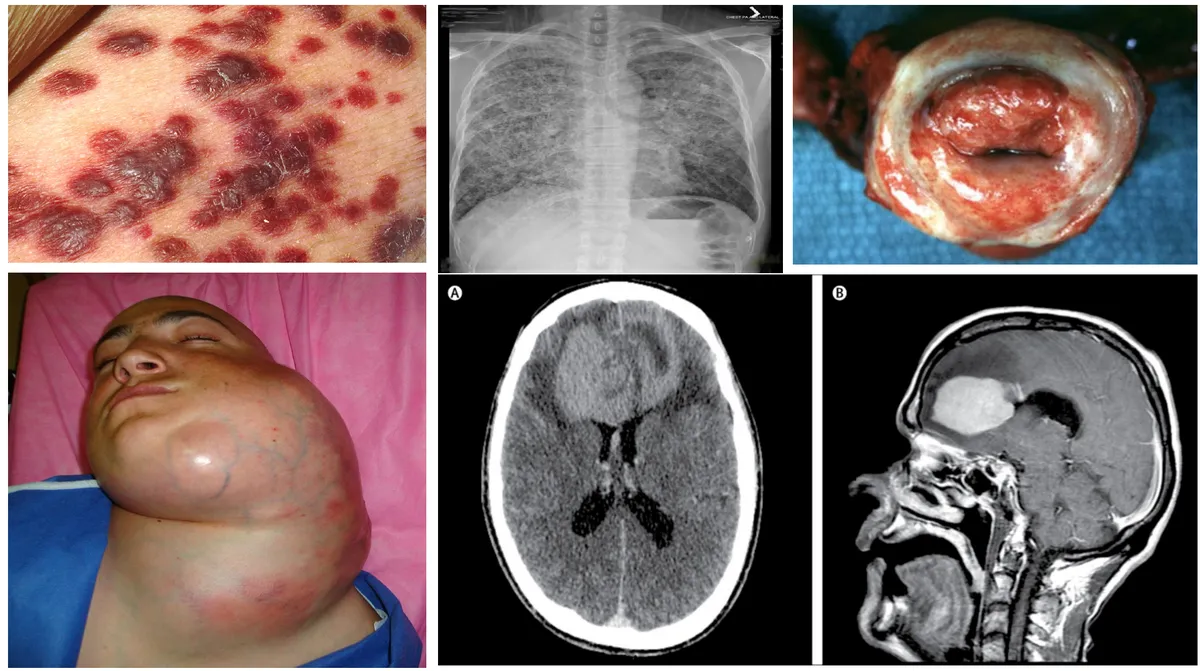
| Any count (risk rises as CD4 falls) | Tuberculosis, bacterial pneumonia | Kaposi sarcoma (HHV-8) |
| < 500 | Oral thrush, oral hairy leukoplakia (EBV), zoster: oral and skin findings, not AIDS-defining | — |
| < 200 | Pneumocystis jirovecii pneumonia** (AIDS-defining) | Invasive cervical carcinoma (HPV), aggressive B-cell lymphomas such as Burkitt (EBV) |
| < 100 | Toxoplasma encephalitis, cryptococcal meningitis, esophageal or systemic candidiasis (AIDS-defining) | — |
| < 50 | Disseminated CMV (retinitis, colitis), disseminated **Mycobacterium avium complex**: common causes of death | Primary CNS lymphoma (EBV) |
Any count (risk rises as CD4 falls)
- Infections
- Tuberculosis, bacterial pneumonia
- Tumors
- Kaposi sarcoma (HHV-8)
< 500
- Infections
- Oral thrush, oral hairy leukoplakia (EBV), zoster: oral and skin findings, not AIDS-defining
- Tumors
- —
< 200
- Infections
- Pneumocystis jirovecii pneumonia** (AIDS-defining)
- Tumors
- Invasive cervical carcinoma (HPV), aggressive B-cell lymphomas such as Burkitt (EBV)
< 100
- Infections
- Toxoplasma encephalitis, cryptococcal meningitis, esophageal or systemic candidiasis (AIDS-defining)
- Tumors
- —
< 50
- Infections
- Disseminated CMV (retinitis, colitis), disseminated **Mycobacterium avium complex**: common causes of death
- Tumors
- Primary CNS lymphoma (EBV)
Lecture image
Complement deficiencies: early, central, terminal and regulators
L3 p.54Complement has three jobs: opsonization (C3b on a microbe lets phagocytes eat it), inflammation (C3a and C5a), and lysis (C5b–C9, the membrane attack complex). It also clears immune complexes and apoptotic debris. Which component is missing decides which job fails, and so which disease follows.
Teaching schematic
Classical
C1q binds IgM/IgG on antigen → C1r, C1s → C4, C2✕ C1q, C2, C4poor complex clearance → SLE; C2: pneumococcus✕ C1-INH (regulator)kallikrein unchecked → bradykinin → HAELectin
Mannose-binding lectin on microbial sugars → MASPs → C4, C2Alternative
Spontaneous C3 activation on microbial surfaces; factors B, D, properdinC3 convertase → C3a (inflammation) + C3b (opsonin, clears complexes)
✕ C3severe pyogenic infections; GNC5 convertase → C5a (chemotaxis) + C5b–C9 = membrane attack complex (lysis)
✕ C5–C9recurrent Neisseria✕ DAF (CD55) + CD59 lost (PNH)own red cells lysed: hemoglobinuria, thrombosisDeficiency → what fails → diseaseL3 p.54
| Missing | Job that fails | Disease |
|---|---|---|
| Early classical (C1q, C2, C4) | Clearance of immune complexes and apoptotic cells; some opsonization | SLE-like disease; C2 (the most common deficiency) also pneumococcal septicemia |
| C3 | Opsonization and everything downstream | Severe recurrent pyogenic infections with encapsulated bacteria from infancy; immune-complex glomerulonephritis |
| Terminal (C5–C9) | Membrane attack complex | Recurrent, disseminated *Neisseria meningitidis and N. gonorrhoeae* infections (thin-walled organisms killed best by lysis); the same risk follows drugs that block C5 |
| C1 esterase inhibitor (regulator) | Control of C1 and of kallikrein | Hereditary angioedema (bradykinin; next topic) |
| DAF (CD55) and CD59 (regulators on cell membranes) | Protection of our own cells from complement | Paroxysmal nocturnal hemoglobinuria: an acquired PIGA mutation in a stem cell removes the GPI anchor that holds DAF and CD59, so red cells are lysed by complement (hemoglobinuria, thrombosis; diagnosed by flow cytometry) |
Early classical (C1q, C2, C4)
- Job that fails
- Clearance of immune complexes and apoptotic cells; some opsonization
- Disease
- SLE-like disease; C2 (the most common deficiency) also pneumococcal septicemia
C3
- Job that fails
- Opsonization and everything downstream
- Disease
- Severe recurrent pyogenic infections with encapsulated bacteria from infancy; immune-complex glomerulonephritis
Terminal (C5–C9)
- Job that fails
- Membrane attack complex
- Disease
- Recurrent, disseminated *Neisseria meningitidis and N. gonorrhoeae* infections (thin-walled organisms killed best by lysis); the same risk follows drugs that block C5
C1 esterase inhibitor (regulator)
- Job that fails
- Control of C1 and of kallikrein
- Disease
- Hereditary angioedema (bradykinin; next topic)
DAF (CD55) and CD59 (regulators on cell membranes)
- Job that fails
- Protection of our own cells from complement
- Disease
- Paroxysmal nocturnal hemoglobinuria: an acquired PIGA mutation in a stem cell removes the GPI anchor that holds DAF and CD59, so red cells are lysed by complement (hemoglobinuria, thrombosis; diagnosed by flow cytometry)
Angioedema: bradykinin or histamine?
L3 p.54–55Ch. III cases p.9, 13Angioedema is swelling of the deep dermis, subcutaneous tissue or submucosa: lips, eyelids, tongue, larynx, bowel wall. Two different mediators cause it, and they are treated differently, so the first question is always which mediator.
Lecture image
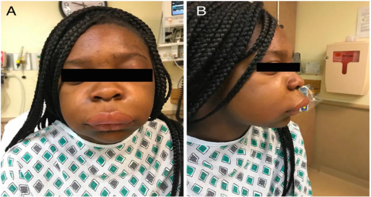
- Hereditary angioedema (autosomal dominant). C1 esterase inhibitor normally restrains C1 and also kallikrein, the enzyme that releases bradykinin from kininogen. When it is deficient, kallikrein runs free, bradykinin accumulates and opens endothelial junctions. Attacks of nonpitting, non-itchy swelling of the face, limbs, upper airway and gut (abdominal pain, vomiting, diarrhea) come on over hours and resolve in 2–3 days; laryngeal swelling can kill. Trauma, dental work and estrogens trigger attacks; onset is usually in childhood or adolescence. There are no hives, and antihistamines and corticosteroids do not work. Because C1 is uncontrolled, C4 is low even between attacks, the best screening test; C1-INH level or function confirms. Treatment: C1-INH concentrate or a bradykinin B2-receptor antagonist.
- ACE-inhibitor angioedema. ACE is the enzyme that degrades bradykinin (kininase II). Block it and bradykinin accumulates: the same swelling of lips, tongue, face, airway or bowel, without itching or hives, in someone recently started on (or long taking) an ACE inhibitor. C4 and C1-INH are normal. Stop the drug.
- Mast-cell (histamine) angioedema. IgE-mediated allergy (foods, stings, drugs) or direct mast-cell activation releases histamine: swelling with itch and urticaria, sometimes wheeze and hypotension (anaphylaxis, 3.3). Raised tryptase; responds to antihistamines, corticosteroids and epinephrine.
Core activity · Almost Twins
Almost Twins
Look-alike A
Hereditary angioedema
- Nonpitting swelling of lips, face, larynx
- No itch, no hives; mediator is bradykinin (C1 inhibitor deficient)
- Abdominal attacks; family history (autosomal dominant)
- Low C4; antihistamines and steroids do not help
Look-alike A
Hereditary angioedema
- Nonpitting swelling of lips, face, larynx
- No itch, no hives; mediator is bradykinin (C1 inhibitor deficient)
- Abdominal attacks; family history (autosomal dominant)
- Low C4; antihistamines and steroids do not help
Look-alike B
Allergic (mast-cell) angioedema
- Nonpitting swelling of lips, face, larynx
- Itchy wheals (hives); mediator is histamine from IgE-triggered mast cells
- Minutes after food, sting or drug; may have wheeze and hypotension
- Raised tryptase; responds to epinephrine and antihistamines
Step 1 · What they share
What do the two share?
Select every feature that both show.
Core activity · One Fact Changes
One Fact Changes
1 · The original case
A 72-year-old man has sudden swelling of his lips and tongue with abdominal pain and diarrhea. There is no itching and no rash anywhere on his skin. Three weeks ago he started lisinopril for hypertension.
Which mediator is responsible?
Which fact decided it?
Amyloidosis from the immune system: AA and AL
L1 p.46–48SyllabusAmyloid is protein that misfolds into β-pleated sheets and aggregates outside cells, staining with Congo red and showing apple-green birefringence under polarized light. Chapter 1 holds the full account (amyloid and amyloid types). Two of the systemic forms are products of the immune system:
Chronic inflammation to AA amyloid
- Chronic inflammation (rheumatoid arthritis, IBD, tuberculosis, bronchiectasis, osteomyelitis, familial Mediterranean fever)
- Macrophages release IL-1, IL-6, TNF
- Liver makes serum amyloid A (acute-phase protein) for years
- Incomplete breakdown → AA fragments misfold
- AA amyloid in kidney (nephrotic syndrome), liver, spleen, adrenals
- AA amyloid follows years of raised SAA. Familial Mediterranean fever is an inherited inflammasome disorder (3.1): untreated attacks keep SAA high, and colchicine prevents the amyloid.
- AL amyloid comes from immunoglobulin light chains (λ more than κ) secreted by a monoclonal plasma cell clone, in multiple myeloma or a smaller plasma-cell dyscrasia. Heart (restrictive cardiomyopathy), kidney, nerves (carpal tunnel), tongue (macroglossia).
- Aβ₂M amyloid is β₂-microglobulin, the light chain of MHC class I (3.2), which dialysis membranes do not clear: joints and carpal tunnel.
- Localized forms stay in one organ: Aβ in Alzheimer disease, calcitonin in medullary thyroid carcinoma, amylin in type 2 diabetes islets.
Knowledge check · 3.7
Answer, read why, and try again whenever you like. Answers are kept in both study modes.
- Q1HIV entry: gp120 attachmentL3 p.48–50Ch. III cases p.2
Investigators mix HIV-infected and uninfected CD4⁺ T-cell lines with a synthetic peptide triazole complex. The complex binds gp120 with high affinity and irreversibly inhibits its function, reducing infection of the CD4⁺ T cells.
Which step of HIV infection does this experimental drug most likely inhibit?
- Q2ACE-inhibitor angioedemaL3 p.54Ch. III cases p.9
A 72-year-old man recently diagnosed with heart disease has sudden abdominal pain, diarrhea and vomiting, with painful swelling of his face, lips and mouth. He denies itching. His physician recently started drugs to control his blood pressure and cholesterol. He sometimes gets a rash after antibiotics and has 'hives' every spring.
Accumulation of which substance most likely causes his symptoms?
- Q3Hereditary angioedemaL3 p.54Ch. III cases p.13
A 23-year-old woman with past episodes of facial swelling attributed to 'allergies' arrives with marked difficulty breathing, abdominal pain and swelling of the face, neck and limbs, and is intubated. Her father has had similar attacks and was prescribed antihistamines.
Which laboratory finding is most likely?
- Q4HIV window period and testsL3 p.51Ref.
Twelve days after a condomless sexual exposure to a partner with untreated HIV, a 24-year-old man has fever, sore throat, a diffuse rash and tender lymph nodes. A 4th-generation antigen/antibody test is negative.
Which test is most likely to establish acute HIV infection now?
- Q5Terminal complement deficiencyL3 p.54
A 19-year-old college student has his second episode of meningococcal meningitis; his older brother had disseminated gonococcal infection. Total hemolytic complement (CH50) is undetectable.
Which deficiency best explains this pattern?
- Q6AA amyloid from chronic inflammationL1 p.47
A 61-year-old woman has had poorly controlled rheumatoid arthritis for 20 years. She now has heavy proteinuria and edema. A renal biopsy shows amorphous eosinophilic material in the glomeruli that is apple-green under polarized light after Congo red staining.
What is the precursor of the deposited protein?
- Q7Image: Kaposi sarcomaL3 p.51–52
A 38-year-old man with untreated HIV has the purple-red nodular skin lesions shown in the top-left panel and a CD4 count of 140 cells/mm³.
Lecture image
Which statement about the skin lesions is correct?