Transplantation immunology
A graft is a large, foreign HLA antigen. Whether it is destroyed in minutes, weeks or years depends on what the recipient already has (preformed antibody), what it makes next (T cells and new antibody), and in which direction the attack runs.
Graft types
L3 p.29| Graft | Donor → recipient | Rejection risk | Example |
|---|---|---|---|
| Autograft | Self → self | None; best survival | Skin graft from the thigh to a burn; saphenous vein for bypass; autologous stem cells |
| Isograft (syngeneic) | Identical twin → twin | None (identical HLA) | Kidney between monozygotic twins |
| Allograft | Another person of the same species | Yes: HLA and ABO differences | Almost all organ and marrow transplants |
| Xenograft | Another species | Severe: preformed antibodies against foreign carbohydrates | Experimental pig organs; (decellularized porcine valves are processed to remove living cells) |
Autograft
- Donor → recipient
- Self → self
- Rejection risk
- None; best survival
- Example
- Skin graft from the thigh to a burn; saphenous vein for bypass; autologous stem cells
Isograft (syngeneic)
- Donor → recipient
- Identical twin → twin
- Rejection risk
- None (identical HLA)
- Example
- Kidney between monozygotic twins
Allograft
- Donor → recipient
- Another person of the same species
- Rejection risk
- Yes: HLA and ABO differences
- Example
- Almost all organ and marrow transplants
Xenograft
- Donor → recipient
- Another species
- Rejection risk
- Severe: preformed antibodies against foreign carbohydrates
- Example
- Experimental pig organs; (decellularized porcine valves are processed to remove living cells)
Matching: ABO, HLA and the crossmatch
L3 p.29The lecture lists three requirements for graft viability, in order of importance. Each one removes a mechanism of rejection before it can start.
- ABO compatibility. Graft endothelium carries A and B antigens, and everyone has preformed IgM against the ABO antigens they lack. An ABO-incompatible organ is attacked at once, the same way a mismatched transfusion is.
- No preformed anti-HLA antibodies against the donor. Pregnancy, transfusion or a previous transplant can leave a recipient with antibodies against HLA molecules. The crossmatch mixes the recipient's serum with the donor's lymphocytes (which carry the donor's HLA); if the cells are killed or bound, the recipient has donor-specific antibodies and would reject the organ in minutes. A positive crossmatch normally stops the transplant.
- Close HLA matching. Matching at HLA-A, -B and especially -DR lowers T-cell rejection; HLA-C matters less. With modern immunosuppression, mismatches are tolerated for many organs, but marrow transplants need close matching because the donor's T cells can attack the recipient.
Rejection: minutes, weeks or years
L3 p.30–31Teaching schematic
Minutes–hours
Hyperacute
- Preformed anti-ABO/HLA antibody + complement on endothelium (type II)
- Thrombi, neutrophils, fibrinoid necrosis → infarction
- Irreversible: remove the organ
Days–months
Acute cellular
- CD8 killing + CD4 Th1/macrophages (type IV)
- Interstitial lymphocytes, tubulitis, endarteritis
- Reversible with immunosuppression
Days–months
Acute antibody-mediated
- New donor-specific antibody + complement
- Capillaritis; C4d in peritubular capillaries
- Often reversible (plasmapheresis, IVIG)
Months–years
Chronic
- Repeated T-cell and antibody injury; growth factors
- Graft arteriosclerosis (narrowing), fibrosis, tubular atrophy
- Irreversible; main cause of late loss
- Hyperacute rejection (minutes to hours; type II). Preformed anti-ABO or anti-HLA antibodies bind graft endothelium as soon as blood flows. Complement and neutrophils injure the endothelium, platelets aggregate, and thrombi fill glomerular capillaries and arterioles. The kidney turns from pink to mottled and cyanotic: infarction, with fibrinoid necrosis of vessel walls. It is irreversible and the organ must be removed. ABO typing and the crossmatch now prevent it.
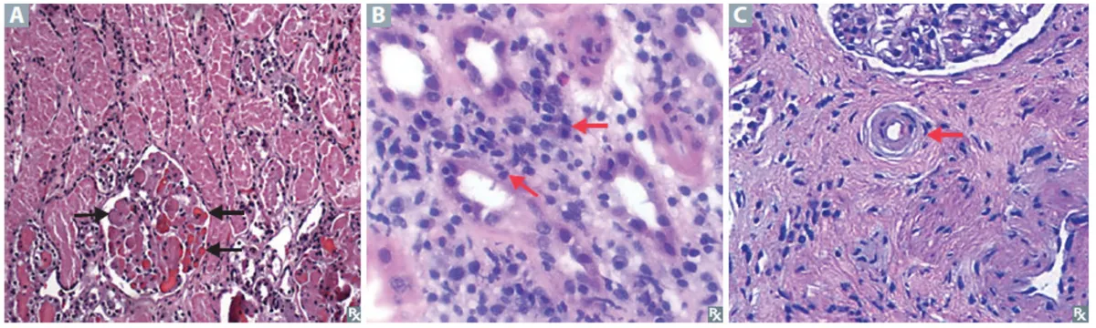
- Acute cellular (T cell–mediated) rejection (days to weeks or months; type IV). Recipient CD8 cells kill donor tubular and endothelial cells that show foreign class I, and CD4 Th1 cells recognize class II on donor dendritic cells and activate macrophages through IFN-γ. The biopsy shows an interstitial lymphocytic infiltrate with lymphocytes invading tubular epithelium (tubulitis) and, when severe, the intima of arteries (endarteritis). It is the most common form and is reversible with stronger immunosuppression.
- Acute antibody-mediated rejection (days to months). Donor-specific anti-HLA antibodies, usually made after transplantation, bind the graft's microvessels and fix complement. Neutrophils and monocytes gather in glomerular and peritubular capillaries, and the complement fragment C4d stays bound to the peritubular capillary walls, where it can be stained. Treatment removes or blocks antibody (plasmapheresis, IVIG).
- Chronic rejection (months to years; irreversible). Repeated T-cell and antibody injury, with cytokines and growth factors, makes intimal smooth-muscle cells proliferate and lay down matrix: graft arteriosclerosis, a concentric narrowing of arteries. The downstream tissue becomes ischemic: interstitial fibrosis, tubular atrophy and glomerular sclerosis. It is the main cause of late graft loss, often in patients who survived earlier acute episodes.
Lecture image
Graft-versus-host disease and graft-versus-leukemia
L3 p.32Rejection is the host attacking the graft. Graft-versus-host disease runs the other way: immunocompetent donor T cells in the graft recognize the recipient's tissues as foreign. Three conditions must hold: the graft contains mature T cells, the recipient cannot eliminate them (immunosuppressed, or immunodeficient), and the recipient carries HLA or minor antigens the donor lacks.
Teaching schematic
Host versus graft = rejection
Recipient T cells and antibodiesDonor organSolid-organ transplants; prevented by matching and immunosuppression.
Graft versus host (GVHD)
Donor T cells in marrow, liver or bloodRecipient skin, liver, gut (and marrow after transfusion)Needs T cells in the graft, a recipient who cannot reject them, and an antigen difference.
Graft versus leukemia (GVL)
Donor T and NK cellsRecipient's leftover leukemic cellsThe benefit that comes with GVHD; T-cell depletion lowers both.
- Settings: allogeneic bone marrow or stem-cell transplantation (the common setting), liver transplants (which carry many donor lymphocytes), and transfusion of non-irradiated blood into a T-cell–deficient recipient (DiGeorge syndrome, SCID) or a newborn. Transfusion-associated GVHD also attacks the marrow and is usually fatal, so blood for these patients is irradiated to disable donor lymphocytes.
- Targets and findings: skin (maculopapular rash, often palms and soles), liver (bile-duct damage → jaundice), gut (diarrhea), with hepatosplenomegaly. Donor CD8 and CD4 cells do the damage, so it is a type IV reaction.
- Graft-versus-leukemia (GVL): in a marrow transplant for leukemia, the donor T cells also recognize the recipient's leukemic cells. Removing T cells from the graft lowers GVHD but raises relapse; infusions of donor lymphocytes are sometimes given to induce GVL.
Knowledge check · 3.4
Answer, read why, and try again whenever you like. Answers are kept in both study modes.
- Q1Image: acute cellular rejectionL3 p.30–31
Biopsies from three different renal allografts are shown. A 46-year-old man's creatinine rose 5 weeks after his transplant; his biopsy is panel B.
Lecture image
What is the process in panel B and how is it usually managed?
- Q2Hyperacute rejection and crossmatchL3 p.29–30
Minutes after the vascular clamps are released, a newly transplanted kidney turns from pink to mottled and cyanotic. A biopsy shows neutrophils and fibrin thrombi in glomerular capillaries and arterioles.
Which pre-transplant test is designed to prevent exactly this?
- Q3GVHD and GVL directionL3 p.32
A 34-year-old man receives an allogeneic stem-cell transplant from his HLA-matched brother for acute myeloid leukemia. On day 24 he develops a maculopapular rash on his palms and soles, profuse watery diarrhea and a rising bilirubin.
Which statement best describes the process?
- Q4Antibody-mediated rejection (C4d)L3 p.30Ref.
Four months after a kidney transplant, a 39-year-old woman's creatinine rises. Biopsy shows neutrophils and monocytes in glomerular and peritubular capillaries with little tubulitis, and immunostaining shows C4d along the peritubular capillaries. A new donor-specific anti-HLA antibody is detected.
What is the mechanism of this rejection?