Chronic and granulomatous inflammation
When the cause will not go away, the response changes character: macrophages and lymphocytes replace neutrophils, and injury and repair run side by side. When the cause is hard to digest, activated macrophages wall it off as a granuloma.
What makes inflammation chronic
L2 p.40–41Chronic inflammation lasts weeks to years. It most often results from persistence of the injuring agent, though it can also begin quietly without a recognizable acute phase. Its defining feature is that three things happen at the same time: infiltration by mononuclear cells, tissue destruction by those cells and their products, and attempts at repair by angiogenesis and fibrosis.
Causes, as the lecture groups themL2 p.40
| Cause | Why it persists | Examples |
|---|---|---|
| Persistent infection | Organisms resist killing or hide inside macrophages | Tuberculosis, leprosy, syphilis, some fungi and viruses |
| Autoimmune and immune-mediated disease | The antigen is self, so it can never be cleared | Rheumatoid arthritis, SLE, inflammatory bowel disease |
| Hard-to-clear exposures | Material cannot be digested or keeps coming back | Silica (silicosis), urate crystals (gout), silicone from breast implants, suture material, cholesterol in atheromas |
Persistent infection
- Why it persists
- Organisms resist killing or hide inside macrophages
- Examples
- Tuberculosis, leprosy, syphilis, some fungi and viruses
Autoimmune and immune-mediated disease
- Why it persists
- The antigen is self, so it can never be cleared
- Examples
- Rheumatoid arthritis, SLE, inflammatory bowel disease
Hard-to-clear exposures
- Why it persists
- Material cannot be digested or keeps coming back
- Examples
- Silica (silicosis), urate crystals (gout), silicone from breast implants, suture material, cholesterol in atheromas
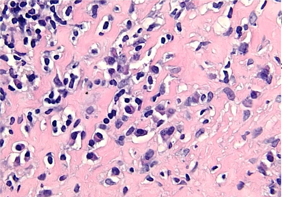
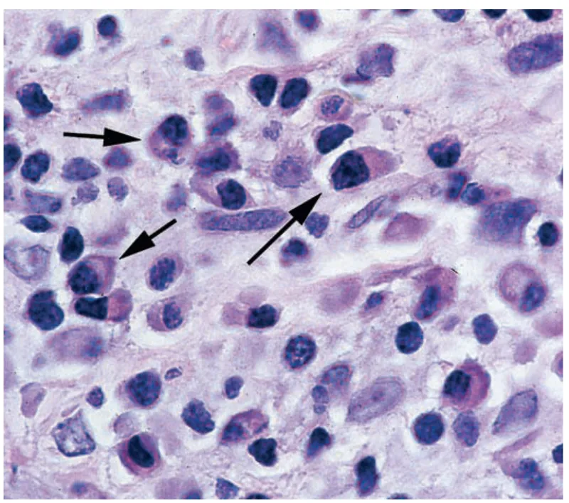
The cells. Macrophages are the centre: blood monocytes that entered tissue. Microbial products and IFN-γ activate them classically (M1): more killing, reactive oxygen species, nitric oxide, more inflammation. IL-4 and IL-13 activate them alternatively (M2): growth factors, TGF-β and collagen deposition, repair. Lymphocytes and macrophages stimulate each other: macrophages present antigen and make IL-12; T cells answer with IFN-γ. Plasma cells (eccentric nucleus with clock-face chromatin, a pale perinuclear Golgi) make antibody, sometimes inside lymphoid follicles. Eosinophils dominate in parasitic and allergic disease (their major basic protein is toxic to helminths and to epithelium). Mast cells take part in both.
Macrophages pull both ways
Classically activated (M1)
- Triggered by microbes, IFN-γ
- ROS, NO, lysosomal enzymes
- IL-1, IL-12, TNF: more inflammation
- Kill microbes; destroy tissue
Alternatively activated (M2)
- Triggered by IL-4, IL-13
- TGF-β, PDGF, IL-10
- Angiogenesis and collagen
- Repair and fibrosis
Lecture image
Case image
Anatomy of a granuloma
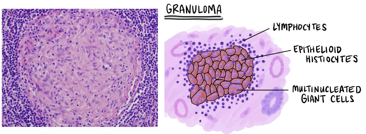
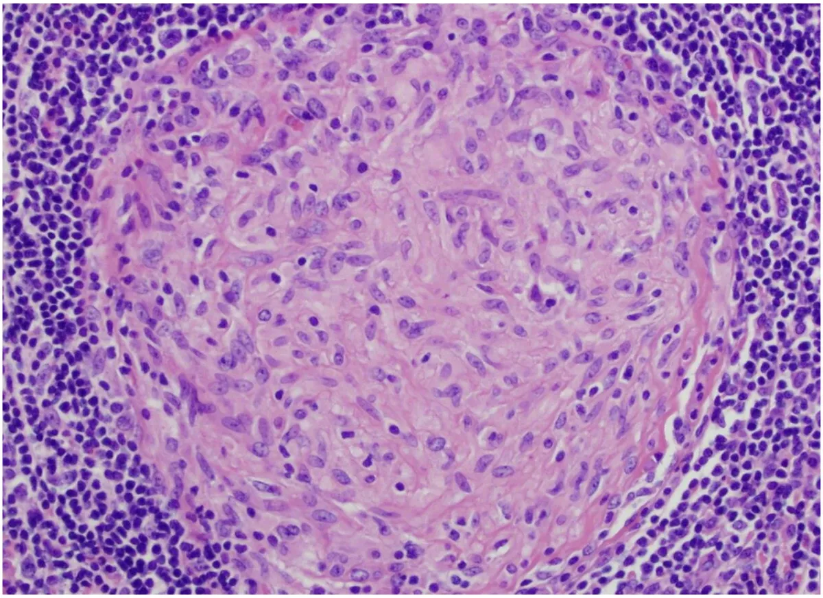
L2 p.22–23, 42A granuloma is a compact nodule of activated macrophages, formed when the body cannot destroy an agent and walls it off instead. To the eye it is a pale, white nodule; under the microscope it has three or four layers.
Teaching schematic
- Epithelioid cells (histiocytes): activated macrophages with abundant pale-pink, granular cytoplasm and indistinct borders, packed together like an epithelium. Their nuclei are oval and pale, often elongated like the sole of a slipper. They secrete more than they engulf.
- Multinucleated giant cells: several macrophages fuse into one cell up to 50 µm across, with 20 or more nuclei.
- Lymphocytes, mainly CD4⁺ Th1 cells, cuff the nodule; older granulomas gain a rim of fibroblasts and collagen.
- ± central necrosis: caseous in tuberculosis.
Lecture image
Teaching schematic
Caseating, non-caseating and foreign-body granulomas
L2 p.44–46Immune granulomas form around persistent antigens that drive a T-cell response. Foreign-body granulomas form around inert material too large for one macrophage to engulf (sutures, talc, silica) and need no specific immune response. The centre then tells you most about the cause.
Reading the centreL2 p.44Ch. II cases p.4, 7, 13
| Type | Centre | Classic causes | Clues |
|---|---|---|---|
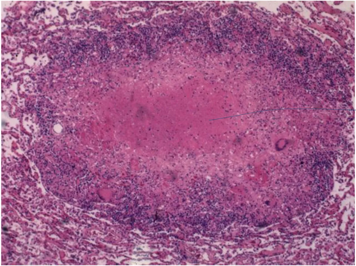
| Caseating | Cheese-like necrosis | Tuberculosis, systemic fungi (Histoplasma, Coccidioides) | Acid-fast or fungal stains; upper-lobe disease, night sweats |
| Non-caseating, tight | No necrosis | Sarcoidosis, Crohn disease, berylliosis | Stains and cultures negative; hilar nodes, hypercalcemia (sarcoid) |
| Non-caseating, loose | No necrosis | Hypersensitivity pneumonitis (bird, farmer's, flour dust) | Small, poorly formed, near bronchioles; exposure history |
| Foreign-body | Visible material | Sutures, talc, silica, urate (gout tophus), silicone | Material often polarizes; foreign-body giant cells |
Caseating
- Centre
- Cheese-like necrosis
- Classic causes
- Tuberculosis, systemic fungi (Histoplasma, Coccidioides)
- Clues
- Acid-fast or fungal stains; upper-lobe disease, night sweats
Non-caseating, tight
- Centre
- No necrosis
- Classic causes
- Sarcoidosis, Crohn disease, berylliosis
- Clues
- Stains and cultures negative; hilar nodes, hypercalcemia (sarcoid)
Non-caseating, loose
- Centre
- No necrosis
- Classic causes
- Hypersensitivity pneumonitis (bird, farmer's, flour dust)
- Clues
- Small, poorly formed, near bronchioles; exposure history
Foreign-body
- Centre
- Visible material
- Classic causes
- Sutures, talc, silica, urate (gout tophus), silicone
- Clues
- Material often polarizes; foreign-body giant cells
Lecture image
Lecture image
Core activity · Almost Twins
Almost Twins
Look-alike A
Caseating granuloma
Lecture image
- Epithelioid macrophages and a lymphocyte rim
- Central caseous necrosis: amorphous pink debris, cell outlines lost
- Tuberculosis, systemic fungi (Histoplasma, Coccidioides)
- Acid-fast or fungal stains often find organisms
Look-alike A
Caseating granuloma
Lecture image
- Epithelioid macrophages and a lymphocyte rim
- Central caseous necrosis: amorphous pink debris, cell outlines lost
- Tuberculosis, systemic fungi (Histoplasma, Coccidioides)
- Acid-fast or fungal stains often find organisms
Look-alike B
Non-caseating granuloma
Lecture image
- Epithelioid macrophages and a lymphocyte rim
- No central necrosis: epithelioid cells to the very centre
- Sarcoidosis, Crohn disease, berylliosis, foreign bodies
- Stains and cultures negative
Step 1 · What they share
What do the two granulomas share?
Select every feature that both show.
Core activity · One Fact Changes
One Fact Changes
1 · The original case
A 40-year-old woman has two months of dry cough and fatigue, and her chest radiograph shows bilateral hilar lymphadenopathy. A node biopsy shows tight granulomas with giant cells and no necrosis; acid-fast and fungal stains are negative. Her serum calcium is raised.
What is the most likely diagnosis?
Which fact decided it?
The granuloma loop: IL-12, IFN-γ and TNF
L2 p.42–43Take Mycobacterium tuberculosis. An alveolar macrophage engulfs it, but the bacterium blocks phagolysosome maturation and keeps multiplying. The macrophage carries antigen to a lymph node and, through the steps on the lecture slide, builds the granuloma:
Teaching schematic
- The macrophage presents antigen on MHC class II to naive CD4⁺ T cells.
- It secretes IL-12, which drives the T cells to become Th1 cells.
- Th1 cells return to the site and secrete IFN-γ, which activates macrophages: phagolysosomes mature, nitric oxide and reactive oxygen species are made, MHC II rises. Activated macrophages become epithelioid cells and fuse into giant cells.
- Activated macrophages make more IL-12, so the loop sustains itself while antigen persists.
- TNF from macrophages and T cells recruits and organizes cells and maintains the granuloma.
This is a type IV (delayed-type) hypersensitivity reaction. The tuberculin skin test reads the same loop in miniature: injected antigen, Th1 cells and IFN-γ produce induration 48–72 hours later. People with inherited defects in the IL-12 or IFN-γ receptors cannot complete the loop and develop disseminated mycobacterial infections.
Core activity · The Missing Arrow
The Missing Arrow
Mycobacterium tuberculosis survives inside an alveolar macrophage. Complete the loop that builds a granuloma.
- A macrophage harbours surviving mycobacteria
- A stable granuloma contains the bacilli, with caseation in its centre
Cards
Tap or drag a card. Placing a used card moves it.
Anti-TNF therapy and TB reactivation
L2 p.42In latent tuberculosis, living bacilli are contained inside granulomas for decades. TNF keeps those granulomas organized. Drugs that block TNF, the antibodies infliximab, adalimumab, golimumab and certolizumab and the receptor fusion protein etanercept, are used for rheumatoid arthritis, Crohn disease, ulcerative colitis, psoriasis and ankylosing spondylitis. Blocking TNF loosens the granulomas, and the bacilli escape: reactivation tuberculosis, often extrapulmonary or disseminated. Histoplasmosis and coccidioidomycosis can reactivate the same way.
Worked case · A Crohn patient on a new drug
A 34-year-old woman with Crohn disease starts infliximab. Her family emigrated from a country where tuberculosis is common. Five months later she has fever, night sweats and weight loss, and her chest radiograph shows innumerable tiny nodules.
What happened, and what should have been done first?
L2 p.42Knowledge check · 2.6
Answer, read why, and try again whenever you like. Answers are kept in both study modes.
- Q1Tuberculosis: caseating granulomaL2 p.44–45Ch. II cases p.4
A 33-year-old man has had productive cough, fever, night sweats and a 9-kg weight loss over 4 months. His chest radiograph is shown.
Case image
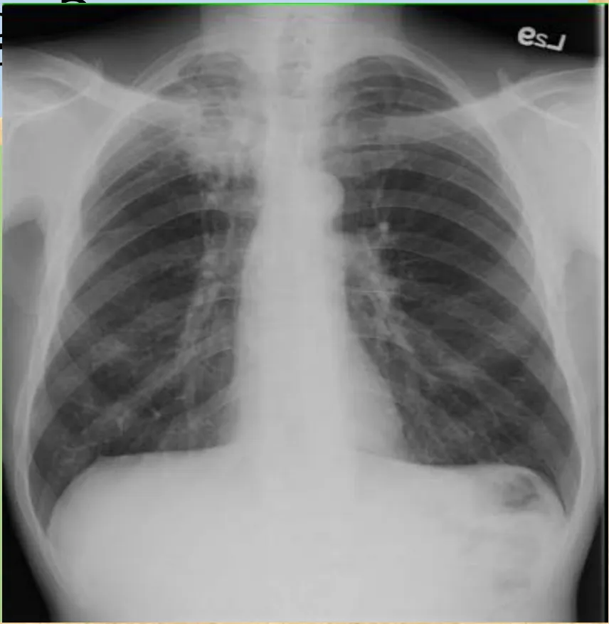
Which best describes his most likely lung histopathology?
- Q2Sarcoidosis: Th1 responseL2 p.43–44, 46Ch. II cases p.7
A 30-year-old woman has fever (38 °C) and dyspnea, erythematous patches and plaques over the front of her lower legs, increased serum calcium and prominent bilateral hilar opacities. A skin biopsy is shown.
Case image
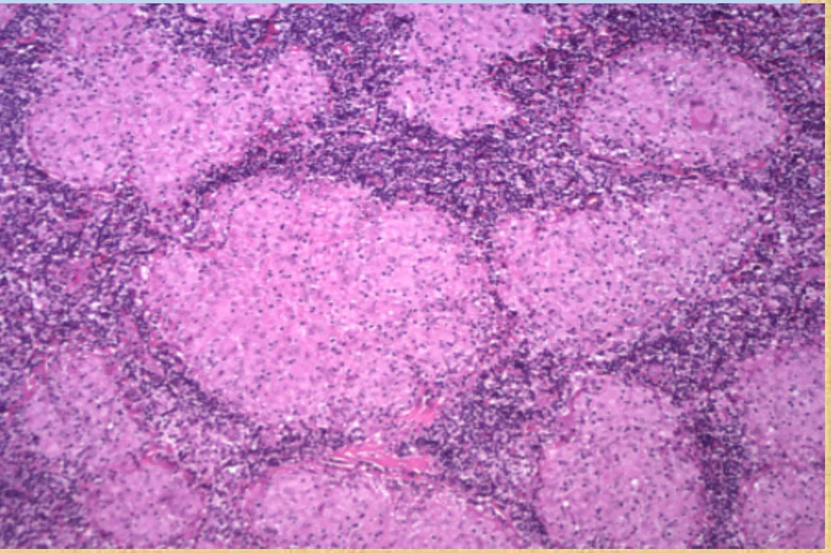
Which pathologic mechanism is most associated with her likely condition?
- Q3Granuloma cytokines: IL-12 and IFN-γL2 p.43Ch. II cases p.13
A 38-year-old non-smoker who works at a flour mill has two months of fever, malaise, cough, dyspnea and a 4-kg weight loss. Diffuse fine crackles are heard, and the radiograph shows patchy reticulonodular infiltrates in the mid and upper zones. A lung biopsy is shown.
Case image
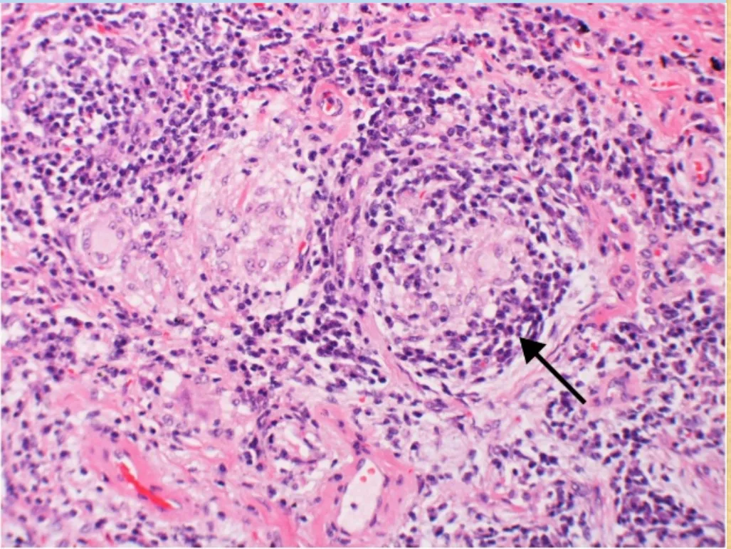
Which cytokines are most involved in forming the lesion at the arrow?
- Q4Giant cells: IFN-γ and IL-12L2 p.43Midterm sim. p.4–5
A 37-year-old man with AIDS has three weeks of chest pain and dyspnea, and bilateral nodules on chest radiography. A CT-guided lung biopsy is shown.
Case image
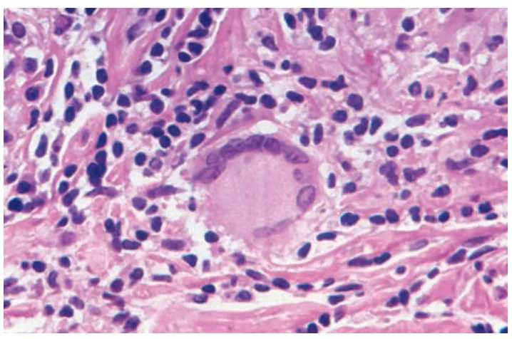
The multinucleated cell in the centre most likely formed through the action of which mediators?
- Q5Chronic inflammation: plasma cellsL2 p.40–41Midterm sim. p.3–4
A 41-year-old woman who uses an intrauterine device has 4 months of heavy menstrual bleeding and pelvic pain. Her endometrial biopsy shows many plasma cells (arrows), macrophages and scattered lymphoid follicles in the stroma.
Case image
The presence of these cells is evidence of which condition?
- Q6Anti-TNF and TB reactivationL2 p.42
A 52-year-old man with rheumatoid arthritis has been well on infliximab for six months. He now has fever, weight loss and an enlarging neck lymph node; biopsy shows acid-fast bacilli in poorly formed granulomas.
Why did this infection appear now?