From injury to acute inflammation
Something harmful is recognized, and within minutes the local vessels change: arterioles dilate, venules leak, flow slows. Those vascular changes explain four of the five cardinal signs and set the stage for neutrophils.
What sets inflammation off
L2 p.2, 6Inflammation is the response of vascularized tissue that brings plasma proteins and leukocytes out of the blood to where they are needed, so they can dilute, destroy or wall off a harmful agent and start repair. Acute inflammation is the fast version: it begins within seconds to minutes, lasts hours to days, and is dominated by fluid exudate and neutrophils. It is part of innate immunity, so it responds the same way to the first encounter as to the tenth.
Four kinds of trigger, one kind of alarmL2 p.2, 40Ref. · Robbins, inflammation
| Trigger | What the tissue senses | Example |
|---|---|---|
| Infection | Microbial molecules (PAMPs) such as LPS, flagellin and bacterial nucleic acids, through Toll-like receptors | A thorn carrying Staphylococcus aureus |
| Tissue necrosis | Molecules released by dead cells (DAMPs): ATP, uric acid, HMGB1, mitochondrial DNA | Myocardial infarct, burn |
| Foreign bodies and crystals | Material that cannot be digested; crystals activate the NLRP3 inflammasome | Splinter, suture, urate crystals in gout |
| Immune reactions | Antigen recognized by antibody or T cells | Hypersensitivity and autoimmune disease (Chapter 3) |
Infection
- What the tissue senses
- Microbial molecules (PAMPs) such as LPS, flagellin and bacterial nucleic acids, through Toll-like receptors
- Example
- A thorn carrying Staphylococcus aureus
Tissue necrosis
- What the tissue senses
- Molecules released by dead cells (DAMPs): ATP, uric acid, HMGB1, mitochondrial DNA
- Example
- Myocardial infarct, burn
Foreign bodies and crystals
- What the tissue senses
- Material that cannot be digested; crystals activate the NLRP3 inflammasome
- Example
- Splinter, suture, urate crystals in gout
Immune reactions
- What the tissue senses
- Antigen recognized by antibody or T cells
- Example
- Hypersensitivity and autoimmune disease (Chapter 3)
The first responders are sentinel cells already in the tissue: macrophages, dendritic cells and mast cells. Their receptors recognize the trigger. Toll-like receptors switch on genes for cytokines such as TNF; the inflammasome, a cytosolic complex, activates caspase-1, which cuts pro-IL-1β into active IL-1. Mast cells release preformed histamine within seconds. These mediators act on the nearest small vessels, and everything visible follows from that.
The whole chapter in one line
- Stimulus recognized by sentinel cells
- Mediators released
- Vessels dilate and leak (2.1)
- Leukocytes recruited (2.2)
- Agent killed and removed (2.3)
- Response switched off (2.5)
- Tissue regenerates or scars (2.7)
Acute versus chronic inflammation, as the lecture contrasts them
Acute
- Onset in seconds to minutes; lasts hours to a few days
- Neutrophils, edema
- Usually mild, self-limited tissue injury
- Prominent local signs
Chronic (2.6)
- Slow onset; weeks to years
- Macrophages and lymphocytes, plasma cells
- Tissue destruction, angiogenesis and fibrosis at the same time
- Local signs often subtle
Lecture image
Five cardinal signs, five mechanisms
L2 p.3–4Celsus described four signs of inflammation (rubor, calor, tumor, dolor), and Virchow added a fifth (functio laesa). Each sign is a visible consequence of a specific vascular or neural event, so knowing the mechanism tells you which mediator to blame and which drug will help.
Each sign has its own vessel or nerveL2 p.3–4
| Sign | Mechanism | Main mediators |
|---|---|---|
| Rubor (redness) | Arterioles and precapillary sphincters dilate; more capillaries fill with blood (hyperemia) | Histamine, nitric oxide, PGI₂, PGE₂ |
| Calor (heat) | More warm core blood reaches the surface; systemic fever adds to it | Same vasodilators; IL-1 and TNF for fever |
| Tumor (swelling) | Postcapillary venules leak protein-rich fluid into the interstitium (exudate) | Histamine, bradykinin, LTC₄/D₄/E₄, C3a/C5a |
| Dolor (pain) | PGE₂ sensitizes nerve endings to bradykinin; swelling raises tissue pressure | PGE₂ + bradykinin |
| Functio laesa (loss of function) | Pain and swelling limit use; tissue damage | Consequence of the others |
Rubor (redness)
- Mechanism
- Arterioles and precapillary sphincters dilate; more capillaries fill with blood (hyperemia)
- Main mediators
- Histamine, nitric oxide, PGI₂, PGE₂
Calor (heat)
- Mechanism
- More warm core blood reaches the surface; systemic fever adds to it
- Main mediators
- Same vasodilators; IL-1 and TNF for fever
Tumor (swelling)
- Mechanism
- Postcapillary venules leak protein-rich fluid into the interstitium (exudate)
- Main mediators
- Histamine, bradykinin, LTC₄/D₄/E₄, C3a/C5a
Dolor (pain)
- Mechanism
- PGE₂ sensitizes nerve endings to bradykinin; swelling raises tissue pressure
- Main mediators
- PGE₂ + bradykinin
Functio laesa (loss of function)
- Mechanism
- Pain and swelling limit use; tissue damage
- Main mediators
- Consequence of the others
The vascular sequence
L2 p.5, 7The lecture lists the vascular events in order: vasoconstriction of arterioles → vasodilation of arterioles → increased permeability of venules → edema. Each step causes the next.
Teaching schematic
1. Brief vasoconstriction
Seconds; neurogenic reflex
2. Arteriolar dilation
Histamine, NO, PGs → more capillaries open: red, hot
3. Venular leak
Endothelial contraction opens gaps → exudate: swelling
4. Stasis and margination
Concentrated, slow blood; leukocytes move to the wall
- Transient vasoconstriction (seconds): a neurogenic reflex and endothelin. It is why a scratched patch of skin briefly blanches.
- Arteriolar vasodilation: histamine, nitric oxide and prostaglandins relax arteriolar smooth muscle and precapillary sphincters. Flow increases and the capillary bed floods: redness and heat.
- Increased permeability of postcapillary venules: histamine, bradykinin and leukotrienes make endothelial cells contract and open gaps between them. Protein-rich fluid escapes: swelling. This contraction response is fast and short-lived (15–30 minutes); cytokines (TNF, IL-1) later loosen junctions for longer.
- Stasis: losing fluid concentrates red cells, so blood becomes more viscous and flow slows in the small vessels.
- Margination: in slow flow, leukocytes leave the central stream and settle along the endothelium, ready to adhere (2.2).
Other ways vessels leakRef. · Robbins, vascular changes
| Mechanism | Time course | Example |
|---|---|---|
| Endothelial contraction (histamine, bradykinin, leukotrienes) | Immediate, transient; venules | Insect bite, early inflammation |
| Junction reorganization by cytokines | Delayed (hours), longer | TNF and IL-1 in bacterial infection |
| Direct endothelial injury | Immediate and sustained; all microvessels | Severe burns, toxins |
| Leukocyte-mediated injury | Late | Neutrophil proteases and oxygen radicals |
| Leaky new vessels | During repair | Granulation tissue (VEGF) (2.7) |
Endothelial contraction (histamine, bradykinin, leukotrienes)
- Time course
- Immediate, transient; venules
- Example
- Insect bite, early inflammation
Junction reorganization by cytokines
- Time course
- Delayed (hours), longer
- Example
- TNF and IL-1 in bacterial infection
Direct endothelial injury
- Time course
- Immediate and sustained; all microvessels
- Example
- Severe burns, toxins
Leukocyte-mediated injury
- Time course
- Late
- Example
- Neutrophil proteases and oxygen radicals
Leaky new vessels
- Time course
- During repair
- Example
- Granulation tissue (VEGF) (2.7)
Core activity · The Missing Arrow
The Missing Arrow
A bee stings the back of a hand. Complete the chain from the first mediator to leukocytes lining the venule wall.
Two gaps ask for a mechanism (how one event causes the next); three ask for an event (what you would see).
- Mast cells around the sting degranulate and release histamine
- histamine also makes endothelial cells of postcapillary venules contract
- Flow slows in the small vessels: stasis
- in slow flow, leukocytes drift out of the central stream
Cards
Tap or drag a card. Placing a used card moves it.
Exudate versus transudate
L2 p.4, 10Fluid leaves a capillary when the hydrostatic pressure pushing it out exceeds the oncotic pressure of plasma proteins holding it in; lymphatics return the small normal excess. Swelling can therefore come from two very different faults.
Teaching schematic
How to tell them apartRef. · Robbins; Light criteria
| Feature | Transudate | Exudate |
|---|---|---|
| Cause | Imbalance of pressures across an intact wall | Increased vascular permeability (inflammation) |
| Typical settings | Heart failure (hydrostatic ↑), cirrhosis or nephrotic syndrome (oncotic ↓) | Infection, abscess, pleuritis, burns |
| Protein | Low | High |
| Specific gravity | < 1.012 | > 1.020 |
| Cells and fibrin | Few cells; does not clot | Leukocytes, debris; fibrin may clot |
| Pleural fluid (Light criteria) | Fluid/serum protein ≤ 0.5 and LDH ≤ 0.6 | Protein ratio > 0.5 or LDH ratio > 0.6 |
Cause
- Transudate
- Imbalance of pressures across an intact wall
- Exudate
- Increased vascular permeability (inflammation)
Typical settings
- Transudate
- Heart failure (hydrostatic ↑), cirrhosis or nephrotic syndrome (oncotic ↓)
- Exudate
- Infection, abscess, pleuritis, burns
Protein
- Transudate
- Low
- Exudate
- High
Specific gravity
- Transudate
- < 1.012
- Exudate
- > 1.020
Cells and fibrin
- Transudate
- Few cells; does not clot
- Exudate
- Leukocytes, debris; fibrin may clot
Pleural fluid (Light criteria)
- Transudate
- Fluid/serum protein ≤ 0.5 and LDH ≤ 0.6
- Exudate
- Protein ratio > 0.5 or LDH ratio > 0.6
Once protein leaks, it adds a second force: protein in the interstitium raises interstitial oncotic pressure and draws still more water out. Pus is a purulent exudate, rich in neutrophils, dead cells and microbes.
Worked case · Two pleural effusions
Patient A has pneumonia and a pleural effusion with protein 4.6 g/dL (serum 6.8) and many neutrophils. Patient B has long-standing heart failure and an effusion with protein 1.8 g/dL (serum 6.6) and few cells.
Which effusion comes from a leaky wall, and which from pressure?
Ref. · Light criteriaCore activity · Almost Twins
Almost Twins
Look-alike A
Exudate
- Excess fluid outside the vessels
- High protein, leukocytes, fibrin; specific gravity > 1.020
- Leaky wall: inflammation opens endothelial gaps
Look-alike A
Exudate
- Excess fluid outside the vessels
- High protein, leukocytes, fibrin; specific gravity > 1.020
- Leaky wall: inflammation opens endothelial gaps
Look-alike B
Transudate
- Excess fluid outside the vessels
- Low protein, few cells; specific gravity < 1.012
- Intact wall: hydrostatic pressure up or oncotic pressure down
Step 1 · What they share
What do they share?
Select every feature that both show.
Inflammation without microbes: the infarct
L1 p.31–35Ch. II cases p.9, 11Teaching schematic
Cells in an infarct over timeCh. II cases p.9, 11Midterm sim. p.3Ref. · Robbins
| Time | Heart (coagulative necrosis) | Brain (liquefactive necrosis) |
|---|---|---|
| First hours | Little to see; wavy fibers at the border | Little to see |
| 12–24 h | Coagulative necrosis; neutrophils begin to arrive | Red (eosinophilic) neurons; neutrophils from about 24 h |
| 1–3 days | Nuclei lost, neutrophils peak | Neutrophils, edema |
| 3–7 days | Neutrophils die; macrophages remove dead myocytes | Microglia and macrophages take over |
| 1–2 weeks | Granulation tissue from the margins | Foamy, lipid-laden macrophages; tissue liquefies |
| Weeks to months | Collagen deposition → dense scar by about 2 months | Reactive astrocytes wall off a cyst (gliosis) |
First hours
- Heart (coagulative necrosis)
- Little to see; wavy fibers at the border
- Brain (liquefactive necrosis)
- Little to see
12–24 h
- Heart (coagulative necrosis)
- Coagulative necrosis; neutrophils begin to arrive
- Brain (liquefactive necrosis)
- Red (eosinophilic) neurons; neutrophils from about 24 h
1–3 days
- Heart (coagulative necrosis)
- Nuclei lost, neutrophils peak
- Brain (liquefactive necrosis)
- Neutrophils, edema
3–7 days
- Heart (coagulative necrosis)
- Neutrophils die; macrophages remove dead myocytes
- Brain (liquefactive necrosis)
- Microglia and macrophages take over
1–2 weeks
- Heart (coagulative necrosis)
- Granulation tissue from the margins
- Brain (liquefactive necrosis)
- Foamy, lipid-laden macrophages; tissue liquefies
Weeks to months
- Heart (coagulative necrosis)
- Collagen deposition → dense scar by about 2 months
- Brain (liquefactive necrosis)
- Reactive astrocytes wall off a cyst (gliosis)
Core activity · One Fact Changes
One Fact Changes
1 · The original case
A 64-year-old man with diabetes has crushing chest pain. His occluded left anterior descending artery cannot be opened in time. He dies of an arrhythmia 2 days later. The anterior wall is examined microscopically.
What dominates the infarcted myocardium?
Which fact decided it?
Knowledge check · 2.1
Answer, read why, and try again whenever you like. Answers are kept in both study modes.
- Q1Pain: PGE₂ + bradykininL2 p.4Ch. II cases p.3
A 22-year-old man has marked right lower quadrant pain over the past day, with rebound tenderness. At laparoscopy the appendix is swollen, erythematous and partly covered by yellowish exudate; microscopy shows numerous neutrophils.
The pain is predominantly the result of which two chemical mediators?
- Q2Swelling: increased vascular permeabilityL2 p.4, 7Midterm sim. p.3
A 5-year-old boy punctures his thumb with a rusty nail. Four hours later the thumb is red and swollen.
The initial swelling of the thumb is primarily due to which mechanism?
- Q3Inflammatory edema: endothelial gapsL2 p.4, 15Ch. II cases p.15
A 22-year-old woman cut her foot on a rock three days ago. Her temperature is 37.7 °C. The lateral foot has a fluctuant, erythematous lesion surrounded by edema.
What is most likely the primary mechanism of the edema?
- Q4Infarct timeline: neutrophils at 1–3 daysL1 p.33Ch. II cases p.11
A 62-year-old man is admitted with an hour of severe chest pain radiating to his left arm and is diagnosed with a myocardial infarction. Treatment fails, and he dies 2 days after admission.
What would most likely be seen in the infarcted myocardium at the time of death?
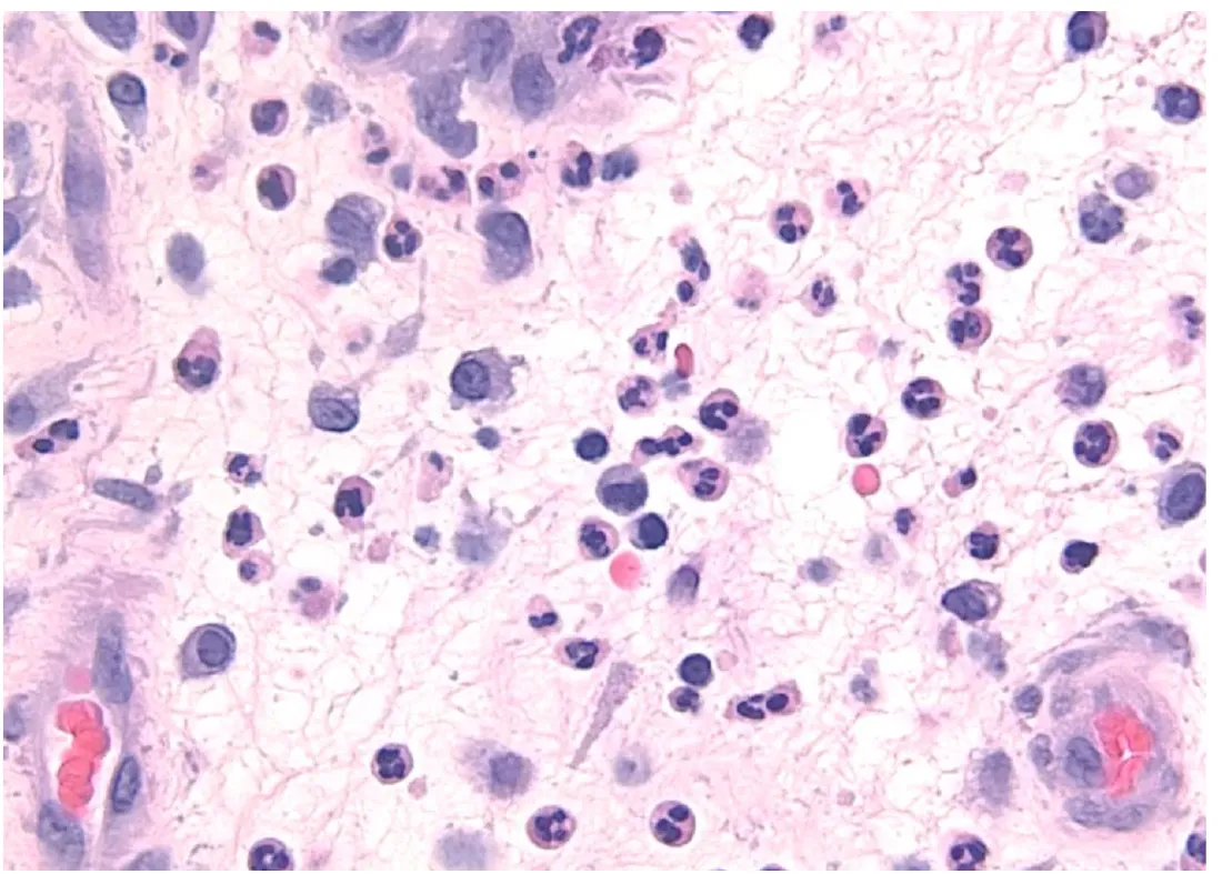
- Q5Neutrophils first: image recognitionL2 p.6
This section is from an appendix removed 18 hours after symptoms began.
Lecture image
Why are the multilobed cells, rather than macrophages, the dominant cell at this time?