Mediators and the systemic response
Every step so far was ordered by a chemical: amines and kinins open vessels in minutes, arachidonic acid products shape pain, leak and chemotaxis, complement tags and attracts, and cytokines carry the alarm to the brain, liver and marrow.
Fast mediators: histamine, bradykinin and nitric oxide
L2 p.4, 27, 29The first mediators are either preformed (histamine sits in mast-cell granules) or made in seconds from precursors already present (bradykinin from plasma kininogen, nitric oxide from arginine). They act within minutes and are destroyed almost as quickly, which is why the earliest swelling of a bee sting or a scratch fades within half an hour.
The fast mediatorsL2 p.27, 29
| Mediator | Source | Released by | Main actions |
|---|---|---|---|
| Histamine | Mast cells (main), basophils, platelets | IgE cross-linking, C3a/C5a, trauma, heat, cold | H₁: arteriolar dilation, venular endothelial contraction (leak), itch, bronchoconstriction |
| Serotonin | Platelets, enterochromaffin cells | Platelet aggregation | Vasoconstriction; contributes to permeability |
| Bradykinin | High-molecular-weight kininogen, cut by kallikrein (after factor XII activation) | Contact of plasma with collagen and negatively charged surfaces | Vasodilation, venular permeability, pain, bronchoconstriction |
| Nitric oxide | Endothelium (eNOS), macrophages (iNOS) | Arginine → citrulline by NO synthase | Smooth-muscle relaxation (vasodilation), less platelet adhesion, microbicidal in macrophages |
Histamine
- Source
- Mast cells (main), basophils, platelets
- Released by
- IgE cross-linking, C3a/C5a, trauma, heat, cold
- Main actions
- H₁: arteriolar dilation, venular endothelial contraction (leak), itch, bronchoconstriction
Serotonin
- Source
- Platelets, enterochromaffin cells
- Released by
- Platelet aggregation
- Main actions
- Vasoconstriction; contributes to permeability
Bradykinin
- Source
- High-molecular-weight kininogen, cut by kallikrein (after factor XII activation)
- Released by
- Contact of plasma with collagen and negatively charged surfaces
- Main actions
- Vasodilation, venular permeability, pain, bronchoconstriction
Nitric oxide
- Source
- Endothelium (eNOS), macrophages (iNOS)
- Released by
- Arginine → citrulline by NO synthase
- Main actions
- Smooth-muscle relaxation (vasodilation), less platelet adhesion, microbicidal in macrophages
Arachidonic acid: two enzymes, many mediators, four drug targets
L2 p.26–27, 39When a cell is stimulated, phospholipase A₂ cuts arachidonic acid out of membrane phospholipids. Two enzyme families then compete for it. Cyclooxygenases (COX-1, always present; COX-2, induced at sites of inflammation) make PGG₂ and PGH₂, which tissue-specific synthases turn into prostaglandins and thromboxane. 5-Lipoxygenase, mainly in leukocytes, makes 5-HPETE, then LTA₄, then LTB₄ or the cysteinyl leukotrienes LTC₄ → LTD₄ → LTE₄. Lipoxins are made when two cells cooperate: a neutrophil's LTA₄ is finished by a platelet's 12-lipoxygenase.
Teaching schematic
Membrane phospholipids
phospholipase A₂ ⊣ glucocorticoids
Cyclooxygenase (COX-1, COX-2) ⊣ aspirin, NSAIDs
PGG₂ → PGH₂ → tissue synthases
- PGE₂ vasodilation, pain, fever
- PGI₂ vasodilation, platelets off
- TXA₂ vasoconstriction, platelets on
- PGD₂ mast cells: leak, bronchospasm
- PGF₂α uterine, bronchial contraction
5-Lipoxygenase ⊣ zileuton
5-HPETE → LTA₄
- LTB₄ neutrophil chemotaxis
- LTC₄ → D₄ → E₄ bronchoconstriction, vasoconstriction, leak⊣ montelukast (receptor)
- Lipoxins stop signal (with platelet 12-LOX)
What each product doesL2 p.26–27, 39
| Product | Main source | Actions |
|---|---|---|
| PGE₂ | Macrophages, many cells | Vasodilation; potentiates edema; pain (sensitizes nerves to bradykinin); fever (hypothalamus) |
| PGI₂ (prostacyclin) | Endothelium | Vasodilation; inhibits platelet aggregation |
| TXA₂ | Platelets | Vasoconstriction; platelet aggregation |
| PGD₂ | Mast cells | Vasodilation, permeability; bronchoconstriction; attracts eosinophils and Th2 cells |
| PGF₂α | Uterus, many tissues | Contracts uterine and bronchial smooth muscle |
| LTB₄ | Neutrophils, macrophages | Neutrophil chemotaxis and activation (adhesion) |
| LTC₄, LTD₄, LTE₄ | Mast cells, eosinophils | Bronchoconstriction, vasoconstriction, venular permeability |
| Lipoxins (LXA₄, LXB₄) | Neutrophil + platelet together | Stop signals: inhibit neutrophil chemotaxis and transmigration; promote clearance of apoptotic cells |
PGE₂
- Main source
- Macrophages, many cells
- Actions
- Vasodilation; potentiates edema; pain (sensitizes nerves to bradykinin); fever (hypothalamus)
PGI₂ (prostacyclin)
- Main source
- Endothelium
- Actions
- Vasodilation; inhibits platelet aggregation
TXA₂
- Main source
- Platelets
- Actions
- Vasoconstriction; platelet aggregation
PGD₂
- Main source
- Mast cells
- Actions
- Vasodilation, permeability; bronchoconstriction; attracts eosinophils and Th2 cells
PGF₂α
- Main source
- Uterus, many tissues
- Actions
- Contracts uterine and bronchial smooth muscle
LTB₄
- Main source
- Neutrophils, macrophages
- Actions
- Neutrophil chemotaxis and activation (adhesion)
LTC₄, LTD₄, LTE₄
- Main source
- Mast cells, eosinophils
- Actions
- Bronchoconstriction, vasoconstriction, venular permeability
Lipoxins (LXA₄, LXB₄)
- Main source
- Neutrophil + platelet together
- Actions
- Stop signals: inhibit neutrophil chemotaxis and transmigration; promote clearance of apoptotic cells
Where drugs cut inL2 p.26–27
| Drug | Target | Result |
|---|---|---|
| Glucocorticoids | Phospholipase A₂ (via annexin A1) and COX-2/cytokine gene transcription | Both arms reduced at the source |
| Aspirin | Irreversibly acetylates COX-1 and COX-2 | Prostaglandins ↓; platelet TXA₂ ↓ for the platelet's lifespan |
| Other NSAIDs (ibuprofen, naproxen) | Reversibly inhibit COX | Prostaglandins ↓ (pain, fever, swelling) |
| Celecoxib | COX-2 selectively | Less gastric injury; spares platelet TXA₂, so thrombotic risk rises |
| Zileuton | 5-Lipoxygenase | LTB₄ and LTC₄/D₄/E₄ synthesis ↓ |
| Montelukast, zafirlukast | Cysteinyl-leukotriene receptor (CysLT₁) | LTC₄/D₄/E₄ cannot act (asthma) |
Glucocorticoids
- Target
- Phospholipase A₂ (via annexin A1) and COX-2/cytokine gene transcription
- Result
- Both arms reduced at the source
Aspirin
- Target
- Irreversibly acetylates COX-1 and COX-2
- Result
- Prostaglandins ↓; platelet TXA₂ ↓ for the platelet's lifespan
Other NSAIDs (ibuprofen, naproxen)
- Target
- Reversibly inhibit COX
- Result
- Prostaglandins ↓ (pain, fever, swelling)
Celecoxib
- Target
- COX-2 selectively
- Result
- Less gastric injury; spares platelet TXA₂, so thrombotic risk rises
Zileuton
- Target
- 5-Lipoxygenase
- Result
- LTB₄ and LTC₄/D₄/E₄ synthesis ↓
Montelukast, zafirlukast
- Target
- Cysteinyl-leukotriene receptor (CysLT₁)
- Result
- LTC₄/D₄/E₄ cannot act (asthma)
Core activity · The Missing Arrow
The Missing Arrow
Gram-negative bacteria multiply in a kidney (pyelonephritis). Follow the alarm from a tissue macrophage to the fever and the blood tests.
- LPS binds Toll-like receptor 4 on tissue macrophages
- cytokine genes are switched on (NF-κB)
- IL-6 reaches hepatocytes
- The ESR rises over the next days
Cards
Tap or drag a card. Placing a used card moves it.
Complement: split products with three jobs
L2 p.16, 27Complement is a cascade of plasma proteins made by the liver. Three routes start it: the classical pathway (C1q binds antibody on an antigen), the alternative pathway (C3 activates spontaneously and is stabilized on microbial surfaces) and the lectin pathway (mannose-binding lectin binds microbial sugars). All three converge on a C3 convertase that splits C3, and then a C5 convertase that splits C5.
Teaching schematic
Classical (antibody) · alternative (microbe surface) · lectin (MBL) → C3 convertase → C5 convertase
- C3
C3a small, diffuses
anaphylatoxin: mast cells release histamine
C3b large, stays on the microbe
opsonin: coats the microbe
- C5
C5a small, diffuses
anaphylatoxin + neutrophil chemotaxis
C5b → C5b–9 large, stays on the microbe
membrane attack complex: lysis
Three jobsL2 p.27
| Product | Job | Effect |
|---|---|---|
| C3a, C5a (anaphylatoxins) | Inflammation | Mast cells release histamine → vasodilation and leak |
| C5a | Recruitment | Neutrophil chemotaxis, integrin activation, lipoxygenase activation |
| C3b (iC3b) | Opsonization | Coats microbes for phagocytes (CR1, CR3) |
| C5b–C9 (membrane attack complex) | Lysis | Pores in thin-walled microbes, especially Neisseria |
C3a, C5a (anaphylatoxins)
- Job
- Inflammation
- Effect
- Mast cells release histamine → vasodilation and leak
C5a
- Job
- Recruitment
- Effect
- Neutrophil chemotaxis, integrin activation, lipoxygenase activation
C3b (iC3b)
- Job
- Opsonization
- Effect
- Coats microbes for phagocytes (CR1, CR3)
C5b–C9 (membrane attack complex)
- Job
- Lysis
- Effect
- Pores in thin-walled microbes, especially Neisseria
Cytokines: the long-range signals
L2 p.27–29Cytokines are proteins secreted mainly by activated macrophages, lymphocytes and endothelium. Locally they activate endothelium and leukocytes; in blood they act on the brain, liver and marrow. TNF and IL-1 are the central pair of acute inflammation, and IL-6 carries the message to the liver.
The lecture's cytokine table, by functionL2 p.27–29
| Cytokine | Main source | Key actions |
|---|---|---|
| TNF | Macrophages, T cells, mast cells | Endothelial activation (E-selectin, ICAM-1, VCAM-1); neutrophil activation; fever; acute-phase proteins; cachexia; apoptosis; shock at high levels; maintains granulomas (2.6) |
| IL-1 | Macrophages (main), endothelium, epithelium | Endothelial activation; fever via hypothalamic PGE₂; acute-phase proteins; activates fibroblasts. Made by the inflammasome (caspase-1) |
| IL-6 | Macrophages, endothelium, T cells | Main signal for hepatic acute-phase proteins (CRP, fibrinogen, hepcidin); fever; Th17; B-cell growth |
| IL-8 (CXCL8) | Macrophages, endothelium, epithelium | Neutrophil chemotaxis and activation (integrins, burst, degranulation) |
| IL-12 | Macrophages, dendritic cells | Th1 differentiation; IFN-γ from NK and T cells |
| IFN-γ | Th1 cells, NK cells, CD8⁺ T cells | Activates macrophages (killing, MHC II, epithelioid change); granulomas |
| IL-2 | Helper T cells | T-cell (and NK) proliferation; memory and effector cells |
| IL-3 | T cells | Growth of all hematopoietic lineages in the marrow |
| IL-4 | Th2 cells, mast cells | IgE class switching; Th2 differentiation; alternative macrophage activation |
| IL-5 | Th2 cells | Eosinophil production and activation; B-cell growth, IgA |
| IL-10 | Regulatory T cells, macrophages | Anti-inflammatory: suppresses IL-12, MHC II and costimulators on macrophages and dendritic cells |
TNF
- Main source
- Macrophages, T cells, mast cells
- Key actions
- Endothelial activation (E-selectin, ICAM-1, VCAM-1); neutrophil activation; fever; acute-phase proteins; cachexia; apoptosis; shock at high levels; maintains granulomas (2.6)
IL-1
- Main source
- Macrophages (main), endothelium, epithelium
- Key actions
- Endothelial activation; fever via hypothalamic PGE₂; acute-phase proteins; activates fibroblasts. Made by the inflammasome (caspase-1)
IL-6
- Main source
- Macrophages, endothelium, T cells
- Key actions
- Main signal for hepatic acute-phase proteins (CRP, fibrinogen, hepcidin); fever; Th17; B-cell growth
IL-8 (CXCL8)
- Main source
- Macrophages, endothelium, epithelium
- Key actions
- Neutrophil chemotaxis and activation (integrins, burst, degranulation)
IL-12
- Main source
- Macrophages, dendritic cells
- Key actions
- Th1 differentiation; IFN-γ from NK and T cells
IFN-γ
- Main source
- Th1 cells, NK cells, CD8⁺ T cells
- Key actions
- Activates macrophages (killing, MHC II, epithelioid change); granulomas
IL-2
- Main source
- Helper T cells
- Key actions
- T-cell (and NK) proliferation; memory and effector cells
IL-3
- Main source
- T cells
- Key actions
- Growth of all hematopoietic lineages in the marrow
IL-4
- Main source
- Th2 cells, mast cells
- Key actions
- IgE class switching; Th2 differentiation; alternative macrophage activation
IL-5
- Main source
- Th2 cells
- Key actions
- Eosinophil production and activation; B-cell growth, IgA
IL-10
- Main source
- Regulatory T cells, macrophages
- Key actions
- Anti-inflammatory: suppresses IL-12, MHC II and costimulators on macrophages and dendritic cells
A memory aid that fits the slide: “hot T-bone stEAK”
- IL-1: hot (fever)
- IL-2: T cells
- IL-3: bone marrow
- IL-4: IgE
- IL-5: IgA (and eosinophils)
- IL-6: aKute-phase proteins
The acute-phase response
L2 p.27–30When enough IL-1, TNF and IL-6 reach the blood, the whole body responds. Three organs matter most.
Teaching schematic
Hypothalamus · IL-1, TNF
COX-2 → PGE₂ → set point ↑
Fever
Liver · IL-6 (+ IL-1, TNF)
Positive reactants ↑ (CRP, fibrinogen, SAA, hepcidin, ferritin, ceruloplasmin); negative ↓ (albumin, transferrin)
Acute-phase response
Bone marrow · IL-1, TNF, CSFs
Storage pool released; production ↑
Leukocytosis, left shift
Muscle and fat · TNF
Protein and fat breakdown, anorexia
Cachexia (chronic)
- Fever: IL-1 and TNF act on hypothalamic endothelium, which induces COX-2 and makes PGE₂. PGE₂ raises the thermoregulatory set point through cAMP, so the body conserves and generates heat (vasoconstriction, shivering). Aspirin, NSAIDs and acetaminophen lower fever by blocking prostaglandin synthesis.
- Acute-phase proteins: IL-6 (with IL-1 and TNF) reprograms hepatocytes: some plasma proteins rise (positive reactants) and others fall (negative reactants).
- Leukocytosis: IL-1 and TNF release neutrophils from the marrow storage pool, often with immature band forms (left shift); colony-stimulating factors increase production. Bacteria favour neutrophilia, viruses lymphocytosis, parasites and allergy eosinophilia; some infections (typhoid, many viruses) lower the count.
- Also: tachycardia, chills, anorexia, sleepiness and malaise; in severe infection, sepsis and septic shock (Chapter 4: Shock).
Acute-phase reactantsL2 p.29–30Midterm sim. p.5
| Protein | Direction | Function | Why it matters |
|---|---|---|---|
| C-reactive protein | ↑ (fast) | Opsonin; fixes complement | Rises within ~6 h, falls promptly: a monitor of activity |
| Fibrinogen | ↑ | Clotting factor | Promotes rouleaux → raised ESR |
| Serum amyloid A | ↑ | Recruits immune cells; lipid transport | Prolonged elevation → secondary (AA) amyloidosis |
| Hepcidin | ↑ | Degrades ferroportin: less iron absorbed and released from macrophages | Anemia of chronic disease |
| Ferritin | ↑ | Stores iron away from microbes | Can be normal or high despite iron deficiency |
| Haptoglobin | ↑ | Binds free hemoglobin | — |
| Ceruloplasmin | ↑ | Carries copper; ferroxidase; antioxidant | Simulation Q14 |
| α₂-Macroglobulin | ↑ (modest in humans) | Broad protease inhibitor | Simulation Q14 |
| α₁-Antitrypsin | ↑ | Inhibits neutrophil elastase | Limits collateral damage (2.3) |
| Procalcitonin | ↑ | — | High in bacterial, normal in viral infection |
| Albumin | ↓ | Main oncotic protein | Amino acids redirected to positive reactants |
| Transferrin | ↓ | Carries iron | Internalized by macrophages to sequester iron |
| Transthyretin (prealbumin) | ↓ | Carries thyroxine and retinol | Falls in inflammation as well as malnutrition |
C-reactive protein
- Direction
- ↑ (fast)
- Function
- Opsonin; fixes complement
- Why it matters
- Rises within ~6 h, falls promptly: a monitor of activity
Fibrinogen
- Direction
- ↑
- Function
- Clotting factor
- Why it matters
- Promotes rouleaux → raised ESR
Serum amyloid A
- Direction
- ↑
- Function
- Recruits immune cells; lipid transport
- Why it matters
- Prolonged elevation → secondary (AA) amyloidosis
Hepcidin
- Direction
- ↑
- Function
- Degrades ferroportin: less iron absorbed and released from macrophages
- Why it matters
- Anemia of chronic disease
Ferritin
- Direction
- ↑
- Function
- Stores iron away from microbes
- Why it matters
- Can be normal or high despite iron deficiency
Haptoglobin
- Direction
- ↑
- Function
- Binds free hemoglobin
- Why it matters
- —
Ceruloplasmin
- Direction
- ↑
- Function
- Carries copper; ferroxidase; antioxidant
- Why it matters
- Simulation Q14
α₂-Macroglobulin
- Direction
- ↑ (modest in humans)
- Function
- Broad protease inhibitor
- Why it matters
- Simulation Q14
α₁-Antitrypsin
- Direction
- ↑
- Function
- Inhibits neutrophil elastase
- Why it matters
- Limits collateral damage (2.3)
Procalcitonin
- Direction
- ↑
- Function
- —
- Why it matters
- High in bacterial, normal in viral infection
Albumin
- Direction
- ↓
- Function
- Main oncotic protein
- Why it matters
- Amino acids redirected to positive reactants
Transferrin
- Direction
- ↓
- Function
- Carries iron
- Why it matters
- Internalized by macrophages to sequester iron
Transthyretin (prealbumin)
- Direction
- ↓
- Function
- Carries thyroxine and retinol
- Why it matters
- Falls in inflammation as well as malnutrition
CRP versus ESR
L2 p.30–31CRP is measured directly: an acute-phase protein whose level rises within about 6 hours, peaks in 1–2 days and, with a half-life of about a day, falls quickly once the stimulus ends. ESR is indirect: the distance (mm) that red cells fall in one hour in a vertical tube of anticoagulated blood.
Why red cells fall faster in inflammation: their membranes carry a negative surface charge that keeps them apart. Large, asymmetric plasma proteins, chiefly fibrinogen (and immunoglobulins), reduce that repulsion and bridge neighbouring cells, so the red cells stack into rouleaux. A stack has more mass for its surface area, meets less drag and sinks faster. Fibrinogen has a half-life of about 4 days and immunoglobulins of weeks, so the ESR rises slowly and stays high long after CRP has fallen.
Lecture image
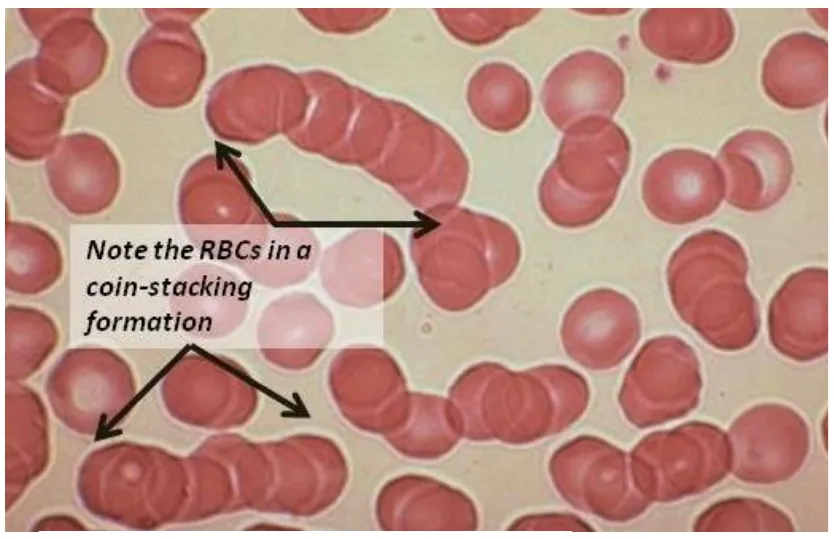
Teaching schematic
Two markers, two time scalesL2 p.30–31
| CRP | ESR | |
|---|---|---|
| What is measured | The protein itself | Speed of red-cell settling |
| Rise | Within ~6 h | Over days |
| Fall after recovery | Within days | Over weeks |
| Affected by red cells and age | No | Yes: anemia, pregnancy, older age and female sex raise it; polycythemia, sickle cells and spherocytes lower it |
| Best for | Acute changes; response to treatment | Slow, chronic disease activity (e.g. polymyalgia rheumatica, giant cell arteritis) |
What is measured
- CRP
- The protein itself
- ESR
- Speed of red-cell settling
Rise
- CRP
- Within ~6 h
- ESR
- Over days
Fall after recovery
- CRP
- Within days
- ESR
- Over weeks
Affected by red cells and age
- CRP
- No
- ESR
- Yes: anemia, pregnancy, older age and female sex raise it; polycythemia, sickle cells and spherocytes lower it
Best for
- CRP
- Acute changes; response to treatment
- ESR
- Slow, chronic disease activity (e.g. polymyalgia rheumatica, giant cell arteritis)
Worked calculation · Is this ESR raised for her age?
- Age
- 70 years (woman)
- Measured ESR
- 36 mm/h
Formula
Approximate upper limit (Westergren): men = age ÷ 2; women = (age + 10) ÷ 2
Ref. · Miller age-adjusted ruleKnowledge check · 2.4
Answer, read why, and try again whenever you like. Answers are kept in both study modes.
- Q1Cysteinyl leukotrienesL2 p.27Midterm sim. p.4
An 8-year-old girl with asthma, allergies and exercise-induced wheeze presents in respiratory distress.
Which mediator of inflammation is the most powerful stimulator of bronchoconstriction and vasoconstriction in this patient?
- Q2Acute-phase reactantsL2 p.29–30Midterm sim. p.5
A 25-year-old woman has two weeks of fever and chest pain, a red macular facial rash, tender wrist and elbow joints, mild anemia and thrombocytopenia. Her serum ceruloplasmin, fibrinogen, α₂-macroglobulin, serum amyloid A and C-reactive protein are all increased.
Together, these markers belong to which family of proteins?
- Q3Leukotriene drug targetsL2 p.26–27
A 35-year-old woman with asthma and nasal polyps wheezes within an hour of taking ibuprofen for a headache.
Which drug would reduce the synthesis of the mediators responsible?
- Q4CRP versus ESR kineticsL2 p.30–31
A 30-year-old woman is treated for pyelonephritis. On day 5 she is afebrile and well. Her CRP has fallen from 180 to 20 mg/L, but her ESR is still 60 mm/h.
What best explains the difference?
- Q5Rouleaux and ESR: image interpretationL2 p.31
A 68-year-old man has back pain and anemia. His ESR is 110 mm/h, and his blood smear shows the pattern in the image.
Lecture image
Which change in his plasma best explains the smear and the ESR?
- Q6Complement split productsL2 p.16, 27
Which complement fragment both attracts neutrophils and makes mast cells release histamine?