Phagocytosis and killing
Once in the tissue, the neutrophil recognizes a coated microbe, swallows it into a phagosome, fuses that with its granules, and kills inside the sealed compartment with an oxygen-driven chain that ends in hypochlorous acid. Each link has an inherited disease that shows what it does.
Recognition and opsonins
L2 p.17–18The lecture asks: what does opsonin mean? The word comes from the Greek for “to prepare food”. An opsonin coats a microbe and gives the phagocyte a handle to grip. Phagocytes can recognize some microbes directly (mannose and scavenger receptors), but coated microbes are engulfed far more efficiently.
Three opsonins and their receptorsL2 p.17–18, 30
| Opsonin | Made by | Receptor on the phagocyte |
|---|---|---|
| IgG | Plasma cells (adaptive immunity) | Fcγ receptors |
| C3b (and iC3b) | Complement activation on the microbe surface | CR1; CR3 (= Mac-1, CD11b/CD18) |
| CRP (and mannose-binding lectin) | Liver, as acute-phase proteins (2.4) | Binds phosphocholine on bacteria, then fixes C1q and complement |
IgG
- Made by
- Plasma cells (adaptive immunity)
- Receptor on the phagocyte
- Fcγ receptors
C3b (and iC3b)
- Made by
- Complement activation on the microbe surface
- Receptor on the phagocyte
- CR1; CR3 (= Mac-1, CD11b/CD18)
CRP (and mannose-binding lectin)
- Made by
- Liver, as acute-phase proteins (2.4)
- Receptor on the phagocyte
- Binds phosphocholine on bacteria, then fixes C1q and complement
After binding, the cell extends pseudopods around the particle and closes them into a phagosome. Granules and lysosomes fuse with it to form a phagolysosome. Killing happens inside that compartment, so the most toxic products are kept away from the neutrophil's own cytoplasm, until the cell dies or regurgitates its contents and damages the surrounding tissue.
The oxidative burst
L2 p.20When a neutrophil is activated, its oxygen consumption jumps, not to make ATP but to make oxidants: the respiratory (oxidative) burst. The enzyme is NADPH oxidase (phagocyte oxidase), whose membrane and cytosolic subunits assemble on the phagosome membrane only when the cell is stimulated.
Teaching schematic
- O₂
- NADPH oxidasefails in CGDO₂•⁻ superoxide
- Superoxide dismutaseH₂O₂
- Myeloperoxidase + Cl⁻fails in MPO deficiencyHOCl (bleach)
- Catalase-positive microbes destroy H₂O₂ (2 H₂O₂ → 2 H₂O + O₂), so a CGD neutrophil cannot borrow theirs.
Oxygen to bleach
- NADPH + O₂
- NADPH oxidase → superoxide (O₂•⁻)
- Superoxide dismutase (or spontaneous) → H₂O₂
- Myeloperoxidase + Cl⁻ → HOCl (hypochlorous acid)
- Halogenation and oxidation of microbial proteins and lipids
NADPH comes from the hexose monophosphate shunt, whose first enzyme is G6PD; very severe G6PD deficiency can weaken the burst. Iron can also turn H₂O₂ into hydroxyl radical (Fenton reaction), the same chemistry that injures cells in Chapter 1. Myeloperoxidase is stored in the azurophilic (primary) granules and gives pus its green tint.
Killing that does not need oxygenRef. · Robbins; Abbas
| Agent | Action |
|---|---|
| Lysozyme | Hydrolyses bacterial cell-wall peptidoglycan |
| Lactoferrin | Binds iron that bacteria need |
| Defensins, cathelicidins | Cationic peptides that punch holes in microbial membranes |
| Elastase and other proteases | Degrade microbial proteins (and, when released, host matrix) |
| Major basic protein (eosinophils) | Toxic to helminths |
| Nitric oxide (macrophages, iNOS) | With superoxide forms peroxynitrite |
Lysozyme
- Action
- Hydrolyses bacterial cell-wall peptidoglycan
Lactoferrin
- Action
- Binds iron that bacteria need
Defensins, cathelicidins
- Action
- Cationic peptides that punch holes in microbial membranes
Elastase and other proteases
- Action
- Degrade microbial proteins (and, when released, host matrix)
Major basic protein (eosinophils)
- Action
- Toxic to helminths
Nitric oxide (macrophages, iNOS)
- Action
- With superoxide forms peroxynitrite
Core activity · The Missing Arrow
The Missing Arrow
A neutrophil has engulfed Staphylococcus aureus. Complete the chain inside the phagolysosome, then notice where each inherited defect would cut it.
The mechanism gaps are enzymes; the event gaps are the molecules they make.
- Phagosome fuses with granules: a phagolysosome
- superoxide dismutase (or spontaneous dismutation)
Cards
Tap or drag a card. Placing a used card moves it.
Lysosomal enzymes, collateral injury and abscesses
L2 p.17, 32–33Ch. II cases p.12Neutrophil granules hold the enzymes that finish the job in the phagolysosome: elastase, collagenases, gelatinase, cathepsins, lysozyme and myeloperoxidase. The same contents also escape into the tissue, which is why inflammation damages the host it protects.
- Leak during feeding: granules fuse with the phagosome before it has fully closed (“regurgitation”).
- Frustrated phagocytosis: the target is too large or flat to swallow (immune complexes on a basement membrane, a prosthesis), so the cell empties its granules onto it.
- Membranolytic material: urate or silica crystals rupture the phagolysosome from inside, releasing its contents and killing the cell.
- Neutrophil death: dying neutrophils release their contents, and extracellular traps (NETs) of chromatin and granule proteins.
Where neutrophils accumulate densely around pyogenic bacteria, these proteases and oxidants digest bacteria, dead neutrophils and the surrounding tissue alike: liquefactive necrosis. The liquid product is pus, and a localized collection of it is an abscess (2.5). Antiproteases in plasma and tissue fluid, such as α₁-antitrypsin and α₂-macroglobulin, normally limit the spread; when they are overwhelmed or deficient, the damage extends (emphysema in α₁-antitrypsin deficiency).
From recruitment to an abscess
- Pyogenic bacteria in tissue
- Neutrophils recruited in large numbers
- Phagocytosis and degranulation; enzymes and oxidants also released outside
- Bacteria, neutrophils and tissue digested (liquefactive necrosis)
- Pus collects and is walled off: an abscess
Chédiak–Higashi syndrome: granules that cannot move
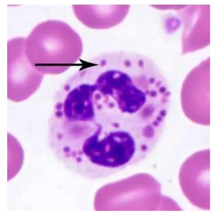
L2 p.19Chédiak–Higashi syndrome (autosomal recessive) is caused by mutations in LYST, a lysosomal trafficking regulator. The lecture calls it a protein-trafficking defect that disturbs microtubule-guided movement, so lysosomes fail to fuse with phagosomes. Lysosome-related organelles instead fuse with each other into giant granules, and every cell type that uses such organelles is affected.
One trafficking defect, many cellsL2 p.19Ref. · Robbins; Abbas
| Finding | Why |
|---|---|
| Recurrent pyogenic infections (S. aureus, streptococci) | Phagolysosome fusion and degranulation are slow; chemotaxis is impaired |
| Giant granules in neutrophils and other leukocytes | Lysosomes fuse with each other instead of with phagosomes |
| Mild neutropenia | Abnormal precursors are destroyed in the marrow |
| Partial oculocutaneous albinism, silvery hair | Melanosomes clump and cannot transfer pigment |
| Bleeding, easy bruising | Platelet dense granules are deficient |
| Peripheral neuropathy | Abnormal trafficking in neurons |
| Risk of an accelerated phase (hemophagocytic lymphohistiocytosis) | NK and cytotoxic T cells cannot release their granules |
Recurrent pyogenic infections (S. aureus, streptococci)
- Why
- Phagolysosome fusion and degranulation are slow; chemotaxis is impaired
Giant granules in neutrophils and other leukocytes
- Why
- Lysosomes fuse with each other instead of with phagosomes
Mild neutropenia
- Why
- Abnormal precursors are destroyed in the marrow
Partial oculocutaneous albinism, silvery hair
- Why
- Melanosomes clump and cannot transfer pigment
Bleeding, easy bruising
- Why
- Platelet dense granules are deficient
Peripheral neuropathy
- Why
- Abnormal trafficking in neurons
Risk of an accelerated phase (hemophagocytic lymphohistiocytosis)
- Why
- NK and cytotoxic T cells cannot release their granules
Lecture image
Chronic granulomatous disease
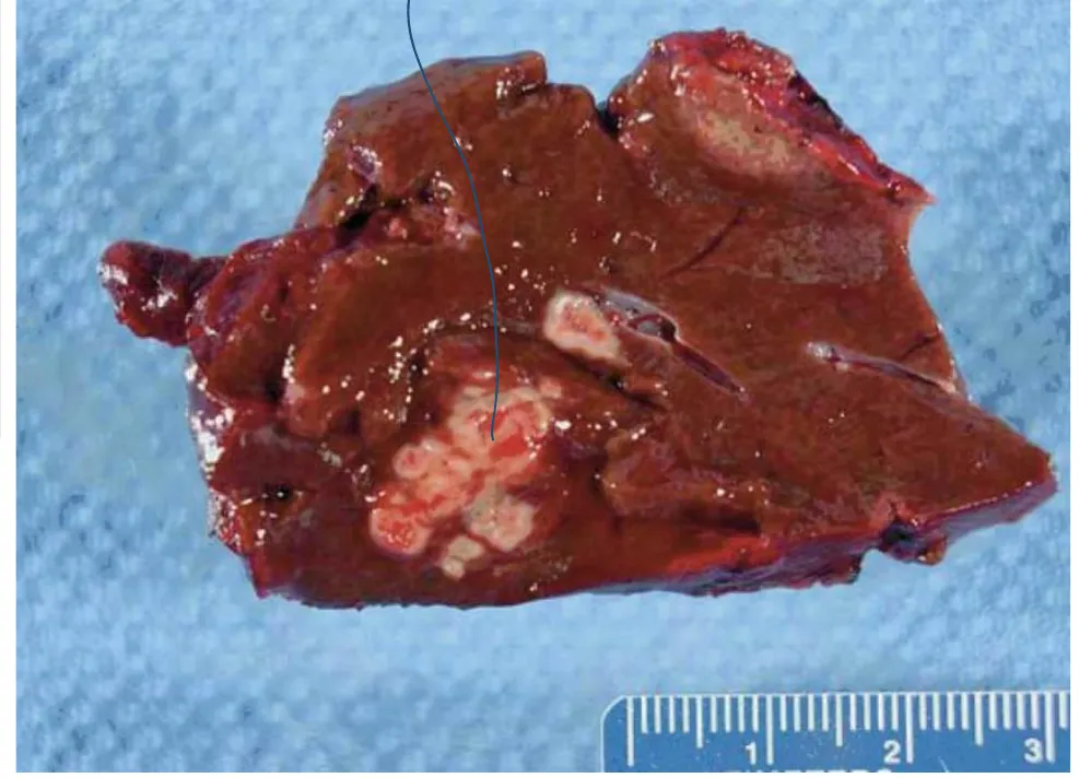
L2 p.20–25In chronic granulomatous disease (CGD), NADPH oxidase does not work. About two-thirds of cases are X-linked (the gp91phox subunit, gene CYBB); the rest are autosomal recessive. Neutrophils are recruited normally, engulf normally and form pus, but cannot make superoxide, so their own burst produces no H₂O₂ and no HOCl.
Why catalase matters. Many bacteria generate H₂O₂ themselves. If a catalase-negative organism (for example a streptococcus) is engulfed, the CGD neutrophil can borrow that H₂O₂, and its intact myeloperoxidase turns it into HOCl. Catalase-positive organisms destroy their own H₂O₂ (2 H₂O₂ → 2 H₂O + O₂), leaving the neutrophil nothing to work with. Those organisms survive: **Staphylococcus aureus, Serratia marcescens, Burkholderia cepacia, Nocardia, Aspergillus* and Candida*.
Organisms that survive inside phagocytes keep stimulating T cells and macrophages, so neutrophils are joined by lymphocytes and macrophages that fuse into giant cells: granulomas, often with abscesses, in lymph nodes, lungs, liver, skin and bone. Pneumonia is the most common presentation; Aspergillus is the leading cause of death.
Lecture image
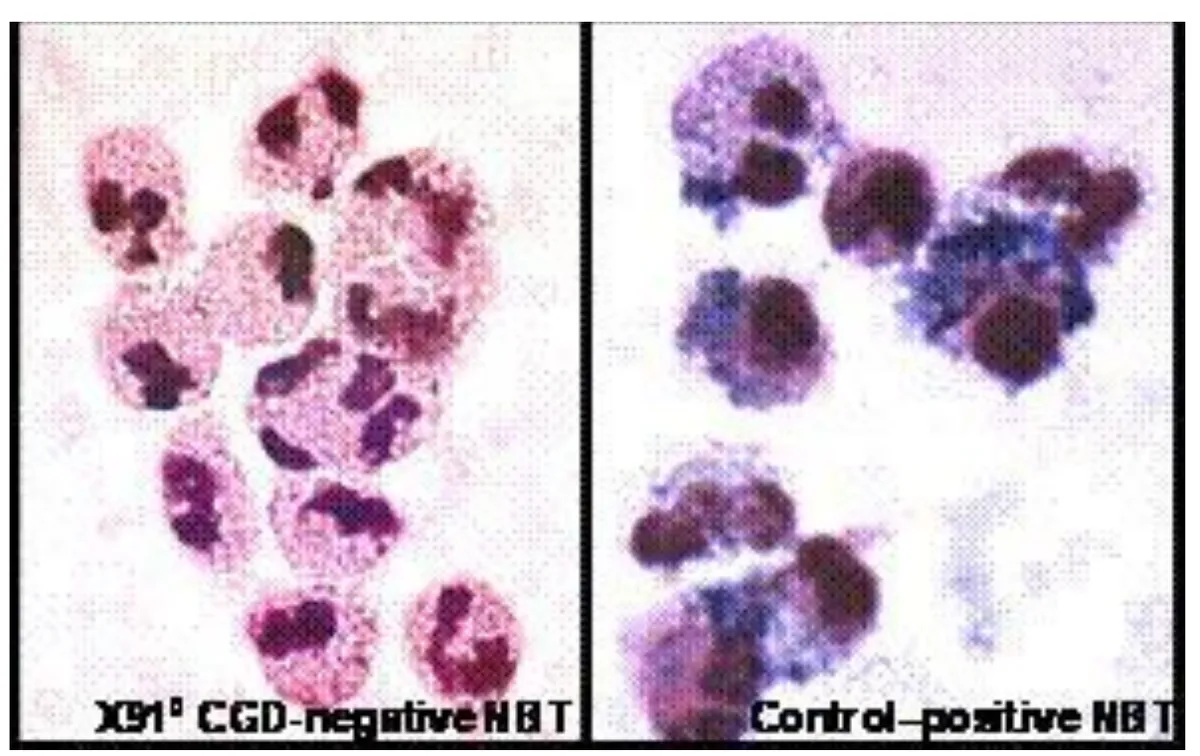
Testing the burst. In the nitroblue tetrazolium (NBT) test, stimulated neutrophils that make superoxide reduce the yellow dye to dark blue formazan; blue cells mean the burst is intact (a positive test). CGD neutrophils stay unstained. The more sensitive dihydrorhodamine (DHR) flow cytometry test measures oxidation of DHR to fluorescent rhodamine after stimulation with PMA, a protein kinase C agonist: CGD cells do not fluoresce. Mothers who carry X-linked CGD show two populations, one normal and one not, because of X inactivation.
Lecture image
Myeloperoxidase deficiency
L2 p.20Myeloperoxidase deficiency (autosomal recessive) is the most common inherited phagocyte defect. The burst runs normally and makes plenty of H₂O₂, which alone kills most bacteria, but no HOCl. Most people never know they have it. The exception is *Candida*: disseminated or persistent candidiasis, especially in people with diabetes, whose neutrophils already work less well.
Same pathway, different step
CGD
- Fails at NADPH oxidase
- No superoxide, no H₂O₂
- NBT negative, DHR negative
- Serious catalase-positive infections, granulomas
MPO deficiency
- Fails at myeloperoxidase
- Superoxide and H₂O₂ made; no HOCl
- NBT normal; MPO stain absent
- Usually healthy; Candida
Find the broken step: recruitment and killing lab
L2 p.10–25Five inherited defects, five places where the same pathway can break. For each one you are told only what is molecularly wrong. Predict the blood and the tissue first; the lab then shows where the neutrophil stops and which clinical clue gives it away.
Centerpiece · Recruitment and killing lab
Find the broken step
0 of 5 defects worked out.
Step 1 · Rolling
E-/P-selectin ↔ sialyl-Lewis X; L-selectin
Selectins on activated endothelium catch sugar ligands on the neutrophil. Bonds form and break, so the cell tumbles slowly along the venule wall.
Tap each step to see its molecules. When the sequence is clear, choose defect 1 above and break it.
Transfer round · same mechanisms, new wording
Work out all five defects above to open five short cases that describe them in different words.
The five defects side by sideL2 p.14, 19–25
| Defect | Failed step | Blood | Tissue | Clinical clue | Test |
|---|---|---|---|---|---|
| LAD II | Rolling (no sialyl-Lewis X) | Neutrophilia | Little pus, milder infections | Bombay phenotype, intellectual disability | CD15s absent |
| LAD I | Tight adhesion (CD18) | Marked neutrophilia | No pus, poor healing | Delayed cord separation, omphalitis | CD18 absent |
| Chédiak–Higashi | Phagolysosome fusion (LYST) | Neutropenia, giant granules | Pyogenic infections | Albinism, neuropathy, bleeding | Smear: giant granules |
| CGD | Killing: NADPH oxidase | Normal | Pus + granulomas | Catalase-positive infections | DHR/NBT abnormal |
| MPO deficiency | Killing: MPO | Normal | Usually normal | Candida | NBT normal; MPO stain absent |
LAD II
- Failed step
- Rolling (no sialyl-Lewis X)
- Blood
- Neutrophilia
- Tissue
- Little pus, milder infections
- Clinical clue
- Bombay phenotype, intellectual disability
- Test
- CD15s absent
LAD I
- Failed step
- Tight adhesion (CD18)
- Blood
- Marked neutrophilia
- Tissue
- No pus, poor healing
- Clinical clue
- Delayed cord separation, omphalitis
- Test
- CD18 absent
Chédiak–Higashi
- Failed step
- Phagolysosome fusion (LYST)
- Blood
- Neutropenia, giant granules
- Tissue
- Pyogenic infections
- Clinical clue
- Albinism, neuropathy, bleeding
- Test
- Smear: giant granules
CGD
- Failed step
- Killing: NADPH oxidase
- Blood
- Normal
- Tissue
- Pus + granulomas
- Clinical clue
- Catalase-positive infections
- Test
- DHR/NBT abnormal
MPO deficiency
- Failed step
- Killing: MPO
- Blood
- Normal
- Tissue
- Usually normal
- Clinical clue
- Candida
- Test
- NBT normal; MPO stain absent
Knowledge check · 2.3
Answer, read why, and try again whenever you like. Answers are kept in both study modes.
- Q1CGD: abnormal DHRL2 p.21–25Ch. II cases p.8
A 2-year-old boy has a red, edematous rash on his shin. He has had several Staphylococcus aureus skin infections and a pulmonary Aspergillus infection. Earlier flow cytometry showed that his neutrophils, stimulated with protein kinase C agonists, could not oxidize a nonfluorescent substrate into a fluorescent product.
The activity of which enzyme is most likely decreased?
- Q2Neutrophil killing: H₂O₂L2 p.20Ch. II cases p.6
A 58-year-old woman has had cough and fever for 3 days. A chest radiograph shows right lower lobe infiltrates, and sputum grows Streptococcus pneumoniae.
Clearance of these organisms would be most effectively accomplished by generation of which substance by the main inflammatory cell responding?
- Q3Myeloperoxidase makes HOClL2 p.20Midterm sim. p.4
A 75-year-old woman has chest pain, fever and a cough with rust-colored sputum. A chest radiograph shows a right middle lobe infiltrate, and sputum grows Streptococcus pneumoniae.
Phagocytes in the affected lung generate bactericidal hypochlorous acid using which enzyme?
- Q4Chédiak–Higashi syndrome: image recognitionL2 p.19
A 4-year-old boy with silvery hair and light-sensitive eyes has had recurrent S. aureus skin abscesses and bruises easily. His blood smear is shown.
Lecture image
Which process is defective?
- Q5NBT test: image interpretationL2 p.22, 25
A 7-year-old boy has had Serratia osteomyelitis and a liver abscess. His stimulated neutrophils (left panel) are compared with a control (right) in the test shown.
Lecture image
Which infection pattern fits this result?