Regeneration and repair
Damaged tissue is restored in one of two ways: surviving cells divide and rebuild the original (regeneration), or new vessels and fibroblasts fill the gap with collagen (repair by scar). Which one happens depends on whether the cells can divide and whether the framework survived.
Regeneration or scar: cells and scaffold
L2 p.47Regeneration needs two things: cells that can still divide, and an intact extracellular-matrix scaffold (basement membranes and stroma) to guide them back into the original architecture.
Cells by their capacity to divideL2 p.47
| Group | Behaviour | Examples (lecture examples in bold) |
|---|---|---|
| Labile | Divide continuously from stem cells | Basal layer of the epidermis (skin cut), gut epithelium, hematopoietic cells (anemia), lining epithelia |
| Stable | Quiescent, but divide when stimulated | Hepatocytes, renal tubular cells, type II pneumocytes (lung injury), fibroblasts, endothelium, smooth muscle |
| Permanent | Cannot divide meaningfully after birth | Neurons, cardiac myocytes (scar after myocardial infarction); skeletal muscle has only limited satellite-cell repair |
Labile
- Behaviour
- Divide continuously from stem cells
- Examples (lecture examples in bold)
- Basal layer of the epidermis (skin cut), gut epithelium, hematopoietic cells (anemia), lining epithelia
Stable
- Behaviour
- Quiescent, but divide when stimulated
- Examples (lecture examples in bold)
- Hepatocytes, renal tubular cells, type II pneumocytes (lung injury), fibroblasts, endothelium, smooth muscle
Permanent
- Behaviour
- Cannot divide meaningfully after birth
- Examples (lecture examples in bold)
- Neurons, cardiac myocytes (scar after myocardial infarction); skeletal muscle has only limited satellite-cell repair
The scaffold decides as much as the cell. After acetaminophen injury, hepatocytes die but the reticulin framework survives, so the liver regenerates its lobules perfectly. In a liver abscess, the framework is destroyed with the cells, so the same stable hepatocytes cannot rebuild the architecture and the defect heals by scar. Repeated injury with framework loss produces cirrhosis: regenerating nodules separated by scar. The lecture's rule follows: repair by connective tissue occurs when injury is severe or persistent, or when the cells are permanent.
The wound-healing sequence
L2 p.47–50Ch. II cases p.5, 16Healing a skin wound runs through overlapping phases. The timing below is for a clean, sutured incision; larger wounds follow the same order more slowly.
Teaching schematic
What a biopsy would show, day by dayRef. · Robbins
| Time | Phase | What is happening |
|---|---|---|
| Minutes–hours | Hemostasis | Platelets and fibrin plug the gap; platelets release PDGF and TGF-β; the clot is a provisional matrix |
| 24 h | Inflammation | Neutrophils at the margins; basal epidermal cells start to migrate and divide (re-epithelialization begins) |
| Days 2–3 | Inflammation → proliferation | Macrophages replace neutrophils, clear fibrin and debris, and release growth factors; granulation tissue begins |
| Day 5 | Proliferation | Granulation tissue fills the incision; neovascularization peaks; type III collagen; epidermis restored to normal thickness |
| Week 2 | Proliferation | Collagen accumulates, fibroblasts proliferate; fewer leukocytes, less edema, fewer vessels (blanching) |
| Month 1 onward | Remodeling | Cellular scar of collagen; type III replaced by type I; cross-linking by lysyl oxidase; collagenases (MMPs) reshape it; contraction by myofibroblasts |
Minutes–hours
- Phase
- Hemostasis
- What is happening
- Platelets and fibrin plug the gap; platelets release PDGF and TGF-β; the clot is a provisional matrix
24 h
- Phase
- Inflammation
- What is happening
- Neutrophils at the margins; basal epidermal cells start to migrate and divide (re-epithelialization begins)
Days 2–3
- Phase
- Inflammation → proliferation
- What is happening
- Macrophages replace neutrophils, clear fibrin and debris, and release growth factors; granulation tissue begins
Day 5
- Phase
- Proliferation
- What is happening
- Granulation tissue fills the incision; neovascularization peaks; type III collagen; epidermis restored to normal thickness
Week 2
- Phase
- Proliferation
- What is happening
- Collagen accumulates, fibroblasts proliferate; fewer leukocytes, less edema, fewer vessels (blanching)
Month 1 onward
- Phase
- Remodeling
- What is happening
- Cellular scar of collagen; type III replaced by type I; cross-linking by lysyl oxidase; collagenases (MMPs) reshape it; contraction by myofibroblasts
Re-epithelialization. Epidermal cells at the wound edge (and from surviving hair follicles) lose their attachments, migrate across the provisional matrix under the scab, and proliferate under EGF, TGF-α and KGF until the layers meet; then they stratify. In a sutured wound this happens within 24–48 hours.
Strength. When skin sutures are removed at about a week, the wound has roughly 10% of normal strength. Strength rises quickly over the next two months as collagen is made and cross-linked, plateaus at about 70–80% by three months, and never fully returns.
Granulation tissue and angiogenesis
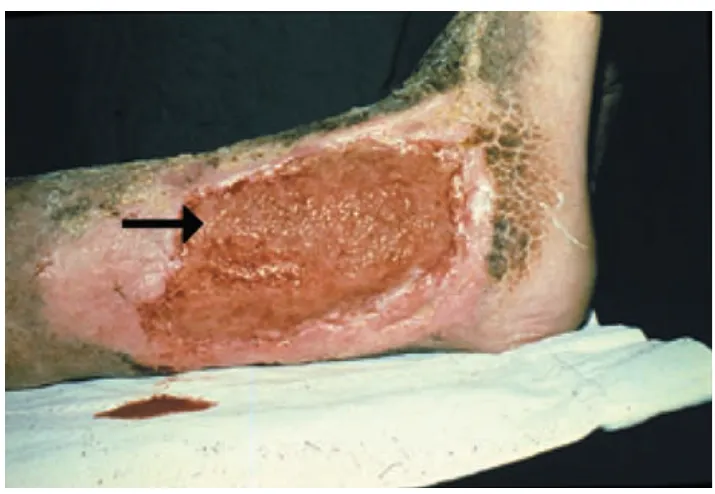
L2 p.47–49Granulation tissue is the initial phase of repair and the precursor of scar. To the eye it is soft, pink and granular, and bleeds easily; each “granule” is a loop of new capillary. Under the microscope it has three components: new, leaky blood vessels, activated fibroblasts and myofibroblasts, and macrophages (with some other inflammatory cells), in an edematous matrix containing type III collagen.
Lecture image
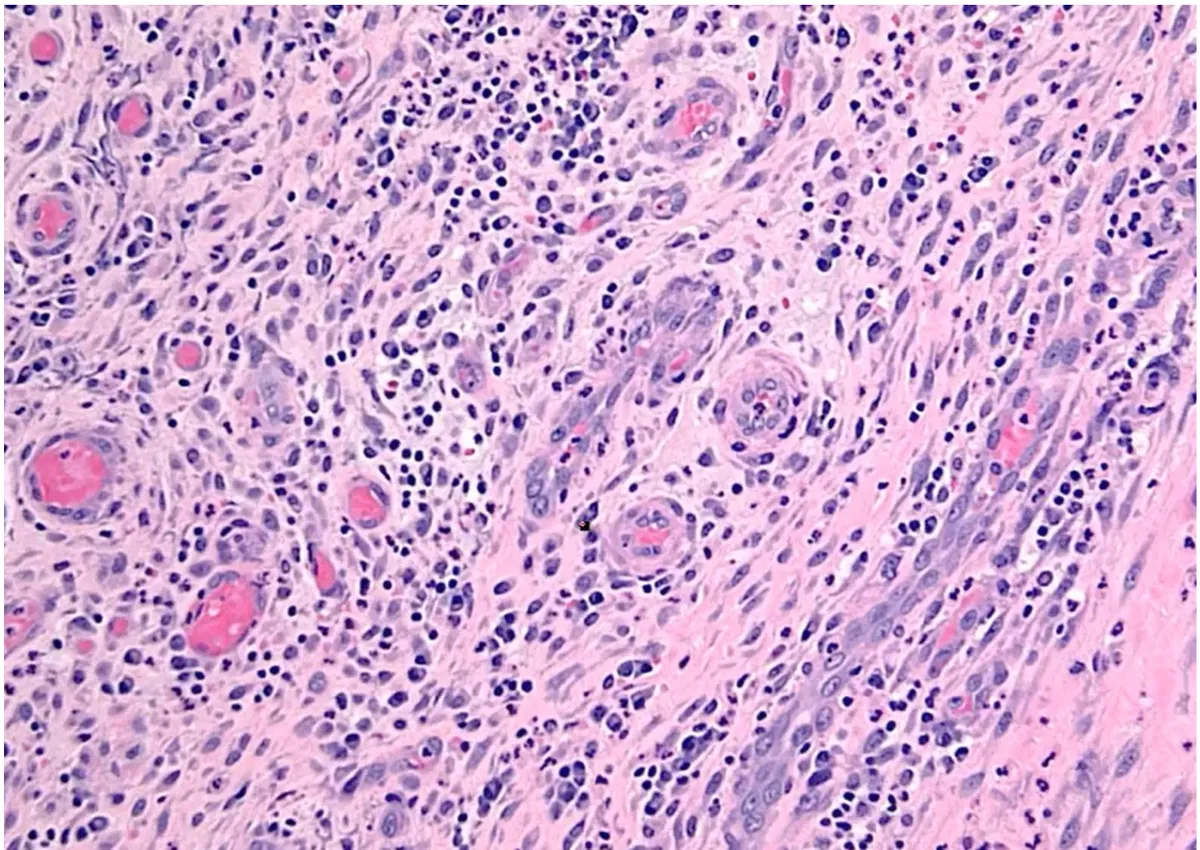
- Vasodilation (nitric oxide) and increased permeability (VEGF).
- Pericytes separate, and matrix metalloproteinases break down the basement membrane.
- Endothelial cells migrate toward the wound and proliferate behind the leading tip cell.
- The sprouts form tubes and connect into loops.
- Pericytes and smooth-muscle cells are recruited and the new vessels stabilize; until then they leak, which is why granulation tissue is edematous.
Case image
Growth factors that drive repair
L2 p.48The lecture's five factorsL2 p.48
| Factor | Main sources | Main actions |
|---|---|---|
| VEGF | Macrophages, many cells under hypoxia (HIF-1α) | Angiogenesis: endothelial migration and proliferation; vascular permeability |
| FGF (FGF-2) | Macrophages, endothelium, mast cells | Angiogenesis; fibroblast proliferation; epithelial migration |
| PDGF | Platelets, macrophages, endothelium | Migration and proliferation of fibroblasts and smooth-muscle cells; attracts monocytes |
| EGF (and TGF-α) | Platelets, macrophages, saliva, keratinocytes | Keratinocyte migration and proliferation (re-epithelialization); fibroblast proliferation |
| TGF-β | Platelets, macrophages, many cells | The main fibrogenic factor: fibroblast migration and proliferation, more collagen and fibronectin, less matrix breakdown (MMPs ↓, TIMPs ↑); also anti-inflammatory |
VEGF
- Main sources
- Macrophages, many cells under hypoxia (HIF-1α)
- Main actions
- Angiogenesis: endothelial migration and proliferation; vascular permeability
FGF (FGF-2)
- Main sources
- Macrophages, endothelium, mast cells
- Main actions
- Angiogenesis; fibroblast proliferation; epithelial migration
PDGF
- Main sources
- Platelets, macrophages, endothelium
- Main actions
- Migration and proliferation of fibroblasts and smooth-muscle cells; attracts monocytes
EGF (and TGF-α)
- Main sources
- Platelets, macrophages, saliva, keratinocytes
- Main actions
- Keratinocyte migration and proliferation (re-epithelialization); fibroblast proliferation
TGF-β
- Main sources
- Platelets, macrophages, many cells
- Main actions
- The main fibrogenic factor: fibroblast migration and proliferation, more collagen and fibronectin, less matrix breakdown (MMPs ↓, TIMPs ↑); also anti-inflammatory
Healing by primary, secondary and tertiary intention
L2 p.50Teaching schematic
Primary intention
Clean incision Sutured Fine scar Secondary intention
Gaping wound Granulation fills it Contracted, larger scar Tertiary intention
Contaminated wound Cleaned first Closed late
Three ways to close a woundL2 p.50
| Intention | What is done | When | Result |
|---|---|---|---|
| Primary | Edges approximated by suture | Clean surgical incisions | Little granulation tissue; thin, fine scar |
| Secondary | Wound left open to fill from the base and close by re-epithelialization | Large or highly contaminated wounds, ulcers, drained abscesses | Abundant granulation tissue, marked contraction by myofibroblasts, larger scar |
| Tertiary (delayed primary) | Repeated débridement and antibiotics for several days, then surgical closure | Contaminated wounds that need cleaning first | Closure once infection is controlled; scar intermediate |
Primary
- What is done
- Edges approximated by suture
- When
- Clean surgical incisions
- Result
- Little granulation tissue; thin, fine scar
Secondary
- What is done
- Wound left open to fill from the base and close by re-epithelialization
- When
- Large or highly contaminated wounds, ulcers, drained abscesses
- Result
- Abundant granulation tissue, marked contraction by myofibroblasts, larger scar
Tertiary (delayed primary)
- What is done
- Repeated débridement and antibiotics for several days, then surgical closure
- When
- Contaminated wounds that need cleaning first
- Result
- Closure once infection is controlled; scar intermediate
Core activity · One Fact Changes
One Fact Changes
1 · The original case
A 25-year-old cook cuts the back of his hand on a clean knife. The edges are sutured the same day. He returns on day 5. A tiny biopsy is taken from the wound.
What dominates the wound now?
Which fact decided it?
What slows healing
L2 p.51Collagen is made in steps, and several nutrients and drugs act at specific steps. That is why the lecture's list of factors that impair healing reads like a map of collagen synthesis.
Teaching schematic
- Pro-α chains made on rough ER (Gly-X-Y repeats)Impaired by Protein malnutrition
- Hydroxylation of proline and lysineImpaired by Vitamin C deficiency; hypoxia
- Triple helix (procollagen) secretedImpaired by Glucocorticoids ↓ synthesis
- Ends cleaved → tropocollagen → fibrils
- Cross-linking by lysyl oxidaseImpaired by Copper deficiency
- Remodeling by collagenases (MMPs); type III → type IImpaired by Zinc deficiency
Factors that impair wound healingL2 p.51
| Factor | Mechanism |
|---|---|
| Persistent infection | The single most important local factor: prolongs inflammation and tissue destruction |
| Diabetes mellitus | Microangiopathy (poor perfusion), impaired neutrophil function, glycation of matrix, neuropathy and repeated trauma |
| Poor perfusion | Atherosclerosis or venous stasis: too little oxygen and nutrients for fibroblasts (oxygen is needed for prolyl and lysyl hydroxylation) |
| Vitamin C deficiency | Prolyl and lysyl hydroxylases need ascorbate; unhydroxylated procollagen cannot form a stable triple helix (scurvy) |
| Copper deficiency | Lysyl oxidase, a copper enzyme, cross-links collagen; weak cross-linking, weak scar |
| Zinc deficiency | Cofactor of collagenases (matrix metalloproteinases) that remodel matrix, and of enzymes for DNA synthesis and cell proliferation |
| Glucocorticoids | Suppress inflammation and TGF-β, reduce collagen synthesis: weaker scar, lower tensile strength |
| Others | Mechanical stress on the wound, foreign bodies, malnutrition (protein), large wound size |
Persistent infection
- Mechanism
- The single most important local factor: prolongs inflammation and tissue destruction
Diabetes mellitus
- Mechanism
- Microangiopathy (poor perfusion), impaired neutrophil function, glycation of matrix, neuropathy and repeated trauma
Poor perfusion
- Mechanism
- Atherosclerosis or venous stasis: too little oxygen and nutrients for fibroblasts (oxygen is needed for prolyl and lysyl hydroxylation)
Vitamin C deficiency
- Mechanism
- Prolyl and lysyl hydroxylases need ascorbate; unhydroxylated procollagen cannot form a stable triple helix (scurvy)
Copper deficiency
- Mechanism
- Lysyl oxidase, a copper enzyme, cross-links collagen; weak cross-linking, weak scar
Zinc deficiency
- Mechanism
- Cofactor of collagenases (matrix metalloproteinases) that remodel matrix, and of enzymes for DNA synthesis and cell proliferation
Glucocorticoids
- Mechanism
- Suppress inflammation and TGF-β, reduce collagen synthesis: weaker scar, lower tensile strength
Others
- Mechanism
- Mechanical stress on the wound, foreign bodies, malnutrition (protein), large wound size
Hypertrophic scars, keloids and contractures
L2 p.52Ch. II cases p.17, 19Repair can overshoot. Too much collagen gives a raised scar; too much contraction gives a contracture.
Hypertrophic scar versus keloid
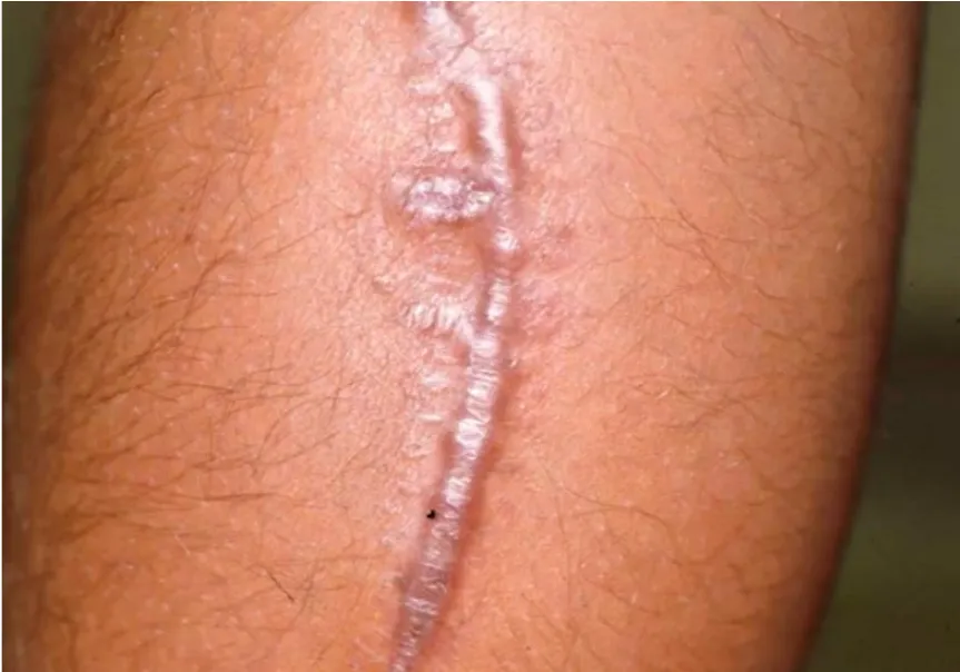
Hypertrophic scar
- Raised, red, stays within the original wound
- Develops within weeks; often over joints or after burns
- Collagen bundles parallel to the epidermis, nodules with myofibroblasts
- Frequently regresses spontaneously
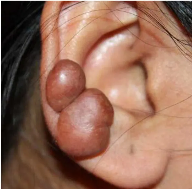
Keloid
- Raised, grows beyond the borders of the wound
- Genetic predisposition: more common with darker skin; earlobes, chest, shoulders
- Thick, glassy, haphazard bundles of type I and III collagen
- Persists; recurs after excision; refractory to treatment
Lecture image
Lecture image
Both are driven by excess TGF-β signalling and fibroblast activity, and an imbalance between collagen synthesis and breakdown.
Myofibroblasts are fibroblasts that express α-smooth-muscle actin. They appear in granulation tissue and pull the wound edges together, which is helpful in secondary intention. When contraction is excessive, especially after burns over joints, palms, soles or the front of the chest, the scar shortens and fixes the joint: a contracture.
Core activity · Almost Twins
Almost Twins
Look-alike A
Hypertrophic scar
Lecture image
- Raised, firm scar
- Stays within the borders of the original wound
- Often regresses over months
- Collagen bundles parallel to the epidermis
Look-alike A
Hypertrophic scar
Lecture image
- Raised, firm scar
- Stays within the borders of the original wound
- Often regresses over months
- Collagen bundles parallel to the epidermis
Look-alike B
Keloid
Lecture image
- Raised, firm scar
- Grows beyond the borders of the original wound
- Persists; recurs after excision
- Thick, glassy, haphazard collagen bundles; genetic predisposition
Step 1 · What they share
What do the two scars share?
Select every feature that both show.
Granuloma versus granulation tissue
L2 p.42, 47–49The words look alike and both appear in inflamed tissue, but they are different structures with different purposes. A granuloma is a defence: macrophages walling off something they cannot destroy. Granulation tissue is repair: vessels and fibroblasts filling a gap.
Make it unmistakableL2 p.42–44, 47–48
| Granuloma | Granulation tissue | |
|---|---|---|
| Purpose | Contain a persistent agent | Fill a defect and build a scar |
| Key cells | Epithelioid macrophages, giant cells, lymphocyte cuff | New capillaries, fibroblasts and myofibroblasts, macrophages |
| Shape | Compact, round nodule | Diffuse layer filling a wound bed |
| Driven by | IL-12 → Th1 → IFN-γ; TNF | VEGF, FGF, PDGF, TGF-β |
| Matrix | Little; fibrous rim when old | Edematous, type III collagen |
| Gross look | Pale, white nodule | Red, moist, granular, bleeds easily |
| Examples | TB, sarcoidosis, Crohn disease, foreign body | Healing wound or ulcer, organizing infarct, abscess wall |
| Becomes | Fibrosis and calcification, or reactivation | Scar (type I collagen) |
Purpose
- Granuloma
- Contain a persistent agent
- Granulation tissue
- Fill a defect and build a scar
Key cells
- Granuloma
- Epithelioid macrophages, giant cells, lymphocyte cuff
- Granulation tissue
- New capillaries, fibroblasts and myofibroblasts, macrophages
Shape
- Granuloma
- Compact, round nodule
- Granulation tissue
- Diffuse layer filling a wound bed
Driven by
- Granuloma
- IL-12 → Th1 → IFN-γ; TNF
- Granulation tissue
- VEGF, FGF, PDGF, TGF-β
Matrix
- Granuloma
- Little; fibrous rim when old
- Granulation tissue
- Edematous, type III collagen
Gross look
- Granuloma
- Pale, white nodule
- Granulation tissue
- Red, moist, granular, bleeds easily
Examples
- Granuloma
- TB, sarcoidosis, Crohn disease, foreign body
- Granulation tissue
- Healing wound or ulcer, organizing infarct, abscess wall
Becomes
- Granuloma
- Fibrosis and calcification, or reactivation
- Granulation tissue
- Scar (type I collagen)
Core activity · Almost Twins
Almost Twins
Look-alike A
Granuloma
Lecture image
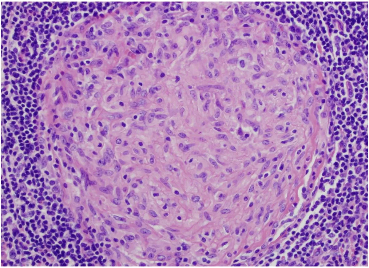
- Contains macrophages
- Compact nodule of epithelioid macrophages, giant cells, lymphocyte cuff
- Driven by IL-12 → Th1 → IFN-γ; TNF holds it together
- Purpose: contain an agent that cannot be destroyed
Look-alike A
Granuloma
Lecture image
- Contains macrophages
- Compact nodule of epithelioid macrophages, giant cells, lymphocyte cuff
- Driven by IL-12 → Th1 → IFN-γ; TNF holds it together
- Purpose: contain an agent that cannot be destroyed
Look-alike B
Granulation tissue
Lecture image
- Contains macrophages
- Diffuse layer of new capillaries with fibroblasts and myofibroblasts
- Driven by VEGF, FGF, PDGF and TGF-β
- Purpose: fill a defect; becomes scar
Step 1 · What they share
What do these two share?
Select every feature that both show.
Knowledge check · 2.7
Answer, read why, and try again whenever you like. Answers are kept in both study modes.
- Q1Day 5: granulation tissueL2 p.47–48Ch. II cases p.5
A 36-year-old man was cut on the lower leg by a machine at work 5 days ago. He cleaned and bandaged the shallow 5-cm laceration, which now shows slight redness, minimal swelling and no drainage.
Which finding is most likely on pathologic examination of the wound?
- Q2Granulation tissue: angiogenesis + type III collagenL2 p.47–48Ch. II cases p.16
A 3-year-old boy is seen 5 days after a forehead laceration. The linear wound is well approximated, clean and dry, with a small amount of pink granulation tissue.
Microscopic examination of the wound would most likely show:
- Q3Granulation tissue cellsL2 p.47, 49Midterm sim. p.5–6
A 70-year-old woman with diabetes develops an ulcer on her right leg. The ulcer bed (arrow) is covered with granulation tissue.
Case image
Which are the principal cellular components in the bed of this wound?
- Q4Zinc and wound healingL2 p.51Ch. II cases p.10
A 38-year-old man has two months of hair loss, a rash and a metallic taste. He has Crohn disease treated with several small-bowel resections and no regular care. There is a red rash around his mouth, excoriated erythema around the anus and buttocks, and patchy scalp alopecia.
He is deficient in which element, which would also impair his wound healing?
- Q5Hypertrophic scar: TGF-βL2 p.48, 52Ch. II cases p.17
Six months after a lower-segment cesarean section, a 26-year-old woman has a linear, red, raised suprapubic scar confined within the original wound margins. Biopsy shows dense collagen fibres arranged parallel to the epidermis.
Increased production of which mediator is most likely responsible?
- Q6Contracture: myofibroblastsL2 p.47Ch. II cases p.19
A 30-year-old man is seen a month after a chemical burn over the back of his right hand and forearm. The hand is hyperextended at the wrist; the skin over the dorsum is tense, with a thick, epithelialized scar, and wrist motion is restricted.
The contracture is most likely due to the activity of which cells?
- Q7Granulation tissue vs granuloma: image recognitionL2 p.47–49
A biopsy is taken from the base of a skin ulcer that has been healing for two weeks.
Lecture image
What is this tissue?