Patterns and outcomes of acute inflammation
What the exudate looks like depends on how much leaks, whether fibrinogen gets out, whether neutrophils pile up and whether a surface is lost. How the episode ends depends on how much tissue was destroyed and whether the cause is gone.
Morphologic patterns: what the exudate looks like
L2 p.32–38The lecture names five patterns. Each one tells you something about the cause: the size of the vascular leak, whether large proteins escape, which organism is involved and which surface is hit.
Five patterns and what produces themL2 p.32–38
| Pattern | What forms | Classic examples | Clue |
|---|---|---|---|
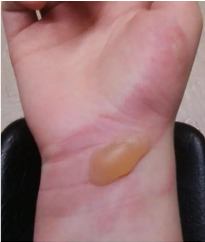
| Serous | Thin, watery fluid with too little fibrinogen to make fibrin | Blister of a second-degree burn; viral pleuritis; effusions | Clear fluid, few cells |
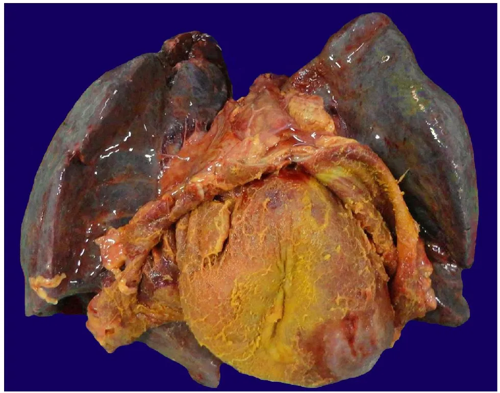
| Fibrinous | A larger leak lets fibrinogen out; it polymerizes on the surface | Pericarditis, pleuritis, peritonitis | Friction rub; shaggy “bread-and-butter” surface |
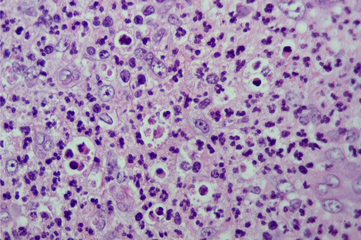
| Purulent (suppurative) | Pus: neutrophils, necrotic cells, edema fluid | S. aureus abscess, appendicitis | Localized, fluctuant collection |
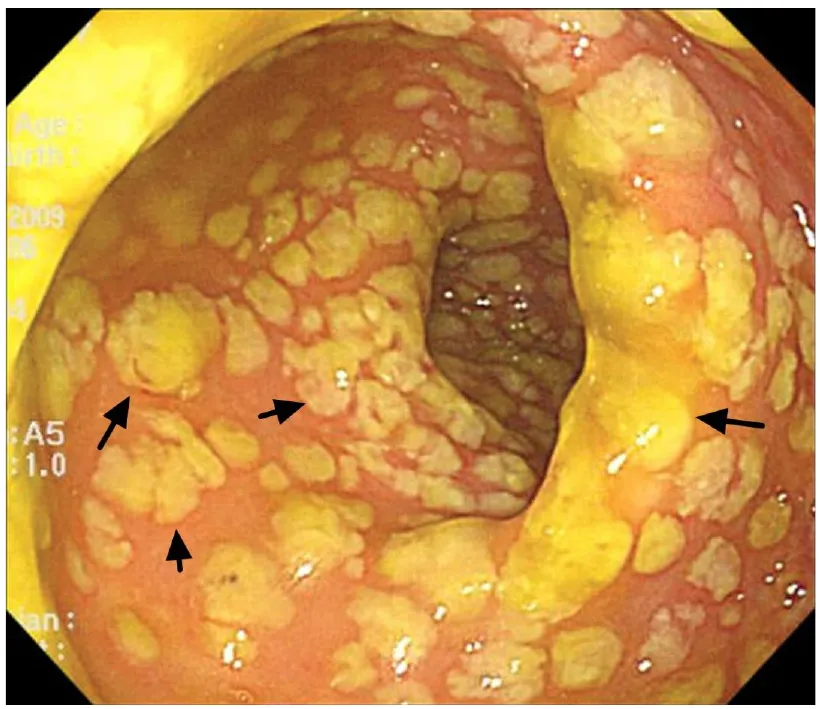
| Pseudomembranous | Toxin kills mucosa; fibrin, necrotic epithelium and neutrophils form a membrane | Clostridioides (Clostridium) difficile colitis; diphtheria (pharynx, trachea) | Yellow plaques; diphtheria membrane bleeds when lifted |
| Ulcerative | Local loss of epithelium, extending into deeper tissue, with an inflamed base | Peptic ulcer, ulcerative colitis, herpes ulcers | Defect in a surface |
Serous
- What forms
- Thin, watery fluid with too little fibrinogen to make fibrin
- Classic examples
- Blister of a second-degree burn; viral pleuritis; effusions
- Clue
- Clear fluid, few cells
Fibrinous
- What forms
- A larger leak lets fibrinogen out; it polymerizes on the surface
- Classic examples
- Pericarditis, pleuritis, peritonitis
- Clue
- Friction rub; shaggy “bread-and-butter” surface
Purulent (suppurative)
- What forms
- Pus: neutrophils, necrotic cells, edema fluid
- Classic examples
- S. aureus abscess, appendicitis
- Clue
- Localized, fluctuant collection
Pseudomembranous
- What forms
- Toxin kills mucosa; fibrin, necrotic epithelium and neutrophils form a membrane
- Classic examples
- Clostridioides (Clostridium) difficile colitis; diphtheria (pharynx, trachea)
- Clue
- Yellow plaques; diphtheria membrane bleeds when lifted
Ulcerative
- What forms
- Local loss of epithelium, extending into deeper tissue, with an inflamed base
- Classic examples
- Peptic ulcer, ulcerative colitis, herpes ulcers
- Clue
- Defect in a surface
Lecture image
Lecture image
Lecture image
Lecture image
Abscess versus cellulitis
L2 p.32–34Ch. II cases p.12The organism decides whether purulent inflammation stays put. Staphylococcus aureus carries coagulase, which converts fibrinogen to fibrin: the fibrin mesh traps bacteria and neutrophils and keeps the infection localized. Inside, neutrophil lysosomal enzymes liquefy everything into pus (2.3). Over days a wall of granulation tissue and later fibrous tissue forms around it: an abscess. Antibiotics penetrate the cavity poorly, so abscesses usually need drainage.
Group A Streptococcus makes hyaluronidase (and streptokinase), which dissolve the ground substance and fibrin, so infection spreads through subcutaneous tissue planes: cellulitis. The lecture's signs are an expanding area of redness, warmth, tenderness and swelling, fever, painful regional lymph nodes and red streaks running toward them, which are inflamed lymphatics (lymphangitis).
Localized versus spreading
Abscess
- S. aureus: coagulase → fibrin wall
- Localized pus; fluctuant
- Liquefactive necrosis by neutrophil enzymes
- Treat by drainage (± antibiotics)
Cellulitis
- Group A strep: hyaluronidase, streptokinase
- Diffuse, expanding redness; no collection
- Red streaks (lymphangitis), tender nodes, fever
- Treat with antibiotics
Four ways acute inflammation can end
L2 p.39Teaching schematic
Complete resolution
Little destruction; cells regenerate; exudate cleared
Scar (fibrosis)
Framework destroyed, permanent cells, or fibrin not cleared
Abscess
Pyogenic bacteria; pus walled off
Fistula
Tract between two epithelial surfaces
Chronic inflammation
Agent persists; macrophages and lymphocytes take over
An abscess can drain through a fistula or a sinus; any outcome with destroyed framework ends in some scar.
The lecture's five outcomesL2 p.39
| Outcome | When it happens | Example |
|---|---|---|
| Complete resolution | Injury limited and short-lived; little tissue destroyed; cells can regenerate | Lobar pneumonia: alveolar walls survive, exudate is cleared |
| Scar (fibrosis) | Substantial destruction, especially of the supporting framework; permanent cells; a large fibrin exudate that cannot be cleared | Healed abscess cavity; organized pleural fibrin; myocardial infarct |
| Abscess | Pyogenic organisms; pus walled off | Diverticular or lung abscess |
| Fistula | Inflammation erodes through two epithelium-lined surfaces and connects them (a sinus is a tract that ends blindly) | Diverticulitis → colovesical fistula (air in the urine); Crohn disease |
| Progression to chronic inflammation | The agent persists or the response keeps re-igniting | Chronic osteomyelitis; peptic ulcer that will not heal |
Complete resolution
- When it happens
- Injury limited and short-lived; little tissue destroyed; cells can regenerate
- Example
- Lobar pneumonia: alveolar walls survive, exudate is cleared
Scar (fibrosis)
- When it happens
- Substantial destruction, especially of the supporting framework; permanent cells; a large fibrin exudate that cannot be cleared
- Example
- Healed abscess cavity; organized pleural fibrin; myocardial infarct
Abscess
- When it happens
- Pyogenic organisms; pus walled off
- Example
- Diverticular or lung abscess
Fistula
- When it happens
- Inflammation erodes through two epithelium-lined surfaces and connects them (a sinus is a tract that ends blindly)
- Example
- Diverticulitis → colovesical fistula (air in the urine); Crohn disease
Progression to chronic inflammation
- When it happens
- The agent persists or the response keeps re-igniting
- Example
- Chronic osteomyelitis; peptic ulcer that will not heal
How the response switches off
L2 p.39Inflammation is too destructive to run until it burns out; ending it is an active program. The lecture's termination slide gives the main elements: mediators with a short half-life, and lipoxins that inhibit neutrophil transmigration and chemotaxis and signal macrophages to eat apoptotic bodies.
- Mediators decay: histamine, kinins, prostaglandins and cytokines are made in bursts and broken down within minutes to hours. Once the stimulus is cleared, production stops.
- Neutrophils die: they live only hours to a day or two in tissue and then undergo apoptosis.
- A lipid-mediator switch: early prostaglandins induce 15-lipoxygenase in neutrophils, so arachidonic acid is turned into lipoxins instead of LTB₄. Resolvins and protectins, made from omega-3 fatty acids, act the same way. Aspirin acetylates COX-2 so that it makes “aspirin-triggered” lipoxins.
- Macrophages clear the debris (efferocytosis): they recognize phosphatidylserine on apoptotic neutrophils and engulf them without alarm. Eating apoptotic cells switches macrophages to make TGF-β and IL-10, which dampen inflammation and start repair.
- Other brakes: IL-1 receptor antagonist, IL-10, lymphatic drainage of fluid and debris, and a vagal anti-inflammatory reflex that restrains TNF release.
From inflammation to repair
- Cause removed
- Mediators decay; no new ones made
- Neutrophils undergo apoptosis
- Lipoxins and resolvins stop further recruitment
- Macrophages engulf apoptotic cells
- Macrophages release TGF-β and IL-10
- Vessels reseal; lymphatics drain; repair begins (2.7)
Core activity · The Missing Arrow
The Missing Arrow
An abscess has been drained and the bacteria are gone. Complete the chain that ends the response and starts repair.
- Bacteria cleared from the tissue
- no new stimulus; short-lived mediators decay
Cards
Tap or drag a card. Placing a used card moves it.
Knowledge check · 2.5
Answer, read why, and try again whenever you like. Answers are kept in both study modes.
- Q1Abscess: lysosomal enzymesL2 p.32–33Ch. II cases p.12
A 63-year-old man with constipation has four days of lower abdominal pain, fever (39.1 °C) and left lower quadrant tenderness. White count is 19,000/mm³. CT shows a thickened descending colon with multiple diverticula and a 5.0-cm, low-attenuation pelvic lesion with air-fluid levels, which is drained.
Release of which substance is most likely responsible for forming the drained lesion?
- Q2Fibrinous inflammation: image recognitionL2 p.35
A 66-year-old man dies 5 days after a large transmural myocardial infarction. In his last days a scratchy sound was heard with each heartbeat. The heart at autopsy is shown.
Lecture image
What produced the coating on the heart's surface?
- Q3Pseudomembranous inflammation: image recognitionL2 p.37
A 72-year-old woman develops profuse watery diarrhea a week after starting clindamycin. Colonoscopy is shown.
Lecture image
What are the yellow plaques made of?
- Q4Resolution: lipoxinsL2 p.26, 39
Which arachidonic-acid products stop neutrophil transmigration and chemotaxis and signal macrophages to phagocytose apoptotic cells as inflammation ends?
- Q5Outcomes: fistulaL2 p.39
A 67-year-old man treated for diverticulitis two months ago now notices bubbles of air when he urinates, and he has recurrent urinary tract infections with bowel organisms.
Which outcome of acute inflammation explains this?