Leukocyte recruitment
A neutrophil leaves the blood in four steps: it rolls on selectins, stops on integrins, squeezes between endothelial cells, and follows a chemical gradient to the target. Break any step and the cells pile up in the blood instead of the tissue.
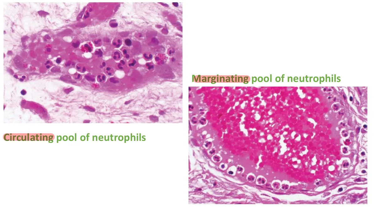
Margination: two pools of neutrophils
L2 p.9, 11In fast flow, cells ride in the centre of the vessel and a cell-poor layer of plasma runs along the wall. In an inflamed venule the picture changes: fluid has leaked out, red cells are concentrated and stack into rouleaux in the central stream, and flow slows (stasis). Leukocytes are pushed out of the axial stream toward the endothelium. This is margination, the precondition for adhesion.
Even in health, roughly half of the neutrophils in the vascular space are not circulating freely: they move slowly along the walls of small vessels, especially in the lungs, spleen and liver. This marginating pool is not counted by a blood count; only the circulating pool is. Any shift between the two pools changes the measured white count without any change in how many neutrophils the body has.
Lecture image
Rolling and tight adhesion
L2 p.10, 12–13Teaching schematic
1.Rolling
E-/P-selectin ↔ sialyl-Lewis X; L-selectin
2.Tight adhesion
LFA-1, Mac-1 (CD11/CD18) ↔ ICAM-1
3.Transmigration
PECAM-1 (CD31); type IV collagenase
4.Chemotaxis
C5a, IL-8, LTB₄, N-formyl peptides
Steps 5 and 6 (phagolysosome and killing) continue in 2.3.
Rolling. Selectins are lectins: they bind sugar groups, chiefly sialyl-Lewis X on leukocyte glycoproteins. The bonds form and break quickly, so the neutrophil tumbles along the wall, bind–detach, bind–detach. P-selectin is stored in the endothelial Weibel–Palade bodies and reaches the surface within minutes of histamine or thrombin; E-selectin is made over 1–2 hours after IL-1 or TNF; L-selectin sits on the leukocyte itself.
Tight adhesion. Rolling slows the cell enough for chemokines (above all IL-8) displayed on the endothelium to signal through its receptors. Within seconds this switches the leukocyte's β₂ integrins into a high-affinity shape: LFA-1 (CD11a/CD18) and Mac-1 (CD11b/CD18). Meanwhile TNF and IL-1 have induced their endothelial ligands, ICAM-1 (for LFA-1 and Mac-1) and VCAM-1 (for the β₁ integrin VLA-4 on monocytes and lymphocytes). Integrin meets ligand, and the cell stops and flattens.
Who binds whomL2 p.12–16
| Step | On the leukocyte | On the endothelium | Switched on by |
|---|---|---|---|
| Rolling | Sialyl-Lewis X (on PSGL-1 and others); L-selectin | P-selectin, E-selectin | Histamine, thrombin (P: minutes); IL-1, TNF (E: hours) |
| Tight adhesion | β₂ integrins: LFA-1 (CD11a/CD18), Mac-1 (CD11b/CD18); VLA-4 (β₁) | ICAM-1, VCAM-1 | Chemokines (IL-8) activate integrins; TNF, IL-1 induce ICAM-1/VCAM-1 |
| Transmigration | PECAM-1 (CD31) | PECAM-1 (CD31) at junctions | Collagenase opens the basement membrane |
| Chemotaxis | Receptors for C5a, IL-8, LTB₄, formyl peptides | — | Gradient from the site of injury |
Rolling
- On the leukocyte
- Sialyl-Lewis X (on PSGL-1 and others); L-selectin
- On the endothelium
- P-selectin, E-selectin
- Switched on by
- Histamine, thrombin (P: minutes); IL-1, TNF (E: hours)
Tight adhesion
- On the leukocyte
- β₂ integrins: LFA-1 (CD11a/CD18), Mac-1 (CD11b/CD18); VLA-4 (β₁)
- On the endothelium
- ICAM-1, VCAM-1
- Switched on by
- Chemokines (IL-8) activate integrins; TNF, IL-1 induce ICAM-1/VCAM-1
Transmigration
- On the leukocyte
- PECAM-1 (CD31)
- On the endothelium
- PECAM-1 (CD31) at junctions
- Switched on by
- Collagenase opens the basement membrane
Chemotaxis
- On the leukocyte
- Receptors for C5a, IL-8, LTB₄, formyl peptides
- On the endothelium
- —
- Switched on by
- Gradient from the site of injury
Transmigration (diapedesis)
L2 p.15The arrested neutrophil crawls to a junction between endothelial cells, where PECAM-1 (CD31) on the leukocyte binds PECAM-1 on the endothelial cells, guiding it through. It then secretes collagenases (type IV) that open the basement membrane, and enters the interstitium. Diapedesis happens mainly in postcapillary venules, the same vessels that leak, in gaps that histamine-driven endothelial contraction has already widened.
Chemotaxis: following the gradient
L2 p.16The four chemoattractants on the lecture slideL2 p.16
| Chemoattractant | Where it comes from |
|---|---|
| C5a | Complement activation on microbes or immune complexes |
| IL-8 (CXCL8) | Macrophages, endothelium and epithelium stimulated by TNF and IL-1 |
| LTB₄ | 5-lipoxygenase products of neutrophils and macrophages |
| Bacterial products | N-formylmethionyl peptides (bacteria start their proteins with formyl-methionine) |
C5a
- Where it comes from
- Complement activation on microbes or immune complexes
IL-8 (CXCL8)
- Where it comes from
- Macrophages, endothelium and epithelium stimulated by TNF and IL-1
LTB₄
- Where it comes from
- 5-lipoxygenase products of neutrophils and macrophages
Bacterial products
- Where it comes from
- N-formylmethionyl peptides (bacteria start their proteins with formyl-methionine)
Each binds a G-protein-coupled receptor. The side of the cell facing the higher concentration polymerizes actin into a leading edge, myosin contracts at the back, and the cell crawls up the gradient. The same signals also activate the neutrophil: integrins switch on, granules are primed, and the oxidative burst becomes possible.
Which cell, when. Neutrophils dominate the first 6–24 hours: there are more of them in blood, they respond faster to chemokines, and they attach firmly to E- and P-selectin. They then die by apoptosis within a day or two, and monocytes, which survive longer and become macrophages, dominate by 24–48 hours. Exceptions: Pseudomonas keeps recruiting neutrophils for days; viral infections bring lymphocytes first; allergic reactions and parasites bring eosinophils.
Leukocyte adhesion deficiency: when cells cannot leave
L2 p.14LAD type I (autosomal recessive) is a defect of CD18, the β₂ chain shared by LFA-1 and Mac-1. Neutrophils can roll, but they cannot stop, so they never leave the blood. Everything follows from that one failed step:
- Very high blood neutrophil count, even between infections: cells cannot hold on to the wall, so the marginating pool joins the counted circulating pool, and the marrow keeps sending more.
- Infections without pus: skin and mucosal infections, necrotic ulcers, omphalitis, severe gingivitis and periodontitis.
- Delayed separation of the umbilical cord (beyond about 3 weeks): the cord stump detaches by an inflammatory process that needs neutrophils in the tissue.
- Poor wound healing: the inflammatory phase that clears debris and recruits repair cells is missing.
Lecture image
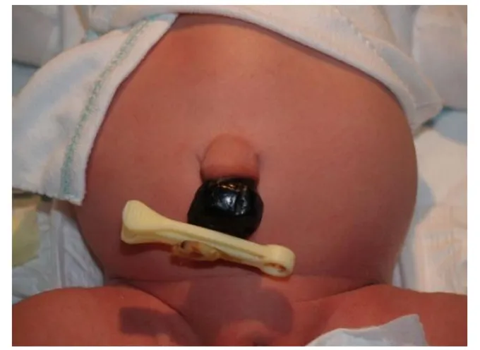
LAD type II (autosomal recessive) is a defect in fucosylation: the transporter that brings GDP-fucose into the Golgi is faulty, so cells cannot build fucose-containing sugars. Leukocytes lack sialyl-Lewis X, the selectin ligand, so rolling fails. Red cells lack the fucose-containing H antigen: the Bombay phenotype. Children also have intellectual disability and short stature; infections are milder than in LAD I.
LAD I versus LAD II
LAD I
- Defect: CD18 (β₂ integrin)
- Failed step: tight adhesion
- Delayed cord separation, omphalitis, no pus, periodontitis, poor healing
- Marked neutrophilia
- Flow cytometry: CD18 absent
LAD II
- Defect: fucosylation → no sialyl-Lewis X
- Failed step: rolling
- Bombay blood group, intellectual disability, short stature
- Neutrophilia; milder infections
- Flow cytometry: CD15s (sialyl-Lewis X) absent
Centerpiece · Recruitment and killing lab
Find the broken step
0 of 5 defects worked out.
Step 1 · Rolling
E-/P-selectin ↔ sialyl-Lewis X; L-selectin
Selectins on activated endothelium catch sugar ligands on the neutrophil. Bonds form and break, so the cell tumbles slowly along the venule wall.
Tap each step to see its molecules. When the sequence is clear, choose defect 1 above and break it.
Transfer round · same mechanisms, new wording
Work out all five defects above to open five short cases that describe them in different words.
Neutrophilia without infection
L2 p.13The adhesion slide warns that catecholamines and corticosteroids inhibit neutrophil adhesion. Their effect on the blood count is a pool shift. Epinephrine (stress, exercise, a seizure) and glucocorticoids loosen marginated neutrophils from the walls, a process called demargination, so they join the circulating pool and the measured count rises within hours. Glucocorticoids also lower adhesion-molecule expression, release neutrophils from the marrow and delay their apoptosis, while lowering eosinophils and lymphocytes.
Two causes of neutrophilia
Infection or tissue injury
- IL-1, TNF and colony-stimulating factors drive the marrow
- Left shift: bands and younger forms
- Toxic granulation, Döhle bodies
- Fever, raised CRP
Demargination (steroids, epinephrine, stress)
- Cells move from the marginating to the circulating pool
- Mature neutrophils, no left shift
- Steroids: eosinophils and lymphocytes fall
- No fever; total body neutrophils unchanged
Core activity · One Fact Changes
One Fact Changes
1 · The original case
A 19-year-old student has a white count of 16,500/µL, 90% neutrophils. He has 24 hours of right lower quadrant pain and a temperature of 38.6 °C, and his smear shows many band forms. He was previously healthy.
What best explains the neutrophilia?
Which fact decided it?
Knowledge check · 2.2
Answer, read why, and try again whenever you like. Answers are kept in both study modes.
- Q1Rolling: selectinsL2 p.12Midterm sim. p.4
A 35-year-old woman has a 1-cm abscess over her left shoulder. Biopsy shows vasodilation and leukocyte margination.
Which glycoprotein mediates the initial tethering of neutrophils to the endothelial cells in this lesion?
- Q2LAD I: adhesion failsL2 p.14Ch. II cases p.2
A 6-month-old girl has a 2-week scalp rash and a week of fever, poor appetite and loose stools. She was hospitalized twice for omphalitis. She is dehydrated; the scalp has an erythematous rash with focal necrosis and no pus. White cell count is 37,000/mm³.
What is the underlying mechanism of her most likely illness?
- Q3Neutrophil chemotaxis: LTB₄L2 p.16, 26Ch. II cases p.14
A 38-year-old man who smokes a pack a day has two years of cough and worsening breathlessness, with accessory-muscle use, a flattened diaphragm and lucent lower lung fields. Testing shows increased activity of an elastase isoform normally inhibited by α₁-antitrypsin.
The cells that make this elastase were most likely stimulated to enter the lung by which substance?
- Q4Demargination by glucocorticoidsL2 p.13
A 58-year-old woman with an asthma exacerbation starts high-dose prednisone. The next morning she is afebrile and feels better. Her white count is 17,000/µL with 92% mature neutrophils, no bands, no eosinophils and few lymphocytes.
What best explains the neutrophilia?
- Q5Pools and LAD: image interpretationL2 p.9, 14
A 5-week-old boy still has his umbilical cord stump, which is red at the base. His white count is 48,000/µL, almost all neutrophils, yet a skin infection on his thigh has formed no pus.
Lecture image
Using the two vessels shown, what explains the high count?