Chromosomal disorders and syndromes
Whole chromosomes or large pieces are gained, lost or moved. The phenotype follows the dose of genes; the recurrence risk follows whether the error was a one-off in one gamete or is carried, balanced, by a parent.
Nondisjunction and mosaicism
L5 p.7–9Nondisjunction is failure of chromosomes to separate during meiosis, so an egg or sperm ends up with 24 or 22 chromosomes instead of 23. Fertilized by a normal gamete, it gives a trisomic (47) or monosomic (45) conceptus. Most autosomal monosomies and many trisomies are lost as miscarriages; the survivors are trisomy 21, 18 and 13, and the sex-chromosome aneuploidies (45,X; 47,XXY; 47,XXX; 47,XYY).
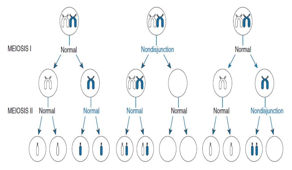
The error can happen in either division. In meiosis I the two homologs fail to part: the disomic gamete receives one of each homolog (one originally from the parent's father, one from the parent's mother). In meiosis II the sister chromatids fail to part: the disomic gamete receives two copies of the same homolog. Polymorphic DNA markers next to the centromere tell them apart: two different alleles from one parent mean meiosis I (heterodisomy), two identical alleles mean meiosis II (isodisomy). Most maternal trisomy 21 arises in maternal meiosis I, the step that has been paused in the oocyte since before the mother was born, which is why risk climbs with maternal age.
Teaching schematic
Normal: Every gamete has one copy
Meiosis I error: Disomic gametes carry both homologs (heterodisomy)
Meiosis II error: Disomic gamete carries two copies of one homolog (isodisomy)
Blue and pink = the two homologs of one parent (from that parent's father and mother). DNA markers near the centromere tell which error happened.
Lecture slide
Mosaicism is the presence of two or more cell lines with different karyotypes in one person, derived from a single zygote. It arises from a mitotic error (nondisjunction or anaphase lag) in the early embryo. Because only some cells are abnormal, mosaics are usually milder; mosaicism is most common with sex chromosomes (milder Turner syndrome, 45,X/46,XX) and causes about 1% of Down syndrome.
Core activity · The Missing Arrow
The Missing Arrow
A 42-year-old woman conceives. One of her oocytes, arrested in meiosis I since before her own birth, completes its first division. Complete the chain.
Separate the error (what failed in meiosis) from the event it produces in the embryo.
- Aged oocyte: cohesion between homologs has weakened over decades
- fertilized by a normal sperm (23 chromosomes)
- Down syndrome: hypotonia, flat face, AV canal defect, duodenal atresia, leukemia risk, early Alzheimer disease
Cards
Tap or drag a card. Placing a used card moves it.
Deletions and translocations
L5 p.7, 9Structural changes move or remove pieces. What matters clinically is whether the change is balanced (every gene still present in two doses) or unbalanced (a net gain or loss). A balanced carrier is usually healthy but can produce unbalanced gametes, which show up as recurrent miscarriage or an affected child.
| Change | Definition | Example |
|---|---|---|
| Deletion | Loss of part of a chromosome (unbalanced) | Cri-du-chat, 46,XX,del(5p): high-pitched cat-like cry, microcephaly, intellectual disability, VSD. 22q11.2 deletion (below) |
| Reciprocal translocation | Exchange of segments between two nonhomologous chromosomes | Balanced in the carrier; unbalanced offspring possible |
| Robertsonian translocation | Two acrocentric chromosomes (13, 14, 15, 21, 22) fuse at their centromeres; the short arms are lost | der(14;21): carrier has 45 chromosomes and is healthy; children can have translocation Down syndrome |
| Isochromosome | One arm duplicated as the other is lost | i(Xq) in some Turner syndrome; i(21q) gives Down syndrome in every viable child of a carrier |
Deletion
- Definition
- Loss of part of a chromosome (unbalanced)
- Example
- Cri-du-chat, 46,XX,del(5p): high-pitched cat-like cry, microcephaly, intellectual disability, VSD. 22q11.2 deletion (below)
Reciprocal translocation
- Definition
- Exchange of segments between two nonhomologous chromosomes
- Example
- Balanced in the carrier; unbalanced offspring possible
Robertsonian translocation
- Definition
- Two acrocentric chromosomes (13, 14, 15, 21, 22) fuse at their centromeres; the short arms are lost
- Example
- der(14;21): carrier has 45 chromosomes and is healthy; children can have translocation Down syndrome
Isochromosome
- Definition
- One arm duplicated as the other is lost
- Example
- i(Xq) in some Turner syndrome; i(21q) gives Down syndrome in every viable child of a carrier
Lecture image
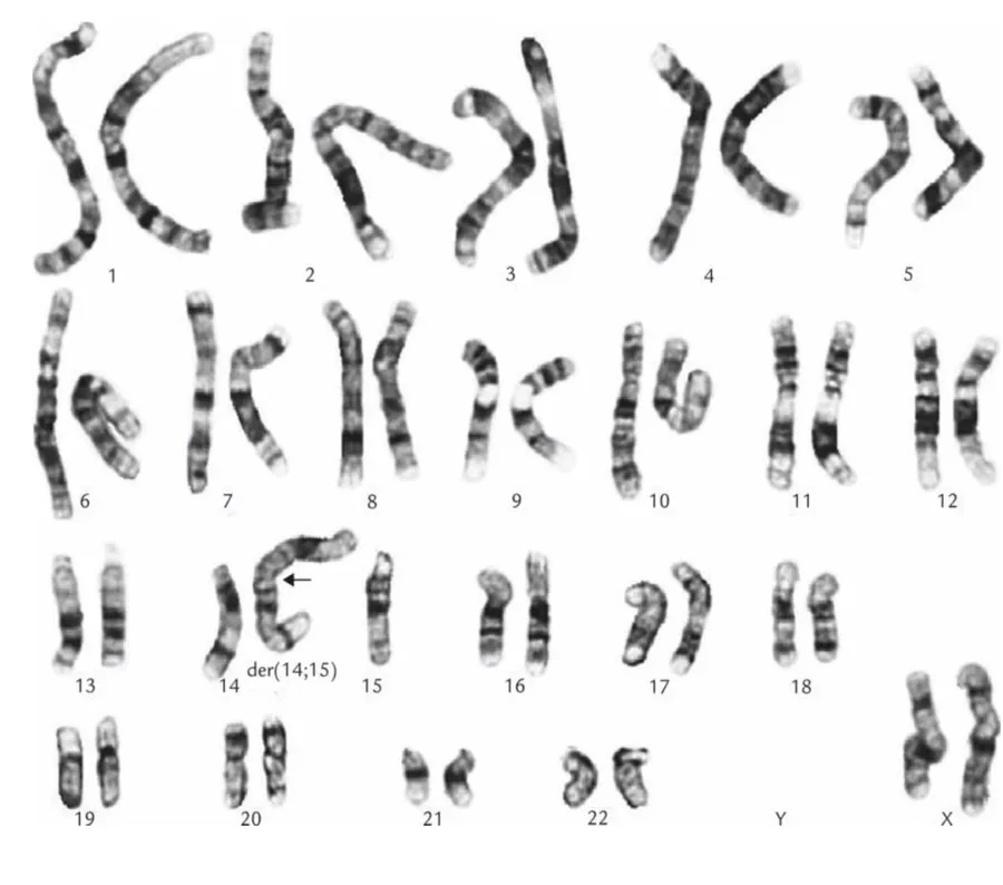
Acrocentric short arms carry only repeated ribosomal RNA genes that also exist on the other acrocentrics, so losing two of them costs nothing. The carrier's problem appears at meiosis: the der chromosome and the two free chromosomes can segregate in several ways, and some gametes carry an extra or a missing long arm.
Teaching schematic
Carrier mother: 45,XX,der(14;21)
Egg 1
Normal
Egg 2
Balanced carrier
Egg 3
Translocation Down (46)
Egg 4
Monosomy 21: lost
Egg 5
Trisomy 14: lost
Egg 6
Monosomy 14: lost
Each egg is fertilized by a normal sperm (14 + 21). Amber = 21q material; indigo = 14q.
Pedigree Lab · exercise
46 chromosomes, still Down syndrome
Step 1
How can a child with 46 chromosomes have Down syndrome?
Step 2
Which parent carries it?
Explain the child's karyotype first.
Step 3
Sort the mother's eggs (each fertilized by a normal sperm: 14 + 21)
Identify the carrier and why she is healthy first.
Step 4
Recurrence risk
Sort the eggs first.
The 22q11.2 deletion (DiGeorge/velocardiofacial syndrome) is a microdeletion of about 3 Mb, too small for a routine karyotype; FISH or microarray finds it. Loss of TBX1 and neighboring genes disturbs the third and fourth pharyngeal pouches and the outflow tract of the heart, giving the CATCH-22 picture: conotruncal heart defects (tetralogy of Fallot, truncus arteriosus, interrupted aortic arch), abnormal face, thymic hypoplasia (T-cell deficiency), cleft palate and hypocalcemia from parathyroid hypoplasia. Most cases are de novo; an affected parent passes it on as an autosomal dominant trait.
X inactivation and Barr bodies
Ch. V cases p.9L5 p.6Females have two X chromosomes but the same X-gene output as males because, early in embryonic life (around the late blastocyst), one X in each cell is silenced at random (Lyon hypothesis). The XIST RNA coats the chosen X; its promoters are silenced by methylation of cytosines in CpG islands, and its histones are deacetylated and methylated into tight heterochromatin. The condensed X is visible at the edge of the interphase nucleus as the Barr body (in neutrophils, a “drumstick”). The choice is permanent and passed to every daughter cell, so every woman is a mosaic of two cell populations.
Teaching schematic
46,XY
0 Barr bodies
46,XX
1 Barr body
45,X
0 Barr bodies
47,XXY
1 Barr body
47,XXX
2 Barr bodies
Barr bodies = number of X chromosomes − 1
If inactivation silenced the whole chromosome, a 45,X woman or a 47,XXY man would have exactly normal X dosage and no syndrome. They do not, because about 15% of X genes escape inactivation, including the pseudoautosomal genes shared with Y such as SHOX. Two active copies of these genes are needed: one copy gives Turner short stature; a third copy contributes to Klinefelter tall stature.
Worked case · A spot at the edge of the nucleus
A 25-year-old infertile man, 189 cm tall, has long limbs, bilateral gynecomastia and small, firm testes. A blood smear shows a dense, dark spot at the periphery of a cell nucleus (Ch. V Q8).
What produced the spot?
Ch. V cases p.9Down syndrome (trisomy 21)
L5 p.10–12Ch. V cases p.10Midterm sim. p.11Three ways to have three doses of 21qL5 p.10
| Cause | Share | Karyotype | Recurrence |
|---|---|---|---|
| Meiotic nondisjunction (free trisomy) | ~95% | 47,+21 | ~1%, or the maternal-age risk if higher |
| Robertsonian translocation | ~4% | 46, with der(14;21) or (21;21) | 10–15% if the mother carries a 14;21, a few percent if the father does, 100% for 21;21 |
| Mosaicism | ~1% | 46/47,+21 | Low; phenotype often milder |
Meiotic nondisjunction (free trisomy)
- Share
- ~95%
- Karyotype
- 47,+21
- Recurrence
- ~1%, or the maternal-age risk if higher
Robertsonian translocation
- Share
- ~4%
- Karyotype
- 46, with der(14;21) or (21;21)
- Recurrence
- 10–15% if the mother carries a 14;21, a few percent if the father does, 100% for 21;21
Mosaicism
- Share
- ~1%
- Karyotype
- 46/47,+21
- Recurrence
- Low; phenotype often milder
Maternal age is the main risk factor for free trisomy: about 1 in 1,500 live births at age 20, rising to about 1 in 25 after 45. Because younger women have most babies, most children with Down syndrome are still born to mothers under 35. Every gene on chromosome 21 is normal; the disease is the 1.5-fold dose. The extra APP gene, for example, explains Alzheimer pathology by 35–40 years.
Findings, groupedL5 p.11
| Domain | Findings |
|---|---|
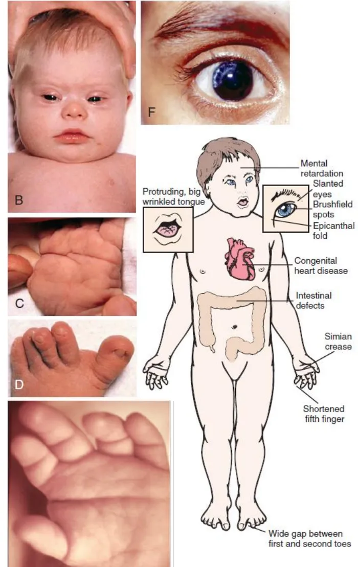
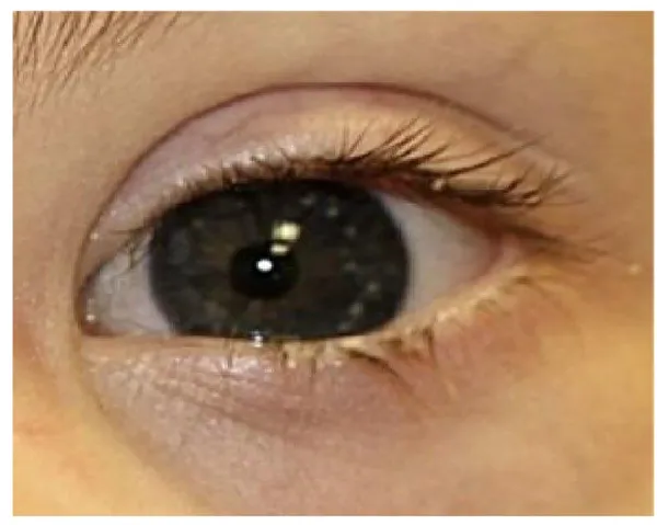
| Newborn and face | Hypotonia (“floppy baby”); flat facial profile, upslanting palpebral fissures, epicanthic folds, macroglossia with protruding tongue, small ears; Brushfield spots; single palmar crease, short 5th finger, wide gap between 1st and 2nd toes |
| Heart (~40%) | Endocardial cushion (atrioventricular septal) defect is the most characteristic; VSD, ASD, tetralogy of Fallot, PDA |
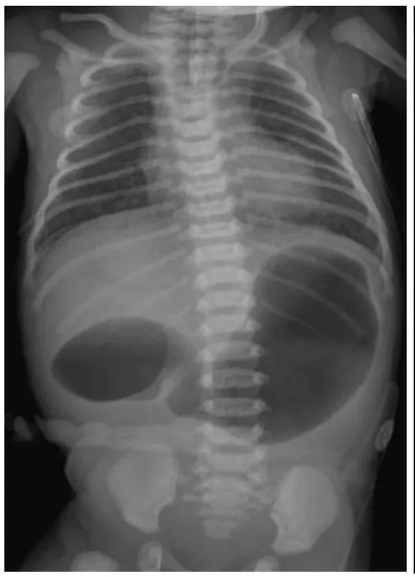
| Gut | Duodenal atresia (double bubble), tracheoesophageal fistula, Hirschsprung disease, umbilical hernia |
| Blood | Leukemia 10–20× (ALL; acute megakaryoblastic leukemia in early childhood, often after a transient myeloproliferative disorder) |
| Brain | Intellectual disability (variable); early Alzheimer disease (APP dose) |
| Reproduction | Males usually infertile; females have reduced fertility; a woman with Down syndrome has a high (theoretically 50%) risk of an affected child |
| Other complications | Atlantoaxial instability (cord compression), hypothyroidism, hearing loss, obstructive sleep apnea, infections |
Newborn and face
- Findings
- Hypotonia (“floppy baby”); flat facial profile, upslanting palpebral fissures, epicanthic folds, macroglossia with protruding tongue, small ears; Brushfield spots; single palmar crease, short 5th finger, wide gap between 1st and 2nd toes
Heart (~40%)
- Findings
- Endocardial cushion (atrioventricular septal) defect is the most characteristic; VSD, ASD, tetralogy of Fallot, PDA
Gut
- Findings
- Duodenal atresia (double bubble), tracheoesophageal fistula, Hirschsprung disease, umbilical hernia
Blood
- Findings
- Leukemia 10–20× (ALL; acute megakaryoblastic leukemia in early childhood, often after a transient myeloproliferative disorder)
Brain
- Findings
- Intellectual disability (variable); early Alzheimer disease (APP dose)
Reproduction
- Findings
- Males usually infertile; females have reduced fertility; a woman with Down syndrome has a high (theoretically 50%) risk of an affected child
Other complications
- Findings
- Atlantoaxial instability (cord compression), hypothyroidism, hearing loss, obstructive sleep apnea, infections
Lecture image
Lecture image
Lecture image
Lecture image
Worked case · A double bubble and 46 chromosomes
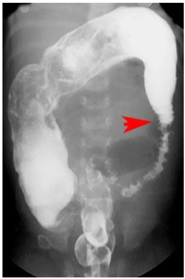
A 2.4-kg term boy of a 26-year-old has a flat nasal bridge, low-set ears, a protruding tongue, a single palmar crease and a sandal gap. His abdomen is distended and an x-ray shows two large air-filled spaces. Karyotype: 46 chromosomes in all cells (Sim. Q28).
What is the underlying cause, and what should happen next?
Midterm sim. p.11Core activity · Almost Twins
Almost Twins
Look-alike A
Free trisomy 21
- Down phenotype: flat face, Brushfield spots, AV canal defect, duodenal atresia
- 47 chromosomes; meiotic nondisjunction, mostly maternal
- Risk rises steeply with maternal age
- Recurrence about 1% (or the age-related risk)
Look-alike A
Free trisomy 21
- Down phenotype: flat face, Brushfield spots, AV canal defect, duodenal atresia
- 47 chromosomes; meiotic nondisjunction, mostly maternal
- Risk rises steeply with maternal age
- Recurrence about 1% (or the age-related risk)
Look-alike B
Translocation Down syndrome
- Down phenotype: indistinguishable
- 46 chromosomes; extra 21q fused to an acrocentric
- Not related to maternal age; ~4% of cases
- High recurrence if a parent is a balanced carrier
Step 1 · What they share
What do the two children share?
Select every feature that both show.
Core activity · One Fact Changes
One Fact Changes
1 · The original case
A 42-year-old woman delivers her third child. The baby has a flat facial profile, upslanting eyes, epicanthal folds, Brushfield spots, a protruding tongue and a loud murmur. The karyotype is pending.
What is the most likely mechanism?
Which fact carried the most weight?
Edwards (trisomy 18) and Patau (trisomy 13)
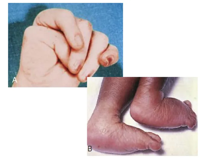
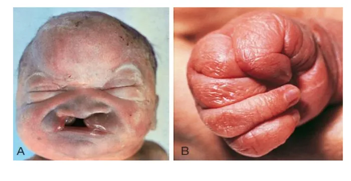
L5 p.13Both are usually free trisomies from maternal nondisjunction, both cause severe intellectual disability and congenital heart disease, and most affected infants die in the first year. The separating clues are where the malformations cluster: Edwards syndrome in the hands, feet and jaw; Patau syndrome in the midline (brain, face) and with extra digits.
Edwards (trisomy 18)
- Clenched fists with overlapping fingers (2nd over 3rd, 5th over 4th)
- Rocker-bottom feet
- Micrognathia, prominent occiput, low-set ears
- VSD; horseshoe kidney
- Second-trimester screen: AFP, estriol and hCG all low
Patau (trisomy 13)
- Midline defects: cleft lip and palate, holoprosencephaly, microphthalmia
- Polydactyly; clenched hands
- Scalp defects (cutis aplasia)
- VSD, cystic kidneys, omphalocele
- Early death
Lecture image
Lecture image
Turner syndrome (45,X)
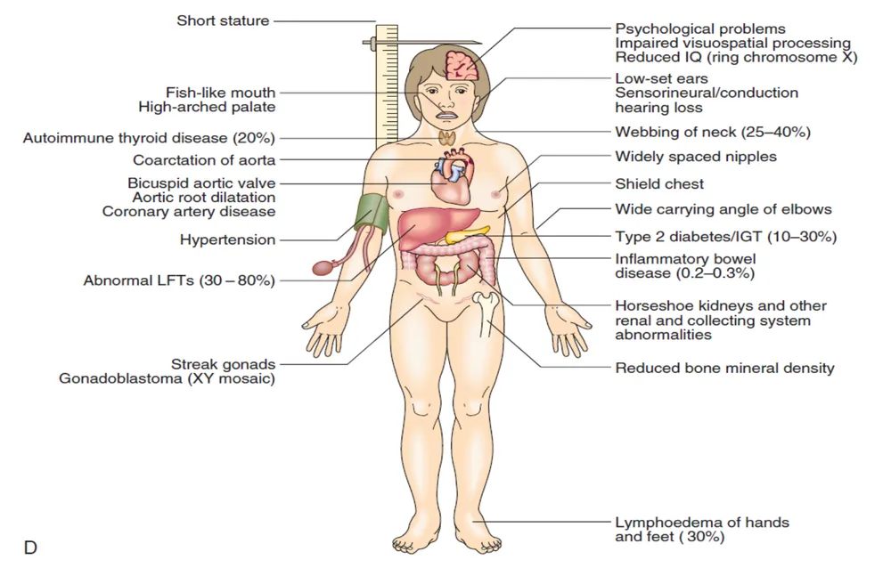
L5 p.14–15Midterm sim. p.11Turner syndrome is complete or partial loss of the second sex chromosome in a phenotypic female: 45,X (about half), mosaics such as 45,X/46,XX, and structural X abnormalities (isochromosome Xq, ring X, Xp deletions). Most 45,X conceptuses miscarry; survivors are often mosaics. Each feature has a mechanism: one copy of SHOX (an escape gene) gives short stature with normal growth hormone; fetal lymphatics fail to drain, giving cystic hygroma that leaves a webbed neck and hand and foot lymphedema at birth; oocytes are lost at an accelerated rate, so the ovaries become streak gonads.
Findings, groupedL5 p.14–15
| Domain | Findings |
|---|---|
| Physical | Short stature; webbed neck, low posterior hairline; shield chest with widely spaced nipples; cubitus valgus (wide carrying angle); short 4th metacarpal; lymphedema of hands and feet at birth |
| Cardiovascular | Bicuspid aortic valve, coarctation of the aorta (delayed, weak femoral pulses; the lecture specifies preductal), aortic dilation and dissection, hypertension |
| Kidney | Horseshoe kidney and other collecting-system anomalies |
| Reproductive and hormones | Streak gonads → primary amenorrhea, absent breast development; ↓ estradiol, ↑ FSH and LH (hypergonadotropic hypogonadism); uterus present; infertility |
| Endocrine and other | Hashimoto hypothyroidism; type 2 diabetes; low bone density; hearing loss; usually normal intelligence (visuospatial difficulty) |
| Tumor risk | Only with Y material (45,X/46,XY): gonadoblastoma, which can progress to dysgerminoma → prophylactic gonadectomy |
Physical
- Findings
- Short stature; webbed neck, low posterior hairline; shield chest with widely spaced nipples; cubitus valgus (wide carrying angle); short 4th metacarpal; lymphedema of hands and feet at birth
Cardiovascular
- Findings
- Bicuspid aortic valve, coarctation of the aorta (delayed, weak femoral pulses; the lecture specifies preductal), aortic dilation and dissection, hypertension
Kidney
- Findings
- Horseshoe kidney and other collecting-system anomalies
Reproductive and hormones
- Findings
- Streak gonads → primary amenorrhea, absent breast development; ↓ estradiol, ↑ FSH and LH (hypergonadotropic hypogonadism); uterus present; infertility
Endocrine and other
- Findings
- Hashimoto hypothyroidism; type 2 diabetes; low bone density; hearing loss; usually normal intelligence (visuospatial difficulty)
Tumor risk
- Findings
- Only with Y material (45,X/46,XY): gonadoblastoma, which can progress to dysgerminoma → prophylactic gonadectomy
Lecture image
Worked case · Leg pain when jogging
A 14-year-old girl has exertional leg pain. She is at the 5th percentile for height, has skin folds from neck to shoulders, a low hairline and ears, an increased carrying angle, and delayed lower-leg pulses (Sim. Q29).
Which additional finding is most likely?
Midterm sim. p.11Core activity · One Fact Changes
One Fact Changes
1 · The original case
A 14-year-old girl is at the 3rd percentile for height and has not begun breast development. She has a webbed neck, a low posterior hairline, widely spaced nipples and an increased carrying angle; her femoral pulses are delayed. Karyotype: 45,X in all cells counted.
Which step should be added to her management?
Which fact decided that gonadectomy is not needed?
Klinefelter (47,XXY) and 47,XYY
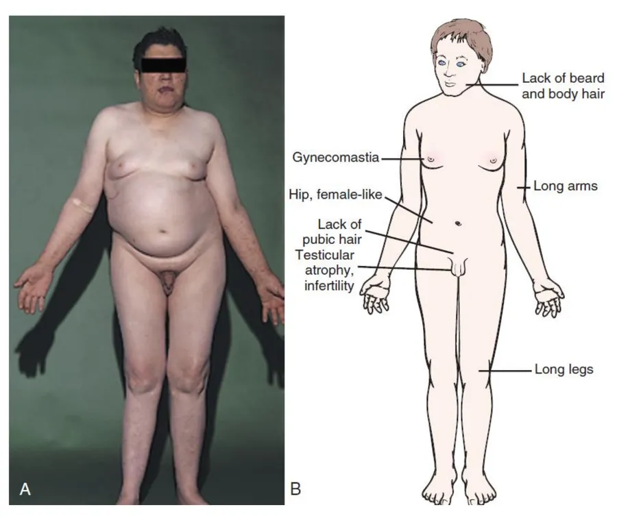
L5 p.16–18Midterm sim. p.11–12Klinefelter syndrome (47,XXY, about 1 in 600 males) is the commonest cause of male hypogonadism. The extra X comes from paternal meiosis I in about half of cases and from maternal meiosis I or II in the rest; about 15% are 46,XY/47,XXY mosaics. The seminiferous tubules hyalinize: few Sertoli and germ cells (low inhibin B, so FSH rises; azoospermia) and dysfunctional Leydig cells (low testosterone, so LH rises). More testosterone is aromatized and sex hormone–binding globulin rises, so the estrogen-to-androgen balance tips toward estrogen: gynecomastia and a female pattern of hair and fat. Late epiphyseal closure plus an extra SHOX copy make them tall with long legs (eunuchoid).
Findings, groupedL5 p.16
| Domain | Findings |
|---|---|
| Physical | Tall, eunuchoid habitus (long legs and arms), gynecomastia, sparse facial and body hair, female-type pubic hair, high-pitched voice |
| Testes and fertility | Small, firm testes (often < 4 mL in adults; the lecture uses < 17 mL at puberty), azoospermia → infertility; penis normal or small |
| Hormones | ↓ testosterone, ↑ LH, ↑ FSH, ↑ estradiol, ↓ inhibin B |
| Brain and behavior | Language and learning difficulties; mean IQ slightly lower; more X chromosomes (XXXY, XXXXY) → lower IQ |
| Associated disease | Mitral valve prolapse; type 2 diabetes and metabolic syndrome; Barr body present |
| Risks | Breast cancer (~20× other men), extragonadal (mediastinal) germ-cell tumors, osteoporosis, autoimmune disease (SLE), venous thromboembolism |
Physical
- Findings
- Tall, eunuchoid habitus (long legs and arms), gynecomastia, sparse facial and body hair, female-type pubic hair, high-pitched voice
Testes and fertility
- Findings
- Small, firm testes (often < 4 mL in adults; the lecture uses < 17 mL at puberty), azoospermia → infertility; penis normal or small
Hormones
- Findings
- ↓ testosterone, ↑ LH, ↑ FSH, ↑ estradiol, ↓ inhibin B
Brain and behavior
- Findings
- Language and learning difficulties; mean IQ slightly lower; more X chromosomes (XXXY, XXXXY) → lower IQ
Associated disease
- Findings
- Mitral valve prolapse; type 2 diabetes and metabolic syndrome; Barr body present
Risks
- Findings
- Breast cancer (~20× other men), extragonadal (mediastinal) germ-cell tumors, osteoporosis, autoimmune disease (SLE), venous thromboembolism
Lecture image
Worked case · A tall boy with poor grades
A 16-year-old is bullied for his high-pitched voice and thin build and struggles at school. He is at the 94th percentile for height, with gynecomastia, sparse pubic hair, long limbs and small, firm testes; LH and FSH are high and testosterone slightly low (Sim. Q30).
Of prostate cancer, breast cancer, aortic dissection and acute leukemia, which is he at greatest risk of?
Midterm sim. p.12L5 p.1647,XYY (about 1 in 1,000 males) arises from paternal meiosis II nondisjunction (the YY sperm). Boys are taller than average with normal testes, hormones and fertility; IQ is on average about 10 points lower, with more speech, learning and attention problems.
Knowledge check · 5.2
Answer, read why, and try again whenever you like. Answers are kept in both study modes.
- Q1Down syndrome: nondisjunctionCh. V cases p.10L5 p.10
A 42-year-old woman delivers a neonate with a flat facial profile, slanted eyes, epicanthal folds, spotted irises, a short neck, dysplastic ears, a large protruding tongue and a loud heart murmur (Ch. V Q9).
What is the most common cause of this condition?
- Q2Translocation Down syndromeMidterm sim. p.11L5 p.10, 12
A term newborn of a 26-year-old has a flat nasal bridge, low-set ears, a protruding tongue, a single palmar crease and a sandal gap. The abdomen is distended; the x-ray is shown. Karyotype: 46 chromosomes in all cells (Sim. Q28).
Lecture image
What is the most likely underlying cause?
- Q3Turner syndrome: streak gonadsMidterm sim. p.11L5 p.14
A 14-year-old girl has exertional leg pain. She is at the 5th percentile for height and the 80th for weight, with excess skin folds from the neck to the shoulders, a low hairline and ears, an increased carrying angle and delayed lower-leg pulses (Sim. Q29).
Which additional finding is most likely?
- Q4Klinefelter: breast cancer riskMidterm sim. p.12L5 p.16
A tall 16-year-old with poor school performance, gynecomastia, sparse pubic hair, long limbs and small firm testes has high LH and FSH and a slightly low testosterone (Sim. Q30).
He is at greatest risk of which condition?
- Q5Barr body: X inactivationCh. V cases p.9
An infertile 25-year-old man, 189 cm tall, has long extremities, gynecomastia and small firm testes. A blood smear shows a dense dark spot at the periphery of a cell's nucleus (Ch. V Q8).
What best explains the spot?
- Q6Meiosis I vs II nondisjunctionL5 p.8
A child has free trisomy 21. At a DNA marker beside the chromosome 21 centromere, the child carries both of the mother's two different alleles and one of the father's.
Normal: Every gamete has one copy
Meiosis I error: Disomic gametes carry both homologs (heterodisomy)
Meiosis II error: Disomic gamete carries two copies of one homolog (isodisomy)
Blue and pink = the two homologs of one parent (from that parent's father and mother). DNA markers near the centromere tell which error happened.
Where did nondisjunction occur?
- Q7Balanced Robertsonian carrier (image)L5 p.7, 9
The healthy mother of a child with Down syndrome and 46 chromosomes is karyotyped. Her chromosomes 14 and 21 are shown.
Chromosomes 14 and 21 only (schematic)
What does her karyotype show?
- Q8Turner with Y mosaicismL5 p.14–15
A 13-year-old girl with short stature, a webbed neck and absent breast development has the karyotype 45,X/46,XY.
Which step is specifically indicated by this karyotype?