Perinatal disease, prenatal testing and aging
Around birth, immaturity is the main enemy: surfactant, gut and brain vessels are not ready. Prenatal testing separates a risk estimate (screening) from an answer (diagnosis). At the other end of life, accumulated damage and failing repair drive aging.
Pregnancy loss, prematurity and growth
L5 p.31–32| Term | Definition | Main causes and clues |
|---|---|---|
| Spontaneous abortion (miscarriage) | Loss before 20 weeks | Mostly first trimester; about half of early losses are chromosomally abnormal (trisomy, 45,X, triploidy); infections (Listeria, group B streptococcus), smoking, alcohol, uterine anomalies |
| Stillbirth | Fetal death at or after 20 weeks | Commonly placental abruption: premature separation of a normally sited placenta by a retroplacental clot (painful bleeding; hypertension, cocaine, smoking, trauma); also infection, cord accidents, malformations |
| Prematurity | Birth before 37 weeks (usually < 2,500 g) | RDS, NEC, IVH, infection, retinopathy; the leading cause of neonatal death with congenital anomalies |
| Intrauterine growth restriction (SGA) | Weight < 10th percentile for gestational age | Preeclampsia and placental insufficiency, poor nutrition, smoking, alcohol and drugs, chromosomal abnormalities, congenital infection |
| Neonatal abstinence syndrome | Withdrawal after in-utero opioid exposure | 1–3 days after birth: tremor, irritability, high-pitched cry, poor feeding and sleep, hypertonia, tachypnea, nasal stuffiness, sneezing, diarrhea |
Spontaneous abortion (miscarriage)
- Definition
- Loss before 20 weeks
- Main causes and clues
- Mostly first trimester; about half of early losses are chromosomally abnormal (trisomy, 45,X, triploidy); infections (Listeria, group B streptococcus), smoking, alcohol, uterine anomalies
Stillbirth
- Definition
- Fetal death at or after 20 weeks
- Main causes and clues
- Commonly placental abruption: premature separation of a normally sited placenta by a retroplacental clot (painful bleeding; hypertension, cocaine, smoking, trauma); also infection, cord accidents, malformations
Prematurity
- Definition
- Birth before 37 weeks (usually < 2,500 g)
- Main causes and clues
- RDS, NEC, IVH, infection, retinopathy; the leading cause of neonatal death with congenital anomalies
Intrauterine growth restriction (SGA)
- Definition
- Weight < 10th percentile for gestational age
- Main causes and clues
- Preeclampsia and placental insufficiency, poor nutrition, smoking, alcohol and drugs, chromosomal abnormalities, congenital infection
Neonatal abstinence syndrome
- Definition
- Withdrawal after in-utero opioid exposure
- Main causes and clues
- 1–3 days after birth: tremor, irritability, high-pitched cry, poor feeding and sleep, hypertonia, tachypnea, nasal stuffiness, sneezing, diarrhea
Keep age and size apart. A preterm baby can be the right size for its age (appropriate for gestational age); a term baby can be small (SGA). They have different problems: the preterm baby's organs are immature (surfactant, gut, germinal matrix), while the growth-restricted term baby has mature organs but low reserves (hypoglycemia, hypothermia, polycythemia). Growth restriction that starts early (chromosomal, infection) shrinks head and body together (symmetric); placental insufficiency later in pregnancy spares the head (asymmetric).
Preterm, appropriate size (e.g., 1,500 g at 30 weeks)
- Immature lungs → RDS
- Immature gut → NEC
- Germinal matrix → IVH
- Problem: development not finished
Term, small for gestational age (e.g., 2,300 g at 40 weeks)
- Mature lungs
- Low glycogen and fat → hypoglycemia, hypothermia
- Chronic hypoxia → polycythemia
- Problem: growth was limited (placenta, toxin, chromosome)
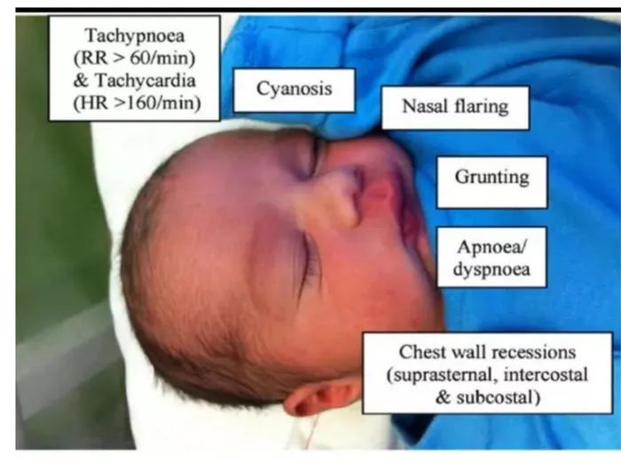
Neonatal respiratory distress syndrome
L5 p.32–33Surfactant, mainly dipalmitoylphosphatidylcholine (lecithin) made by type II pneumocytes, lowers alveolar surface tension so that small alveoli stay open at end-expiration. Production starts late in the second trimester and becomes adequate around 34–35 weeks. In a premature lung, alveoli collapse with each breath (atelectasis); the work of breathing soars and blood passes through unventilated lung, causing hypoxemia. Hypoxia injures epithelium and endothelium; plasma proteins leak and fibrin with necrotic cells forms hyaline membranes in the alveolar ducts.
| Raises risk | Lowers risk |
|---|---|
| Prematurity (the main factor) | Antenatal corticosteroids (betamethasone 24–34 weeks) induce surfactant |
| Maternal diabetes (fetal insulin delays surfactant) | Labor and stress (fetal cortisol surge) |
| Cesarean delivery without labor | Prolonged rupture of membranes, growth restriction (chronic stress) |
| Male sex, second twin, perinatal asphyxia | Exogenous surfactant and CPAP after birth (treatment) |
Prematurity (the main factor)
- Lowers risk
- Antenatal corticosteroids (betamethasone 24–34 weeks) induce surfactant
Maternal diabetes (fetal insulin delays surfactant)
- Lowers risk
- Labor and stress (fetal cortisol surge)
Cesarean delivery without labor
- Lowers risk
- Prolonged rupture of membranes, growth restriction (chronic stress)
Male sex, second twin, perinatal asphyxia
- Lowers risk
- Exogenous surfactant and CPAP after birth (treatment)
Lecture image
Worked calculation · Is the fetal lung mature?
- Setting
- 32 weeks, preterm labor, mother with type 1 diabetes
- Amniotic lecithin
- 2.4 mg/dL
- Amniotic sphingomyelin
- 2.0 mg/dL
Formula
L/S ratio = lecithin ÷ sphingomyelin; ≥ 2.0 suggests maturity (≥ 2.5–3.5, or phosphatidylglycerol present, in diabetic pregnancy)
Ref. · lung maturity testingCore activity · The Missing Arrow
The Missing Arrow
A boy born at 29 weeks by cesarean section, without antenatal steroids, is grunting with nasal flaring and retractions within an hour of birth.
- Birth at 29 weeks: type II pneumocytes immature
- too little surfactant (dipalmitoylphosphatidylcholine) lines the alveoli
- Hypoxemia and acidosis
Cards
Tap or drag a card. Placing a used card moves it.
NEC, intraventricular hemorrhage and SIDS
L5 p.32Necrotizing enterocolitis is the most common gastrointestinal emergency of premature infants. An immature mucosal barrier, reduced blood flow (ischemia) and bacterial colonization, typically after formula feeding has started, combine to injure the bowel wall, usually the terminal ileum and right colon. Infants develop feeding intolerance, abdominal distension and bloody stools; the radiograph shows gas in the bowel wall (pneumatosis intestinalis), sometimes portal venous gas or free air after perforation. Survivors may develop strictures or short-bowel syndrome. Breast milk and antenatal steroids lower the risk.
Intraventricular hemorrhage starts in the germinal matrix, a richly vascular zone beside the lateral ventricles that involutes by about 34 weeks. Its thin-walled vessels rupture when cerebral blood flow fluctuates (hypoxia, hypercapnia, blood-pressure swings) in an infant who cannot autoregulate, usually within the first 3 days in babies born before 32 weeks. Blood can extend into the ventricles and the adjacent white matter; posthemorrhagic hydrocephalus and cerebral palsy can follow. Screening cranial ultrasound finds it.
Sudden infant death syndrome is the sudden death of an infant under 1 year that remains unexplained after a complete autopsy, death-scene investigation and review of the history; it peaks at 2–4 months, usually during sleep. A triple-risk model explains it: a vulnerable infant (often subtle brainstem serotonergic abnormalities that blunt arousal and autoresuscitation), a critical developmental period, and an external stressor (prone sleeping, soft bedding, overheating, bed-sharing, tobacco smoke). Prenatal and postnatal smoking, prematurity and low birth weight add risk. Supine sleep on a firm surface, room-sharing without bed-sharing, breastfeeding and a smoke-free home lower it.
Screening: ultrasound, serum markers and AFP
L5 p.10, 34Ch. V cases p.3Screening tests are offered to every pregnant woman, carry no risk to the fetus and give a probability. Diagnostic tests sample fetal (or placental) cells and give an answer, at a small procedural risk. A positive screen is never a diagnosis; a negative screen does not exclude disease.
Marker patternsL5 p.10, 34
| Condition | 1st trimester (10–13 wk) | 2nd trimester (15–20 wk) | Ultrasound |
|---|---|---|---|
| Down syndrome | ↑ β-hCG, ↓ PAPP-A, ↑ nuchal translucency | ↓ AFP, ↓ estriol, ↑ hCG, ↑ inhibin A | Thick nuchal fold, absent nasal bone, AV canal defect, double bubble |
| Edwards syndrome (18) | ↓ β-hCG, ↓ PAPP-A | ↓ AFP, ↓ estriol, ↓ hCG | Clenched hands, growth restriction, heart defects |
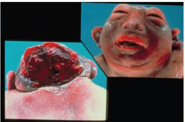
| Open neural tube defect | — | ↑ AFP (others normal) | Anencephaly; spina bifida (lemon and banana signs) |
| Abdominal wall defect | — | ↑ AFP | Omphalocele, gastroschisis |
Down syndrome
- 1st trimester (10–13 wk)
- ↑ β-hCG, ↓ PAPP-A, ↑ nuchal translucency
- 2nd trimester (15–20 wk)
- ↓ AFP, ↓ estriol, ↑ hCG, ↑ inhibin A
- Ultrasound
- Thick nuchal fold, absent nasal bone, AV canal defect, double bubble
Edwards syndrome (18)
- 1st trimester (10–13 wk)
- ↓ β-hCG, ↓ PAPP-A
- 2nd trimester (15–20 wk)
- ↓ AFP, ↓ estriol, ↓ hCG
- Ultrasound
- Clenched hands, growth restriction, heart defects
Open neural tube defect
- 1st trimester (10–13 wk)
- —
- 2nd trimester (15–20 wk)
- ↑ AFP (others normal)
- Ultrasound
- Anencephaly; spina bifida (lemon and banana signs)
Abdominal wall defect
- 1st trimester (10–13 wk)
- —
- 2nd trimester (15–20 wk)
- ↑ AFP
- Ultrasound
- Omphalocele, gastroschisis
Cell-free fetal DNA in maternal blood (from about 10 weeks) detects most trisomy 21 but is still a screen: the DNA is placental.
α-Fetoprotein is made by the yolk sac and fetal liver, enters fetal serum, is excreted in fetal urine into the amniotic fluid and crosses into maternal blood. When the neural tube is open, fetal serum leaks directly into the amniotic fluid, so AFP rises in amniotic fluid and maternal serum (acetylcholinesterase in amniotic fluid confirms an open defect). It also rises with abdominal wall defects, multiple pregnancy, fetal death and underestimated gestational age, and is low in Down syndrome.
Case image
Diagnosis: sampling and the laboratory test
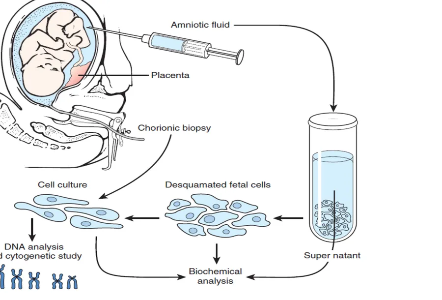
L5 p.34–35Chorionic villus sampling
- 10–13 weeks, transcervical or transabdominal
- Placental (trophoblast) cells: earliest diagnostic result
- Cannot measure amniotic AFP
- Confined placental mosaicism can mislead
Amniocentesis
- 15–20 weeks (earlier carries more risk, as the lecture notes)
- Cultured fetal amniocytes + amniotic fluid
- Measures AFP and acetylcholinesterase
- Small procedure-related loss rate
Lecture image
Which laboratory test answers which questionL5 p.34
| Test | What it sees | Blind spot | Use it for |
|---|---|---|---|
| Karyotype (G-banding) | Every chromosome: number and large structural changes (≳ 5–10 Mb), including balanced rearrangements | Microdeletions, UPD, point mutations; needs dividing cells (days) | Trisomies, Turner, Klinefelter, Robertsonian carriers |
| FISH | Specific targets with fluorescent probes, even in non-dividing cells (rapid) | Only what you probe; no parent of origin | 22q11.2 deletion, rapid 13/18/21/X/Y count, 15q11–q13 deletion |
| Chromosomal microarray | Genome-wide gains and losses down to ~50–100 kb; SNP arrays also detect isodisomy/UPD | Balanced translocations and inversions (no dose change) | First-line for developmental delay or multiple anomalies |
| PCR / Southern blot | Repeat size and methylation | Point changes elsewhere | Fragile X, Huntington, myotonic dystrophy |
| Methylation testing | Parent-of-origin pattern at imprinted loci | UBE3A point mutations (normal methylation) | Prader–Willi, Angelman (deletion, UPD, imprinting defect) |
| Sequencing (gene, panel, exome) | Point mutations and small indels | Large rearrangements, repeats | CFTR, FBN1, PAH, HEXA |
Karyotype (G-banding)
- What it sees
- Every chromosome: number and large structural changes (≳ 5–10 Mb), including balanced rearrangements
- Blind spot
- Microdeletions, UPD, point mutations; needs dividing cells (days)
- Use it for
- Trisomies, Turner, Klinefelter, Robertsonian carriers
FISH
- What it sees
- Specific targets with fluorescent probes, even in non-dividing cells (rapid)
- Blind spot
- Only what you probe; no parent of origin
- Use it for
- 22q11.2 deletion, rapid 13/18/21/X/Y count, 15q11–q13 deletion
Chromosomal microarray
- What it sees
- Genome-wide gains and losses down to ~50–100 kb; SNP arrays also detect isodisomy/UPD
- Blind spot
- Balanced translocations and inversions (no dose change)
- Use it for
- First-line for developmental delay or multiple anomalies
PCR / Southern blot
- What it sees
- Repeat size and methylation
- Blind spot
- Point changes elsewhere
- Use it for
- Fragile X, Huntington, myotonic dystrophy
Methylation testing
- What it sees
- Parent-of-origin pattern at imprinted loci
- Blind spot
- UBE3A point mutations (normal methylation)
- Use it for
- Prader–Willi, Angelman (deletion, UPD, imprinting defect)
Sequencing (gene, panel, exome)
- What it sees
- Point mutations and small indels
- Blind spot
- Large rearrangements, repeats
- Use it for
- CFTR, FBN1, PAH, HEXA
The lecture names karyotyping, PCR and FISH; microarray, methylation testing and sequencing are added as supporting context.
Cellular aging
L5 p.36–37Aging is the gradual decline in a cell's ability to divide, repair and keep its proteins and membranes intact. The lecture lists several theories; they are best read as overlapping mechanisms, each with a disease that exaggerates it.
Teaching schematic
Telomere shortening
TTAGGG ends shorten with each division; critically short telomeres trigger a DNA-damage response (p53/p21) → replicative senescence. Telomerase keeps germ and most cancer cells going.
DNA damage, failed repair
Radiation, replication errors and ROS damage DNA; defective repair accelerates aging. Werner syndrome: WRN RecQ helicase lost (AR) → adult progeria with cancer.
Oxidative stress
Free radicals peroxidize membranes and damage proteins and DNA (Ch. 1.5); antioxidant defenses decline with age.
Cross-linking and glycation
Collagen, elastin and lens crystallin become cross-linked; non-enzymatic glycation (AGEs) stiffens tissue and clouds the lens (cataract).
Nuclear lamina failure
Hutchinson–Gilford progeria: de novo LMNA change → progerin (farnesylated lamin A) → misshapen nuclei, genomic instability.
Genetic programming
Lifespan tracks in families and in identical twins; premature-aging syndromes show genes set the pace.
- Telomere shortening. DNA polymerase cannot copy the very end of a chromosome, so the TTAGGG telomere repeats shorten with each division. Critically short telomeres look like broken DNA, activate p53 and p21, and the cell stops dividing (replicative senescence). Telomerase rebuilds telomeres in germ cells and stem cells and is reactivated in most cancers.
- DNA damage and failing repair (somatic mutation and DNA repair theories). Radiation, replication errors and oxidants damage DNA every day. When repair genes fail, aging accelerates: Werner syndrome (autosomal recessive loss of the WRN RecQ helicase) causes graying, cataracts, skin atrophy, diabetes, atherosclerosis and cancers from early adulthood.
- Oxidative stress (free radical theory). Reactive oxygen species from mitochondria peroxidize membrane lipids and damage proteins and DNA; antioxidant defenses wane with age.
- Cross-linking and glycation. Long-lived extracellular proteins (collagen, elastin, lens crystallin) accumulate cross-links; nonenzymatic glycosylation by glucose forms advanced glycation end-products that stiffen tissues and cloud the lens (cataract), faster in diabetes.
- Genetic programming. Identical twins reach more similar ages than fraternal twins, longevity runs in families, and single-gene premature-aging syndromes exist (progeria; Down syndrome ages early).
Hutchinson–Gilford progeria shows how one base can age a child. A de novo, autosomal dominant point mutation in LMNA (c.1824C>T) does not change its own amino acid, but it activates a cryptic splice site that deletes 50 amino acids, including the cleavage site that normally removes lamin A's farnesyl anchor. The truncated, permanently farnesylated protein, progerin, stays stuck in the inner nuclear membrane, deforms the nucleus (blebbing), and disturbs chromatin, DNA repair and telomeres. Growth falters from 6–18 months; children lose hair and subcutaneous fat, develop scleroderma-like skin, a beaked nose, micrognathia and osteopenia, keep normal intelligence, and die of atherosclerosis (myocardial infarction, stroke) in their teens. A farnesyltransferase inhibitor (lonafarnib) prolongs survival.
Knowledge check · 5.6
Answer, read why, and try again whenever you like. Answers are kept in both study modes.
- Q1AFP in neural tube defectsCh. V cases p.3L5 p.34
A 28-year-old woman delivers a stillborn with the severe neural tube defect shown (Ch. V Q2).
Case image
Which serum marker would have been increased in this pregnancy?
- Q2RDS prevention: antenatal steroidsL5 p.32–33
A boy born at 30 weeks by cesarean section before labor to a mother with poorly controlled diabetes develops grunting, nasal flaring and retractions within an hour. The chest film shows diffuse ground-glass opacity with air bronchograms. No antenatal medication was given.
Which measure before delivery would most have reduced his risk?
- Q3Quadruple screen patternL5 p.10
Which second-trimester serum pattern most suggests Down syndrome?
- Q4Karyotype vs microarrayL5 p.34
A child has translocation Down syndrome. The geneticist wants to know whether either healthy parent carries a balanced Robertsonian translocation.
Which test is needed?
- Q5CVS vs amniocentesisL5 p.34
A 39-year-old woman at 11 weeks has a high-risk first-trimester screen and wants a definitive answer as early as possible.
Which procedure should be offered now?
- Q6Progeria: LMNA/progerinL5 p.37
A 4-year-old with normal intelligence has alopecia, loss of subcutaneous fat, scleroderma-like skin, a beaked nose and growth failure since infancy. Fibroblasts show misshapen, blebbed nuclei.
What is the molecular basis?
- Q7Necrotizing enterocolitisL5 p.32
A 29-week infant started on formula 5 days ago develops abdominal distension, bilious aspirates and bloody stools. The abdominal film shows bubbly gas within the bowel wall.
What is the diagnosis?