Congenital infections and teratogens
A teratogen is anything that crosses into the embryo or fetus and disturbs development. What it does depends on the agent and, as much, on when it arrives.
Congenital infections (TORCH and others)
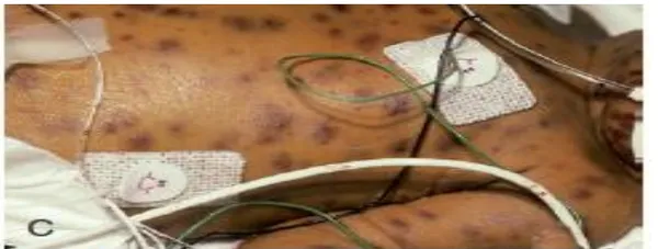
L5 p.25–27Most congenital infections cross the placenta during a maternal infection (often primary, often mild or silent in the mother). Several share a common picture: growth restriction, microcephaly, hepatosplenomegaly, jaundice, thrombocytopenia with petechiae or a blueberry-muffin rash (dermal extramedullary hematopoiesis). What separates them is a small set of specific findings and the timing of transmission.
Transmission and distinguishing findingsL5 p.26
| Infection | Transmission and timing | Distinguishing findings |
|---|---|---|
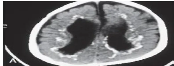
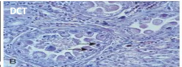
| CMV (commonest) | Transplacental, any trimester; worst after a primary maternal infection | Periventricular calcification, microcephaly, sensorineural hearing loss (leading sequela; can appear later), chorioretinitis, petechiae, hepatosplenomegaly; owl's-eye inclusions in renal tubular cells; urine culture/PCR |
| Toxoplasmosis | Transplacental after a primary infection (cat feces, undercooked meat); transmission ↑ but severity ↓ later in pregnancy | Triad: chorioretinitis, hydrocephalus, diffuse (scattered) intracranial calcification; deafness possible |
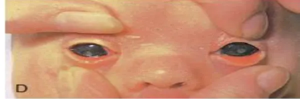
| Rubella | Transplacental; greatest risk in the first trimester (< 12 weeks) | Cataracts, sensorineural deafness, PDA (and peripheral pulmonary stenosis); blueberry-muffin rash; salt-and-pepper retinopathy; microcephaly |
| Syphilis | Transplacental at any time; fetal damage mainly after ~16–20 weeks | Stillbirth, hydrops; early: rhinitis (“snuffles”), rash on palms and soles, hepatosplenomegaly, periostitis; late: saddle nose, notched (Hutchinson) incisors, mulberry molars, saber shins, interstitial keratitis, CN VIII deafness |
| HSV-2 | Mostly at delivery through the birth canal (intrauterine rare) | Vesicles of skin, eye and mouth; encephalitis; disseminated disease with liver failure; cesarean if lesions are active |
| Varicella (VZV) | Transplacental, mostly maternal infection at 8–20 weeks | Dermatomal cicatricial skin scars, limb hypoplasia, chorioretinitis, cataracts, microcephaly |
| Parvovirus B19 | Transplacental; highest risk before 20 weeks | Infects erythroid precursors (P antigen) → severe fetal anemia → high-output heart failure → nonimmune hydrops fetalis; no malformations |
| Zika | Transplacental (mosquito or sexual transmission to the mother) | Severe microcephaly with a collapsed skull, subcortical calcification, eye lesions, contractures |
| HIV | Transplacental, at delivery, and by breastfeeding | Thrush, recurrent bacterial infections, failure to thrive, intracranial calcification; prevented by maternal antiretroviral therapy |
CMV (commonest)
- Transmission and timing
- Transplacental, any trimester; worst after a primary maternal infection
- Distinguishing findings
- Periventricular calcification, microcephaly, sensorineural hearing loss (leading sequela; can appear later), chorioretinitis, petechiae, hepatosplenomegaly; owl's-eye inclusions in renal tubular cells; urine culture/PCR
Toxoplasmosis
- Transmission and timing
- Transplacental after a primary infection (cat feces, undercooked meat); transmission ↑ but severity ↓ later in pregnancy
- Distinguishing findings
- Triad: chorioretinitis, hydrocephalus, diffuse (scattered) intracranial calcification; deafness possible
Rubella
- Transmission and timing
- Transplacental; greatest risk in the first trimester (< 12 weeks)
- Distinguishing findings
- Cataracts, sensorineural deafness, PDA (and peripheral pulmonary stenosis); blueberry-muffin rash; salt-and-pepper retinopathy; microcephaly
Syphilis
- Transmission and timing
- Transplacental at any time; fetal damage mainly after ~16–20 weeks
- Distinguishing findings
- Stillbirth, hydrops; early: rhinitis (“snuffles”), rash on palms and soles, hepatosplenomegaly, periostitis; late: saddle nose, notched (Hutchinson) incisors, mulberry molars, saber shins, interstitial keratitis, CN VIII deafness
HSV-2
- Transmission and timing
- Mostly at delivery through the birth canal (intrauterine rare)
- Distinguishing findings
- Vesicles of skin, eye and mouth; encephalitis; disseminated disease with liver failure; cesarean if lesions are active
Varicella (VZV)
- Transmission and timing
- Transplacental, mostly maternal infection at 8–20 weeks
- Distinguishing findings
- Dermatomal cicatricial skin scars, limb hypoplasia, chorioretinitis, cataracts, microcephaly
Parvovirus B19
- Transmission and timing
- Transplacental; highest risk before 20 weeks
- Distinguishing findings
- Infects erythroid precursors (P antigen) → severe fetal anemia → high-output heart failure → nonimmune hydrops fetalis; no malformations
Zika
- Transmission and timing
- Transplacental (mosquito or sexual transmission to the mother)
- Distinguishing findings
- Severe microcephaly with a collapsed skull, subcortical calcification, eye lesions, contractures
HIV
- Transmission and timing
- Transplacental, at delivery, and by breastfeeding
- Distinguishing findings
- Thrush, recurrent bacterial infections, failure to thrive, intracranial calcification; prevented by maternal antiretroviral therapy
Zika and parvovirus B19 are not in the lecture table; they are added as supporting context.
Lecture image
Lecture image
Lecture image
Lecture image
Lecture image
Lecture image
Core activity · Almost Twins
Almost Twins
Look-alike A
Congenital CMV
- Intracranial calcification, microcephaly
- Periventricular calcification; sensorineural hearing loss is the leading sequela
- Chorioretinitis possible; petechiae, hepatosplenomegaly
- Commonest congenital infection; owl's-eye inclusions in urine/renal tubules
Look-alike A
Congenital CMV
- Intracranial calcification, microcephaly
- Periventricular calcification; sensorineural hearing loss is the leading sequela
- Chorioretinitis possible; petechiae, hepatosplenomegaly
- Commonest congenital infection; owl's-eye inclusions in urine/renal tubules
Look-alike B
Congenital toxoplasmosis
- Intracranial calcification
- Diffuse/scattered calcification with hydrocephalus and prominent chorioretinitis
- Classic triad: chorioretinitis, hydrocephalus, calcifications
- Cat feces and undercooked meat; prevented by avoiding them
Step 1 · What they share
What do they share?
Select every feature that both show.
Drugs and exposures: the lecture's teratogen table
L5 p.25, 29–30Read each row as exposure → mechanism → defect. Many mechanisms repeat: folate antagonism (valproate, methotrexate, phenytoin) gives neural tube and craniofacial defects; vasoconstriction or vascular disruption (cocaine, misoprostol) destroys tissue that had formed; loss of fetal urine (ACE inhibitors) gives the oligohydramnios sequence; hormones (androgenic progestins, DES) act on the genital tract.
Teratogens (lecture Table 6-4, with teaching notes)L5 p.29–30
| Exposure | Defects | Mechanism | Teaching note |
|---|---|---|---|
| ACE inhibitors and ARBs | IUGR, renal failure, Potter-like syndrome, pulmonary hypoplasia, hypoplastic calvaria | Fetal angiotensin II needed for renal perfusion → no fetal urine → oligohydramnios → renal dysplasia | Mainly 2nd–3rd trimester; first-trimester cleft and heart risk is uncertain |
| Alcohol | Microcephaly, intellectual disability, ADHD, VSD/ASD, thin upper lip, smooth philtrum, short palpebral fissures, epicanthal folds, flat nasal bridge | Acetaldehyde disrupts cell differentiation, migration and retinoic acid and hedgehog signaling; apoptosis of neural crest | Leading preventable cause of intellectual disability; no safe dose (see Ch. 6) |
| Amphetamines | Heart defects, IUGR, cleft, limb defects, learning disability | Reactive oxygen and nitrogen species, p53-driven apoptosis, mitochondrial dysfunction in neurons | Often combined with other exposures |
| Aspirin / NSAIDs (prostaglandin inhibitors) | Lecture: orofacial clefts and septal defects | Irreversible COX inhibition; prostaglandins keep the ductus open and maintain placental flow | Main risk is late pregnancy: premature ductus closure and oligohydramnios. Low-dose aspirin is used to prevent preeclampsia. The prostaglandin analog misoprostol causes vascular-disruption defects (limb, Möbius) |
| Carbon monoxide | Cerebral atrophy, microcephaly, seizures | Fetal hemoglobin binds CO avidly → hypoxia, inflammation, lipid peroxidation | Fetal COHb exceeds maternal |
| Cocaine | Microcephaly, low birth weight, renal agenesis, intestinal atresia, heart disease, hydronephrosis, hypospadias | Vasoconstriction → vascular insufficiency (disruption) | Also placental abruption |
| Diethylstilbestrol (DES) | Vaginal/cervical clear cell adenocarcinoma in daughters, T-shaped uterus, cervical incompetence | Disturbs differentiation of Müllerian (paramesonephric) epithelium (vaginal adenosis) | Historical; tumors appear in adolescence |
| Phenytoin | Fetal hydantoin syndrome: distal phalanx and nail hypoplasia, cleft lip/palate, microcephaly; neonatal bleeding | Reactive intermediates and oxidative stress; folate and vitamin K antagonism | Give vitamin K to the newborn |
| Androgenic and progestational exposure | Virilization of a female fetus (clitoromegaly, labial fusion) | Androgenic progestins (19-nortestosterone derivatives), danazol, testosterone act on the genital tubercle | The lecture lists “progesterone”; natural progesterone used to prevent preterm birth does not virilize |
| Retinoids (isotretinoin, high-dose vitamin A) | Microtia, cleft palate, conotruncal heart defects, thymic aplasia, CNS malformations, neural tube defects | Disrupt HOX-gene patterning and neural crest migration | Strict contraception; see vitamin A in Ch. 6 |
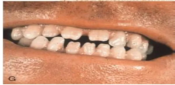
| Tetracyclines | Tooth discoloration, enamel hypoplasia, slowed bone growth | Chelate calcium; deposit in developing teeth and bone | 2nd–3rd trimester |
| Lithium | Ebstein anomaly (tricuspid valve displaced into the right ventricle) | Inhibits GSK-3 in the Wnt pathway | Absolute risk is low; fetal echocardiography |
| Streptomycin (aminoglycosides) | Ototoxicity (eighth-nerve deafness) | Hair-cell death via free radicals and mitochondrial protein synthesis | Also nephrotoxic in the mother |
| Tobacco | IUGR, low birth weight, preterm birth, SIDS | Nicotine vasoconstriction, CO hypoxia, oxidative stress | The slide says no malformations; a modest rise in orofacial clefts is reported |
| Valproate | Neural tube defects (spina bifida), facial, heart and limb defects, autism, lower IQ | Folate antagonism; histone deacetylase inhibition; oxidative stress | Avoid in women who may conceive; high-dose folate |
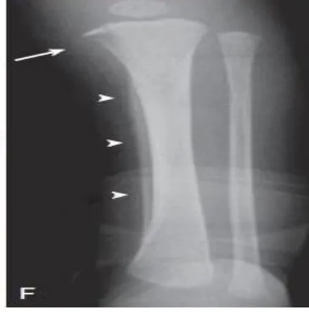
| Warfarin | Nasal hypoplasia, stippled epiphyses (weeks 6–9); CNS defects, fetal hemorrhage, death (later) | Blocks vitamin K–dependent bone proteins; later, hemorrhage | Switch to heparin, which does not cross the placenta |
| Methotrexate | Craniofacial and limb anomalies, neural tube defects | Folate antagonist (dihydrofolate reductase) | Also an abortifacient |
ACE inhibitors and ARBs
- Defects
- IUGR, renal failure, Potter-like syndrome, pulmonary hypoplasia, hypoplastic calvaria
- Mechanism
- Fetal angiotensin II needed for renal perfusion → no fetal urine → oligohydramnios → renal dysplasia
- Teaching note
- Mainly 2nd–3rd trimester; first-trimester cleft and heart risk is uncertain
Alcohol
- Defects
- Microcephaly, intellectual disability, ADHD, VSD/ASD, thin upper lip, smooth philtrum, short palpebral fissures, epicanthal folds, flat nasal bridge
- Mechanism
- Acetaldehyde disrupts cell differentiation, migration and retinoic acid and hedgehog signaling; apoptosis of neural crest
- Teaching note
- Leading preventable cause of intellectual disability; no safe dose (see Ch. 6)
Amphetamines
- Defects
- Heart defects, IUGR, cleft, limb defects, learning disability
- Mechanism
- Reactive oxygen and nitrogen species, p53-driven apoptosis, mitochondrial dysfunction in neurons
- Teaching note
- Often combined with other exposures
Aspirin / NSAIDs (prostaglandin inhibitors)
- Defects
- Lecture: orofacial clefts and septal defects
- Mechanism
- Irreversible COX inhibition; prostaglandins keep the ductus open and maintain placental flow
- Teaching note
- Main risk is late pregnancy: premature ductus closure and oligohydramnios. Low-dose aspirin is used to prevent preeclampsia. The prostaglandin analog misoprostol causes vascular-disruption defects (limb, Möbius)
Carbon monoxide
- Defects
- Cerebral atrophy, microcephaly, seizures
- Mechanism
- Fetal hemoglobin binds CO avidly → hypoxia, inflammation, lipid peroxidation
- Teaching note
- Fetal COHb exceeds maternal
Cocaine
- Defects
- Microcephaly, low birth weight, renal agenesis, intestinal atresia, heart disease, hydronephrosis, hypospadias
- Mechanism
- Vasoconstriction → vascular insufficiency (disruption)
- Teaching note
- Also placental abruption
Diethylstilbestrol (DES)
- Defects
- Vaginal/cervical clear cell adenocarcinoma in daughters, T-shaped uterus, cervical incompetence
- Mechanism
- Disturbs differentiation of Müllerian (paramesonephric) epithelium (vaginal adenosis)
- Teaching note
- Historical; tumors appear in adolescence
Phenytoin
- Defects
- Fetal hydantoin syndrome: distal phalanx and nail hypoplasia, cleft lip/palate, microcephaly; neonatal bleeding
- Mechanism
- Reactive intermediates and oxidative stress; folate and vitamin K antagonism
- Teaching note
- Give vitamin K to the newborn
Androgenic and progestational exposure
- Defects
- Virilization of a female fetus (clitoromegaly, labial fusion)
- Mechanism
- Androgenic progestins (19-nortestosterone derivatives), danazol, testosterone act on the genital tubercle
- Teaching note
- The lecture lists “progesterone”; natural progesterone used to prevent preterm birth does not virilize
Retinoids (isotretinoin, high-dose vitamin A)
- Defects
- Microtia, cleft palate, conotruncal heart defects, thymic aplasia, CNS malformations, neural tube defects
- Mechanism
- Disrupt HOX-gene patterning and neural crest migration
- Teaching note
- Strict contraception; see vitamin A in Ch. 6
Tetracyclines
- Defects
- Tooth discoloration, enamel hypoplasia, slowed bone growth
- Mechanism
- Chelate calcium; deposit in developing teeth and bone
- Teaching note
- 2nd–3rd trimester
Lithium
- Defects
- Ebstein anomaly (tricuspid valve displaced into the right ventricle)
- Mechanism
- Inhibits GSK-3 in the Wnt pathway
- Teaching note
- Absolute risk is low; fetal echocardiography
Streptomycin (aminoglycosides)
- Defects
- Ototoxicity (eighth-nerve deafness)
- Mechanism
- Hair-cell death via free radicals and mitochondrial protein synthesis
- Teaching note
- Also nephrotoxic in the mother
Tobacco
- Defects
- IUGR, low birth weight, preterm birth, SIDS
- Mechanism
- Nicotine vasoconstriction, CO hypoxia, oxidative stress
- Teaching note
- The slide says no malformations; a modest rise in orofacial clefts is reported
Valproate
- Defects
- Neural tube defects (spina bifida), facial, heart and limb defects, autism, lower IQ
- Mechanism
- Folate antagonism; histone deacetylase inhibition; oxidative stress
- Teaching note
- Avoid in women who may conceive; high-dose folate
Warfarin
- Defects
- Nasal hypoplasia, stippled epiphyses (weeks 6–9); CNS defects, fetal hemorrhage, death (later)
- Mechanism
- Blocks vitamin K–dependent bone proteins; later, hemorrhage
- Teaching note
- Switch to heparin, which does not cross the placenta
Methotrexate
- Defects
- Craniofacial and limb anomalies, neural tube defects
- Mechanism
- Folate antagonist (dihydrofolate reductase)
- Teaching note
- Also an abortifacient
Fetal alcohol syndrome
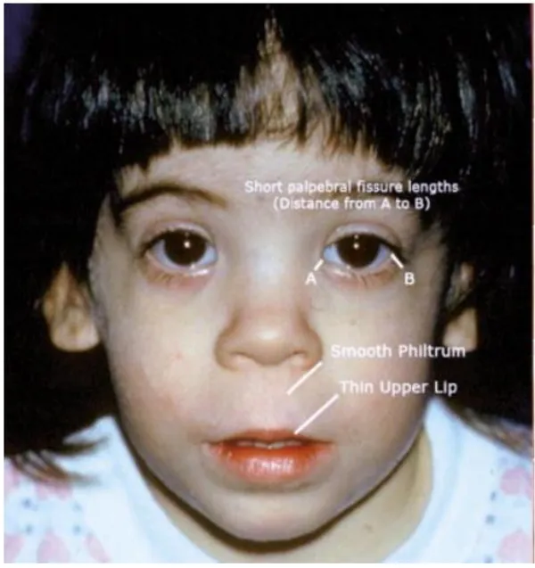
L5 p.29, 31Ch. V cases p.2Alcohol crosses the placenta freely, and the fetus clears it slowly. Ethanol and acetaldehyde disturb proliferation, migration and survival of neural crest and brain cells. The facial features are set by exposure early in the first trimester, while the brain stays vulnerable throughout pregnancy, which is why there is no safe amount or time. Fetal alcohol spectrum disorder is the leading preventable cause of intellectual disability.
| Domain | Findings |
|---|---|
| Face | Short palpebral fissures, smooth philtrum, thin upper lip, epicanthal folds, low nasal bridge, short nose |
| Growth | Prenatal and postnatal growth deficiency |
| Brain | Microcephaly, intellectual disability, attention deficit, poor impulse control and emotional regulation |
| Other | Heart defects (VSD, ASD), joint and renal anomalies |
Face
- Findings
- Short palpebral fissures, smooth philtrum, thin upper lip, epicanthal folds, low nasal bridge, short nose
Growth
- Findings
- Prenatal and postnatal growth deficiency
Brain
- Findings
- Microcephaly, intellectual disability, attention deficit, poor impulse control and emotional regulation
Other
- Findings
- Heart defects (VSD, ASD), joint and renal anomalies
Lecture image
Worked case · An “emotional” 4-year-old
A 4-year-old girl at a preschool check has a small head circumference, a thin upper lip and a low nasal bridge, mild intellectual disability, and her parents say she is often “emotional” (Ch. V Q1).
Which maternal factor best explains the findings?
Ch. V cases p.2Critical periods: when an exposure arrives
L5 p.28Ref. · Moore, The Developing HumanThe same drug can do nothing, kill the embryo, cause a malformation or only slow growth, depending on timing. In the first two weeks after fertilization cells are still interchangeable: an injury either kills enough of them to end the pregnancy or the survivors replace the lost cells (all-or-none). Weeks 3–8 are organogenesis, when each organ has a window of maximal sensitivity to major structural defects. After week 8 (the fetal period) organs grow and mature, so exposures cause growth restriction, functional defects (brain, hearing) and minor anomalies; the brain, palate, ears, teeth and external genitalia remain partly vulnerable.
Teaching schematic
Dark bars: highest sensitivity to major structural defects; light bars: functional and minor defects. Approximate, after Moore's embryology charts.
Core activity · One Fact Changes
One Fact Changes
1 · The original case
A 24-year-old woman takes isotretinoin for severe acne. She stops it on discovering she is pregnant, about 5 weeks after conception. She asks what the drug could have done.
What is the main risk?
Which fact matters most for the type of risk?
Knowledge check · 5.5
Answer, read why, and try again whenever you like. Answers are kept in both study modes.
- Q1Fetal alcohol syndromeCh. V cases p.2L5 p.29, 31
A 4-year-old girl has a small head circumference, a thin upper lip, a low nasal bridge, mild intellectual disability, and is often “emotional” (Ch. V Q1).
Which maternal cause of birth defects most likely accounts for these findings?
- Q2Congenital CMV (image)L5 p.26–27
A 3-week-old with petechiae, hepatosplenomegaly and a failed newborn hearing screen has the head CT shown.
Lecture image
Which congenital infection is most likely?
- Q3Congenital rubellaL5 p.26–27
An unvaccinated woman had a fever and fine rash at 8 weeks of gestation. Her newborn has white pupils, a continuous machinery murmur and fails the hearing screen.
Which infection explains the triad?
- Q4Parvovirus B19 hydropsRef. · parvovirus B19 hydrops
At 18 weeks, ultrasound shows fetal ascites, pleural effusions and skin edema. Three weeks earlier the mother cared for a child with a “slapped-cheek” rash.
What is the mechanism of the fetal findings?
- Q5ACE inhibitor fetopathyL5 p.29
A woman with hypertension continued lisinopril through her second and third trimesters. Her stillborn infant has hypoplastic lungs, a poorly ossified skull and small, dysplastic kidneys; amniotic fluid had been severely reduced.
What is the main mechanism?
- Q6All-or-none periodRef. · Moore, critical periods
A woman learns she is pregnant when her period is late. Dating shows she took a known teratogen for 3 days, from day 6 to day 9 after conception, and none since.
What is the most accurate counseling?
- Q7Valproate and NTDsL5 p.30
A woman with epilepsy who takes valproate plans a pregnancy.
Which defect is her baby at greatest risk of, and what reduces it?