Mutations and Mendelian inheritance
A mutation is a permanent change in DNA. Whether it reaches the next generation, what it does to the protein, and how it travels through a family decide who is affected and what the risk is for the next child.
Germline and somatic mutations
L5 p.2A mutation is a permanent change in the nucleotide sequence or arrangement of DNA. Where it happens decides who can inherit it. A germline mutation is present in an egg or sperm (or the cells that make them), so it can pass to children and is then present in every cell of the child's body. A somatic mutation arises in a body cell after conception; it is copied only into that cell's descendants and is not transmitted to offspring.
Germline
- In gametes or their precursors
- Every cell of the offspring carries it
- Causes inherited (familial) disease
- Example: a CFTR allele passed from a carrier parent
Somatic
- Arises in one body cell after conception
- Only that cell's clone carries it (a mosaic)
- Not passed to children
- Example: most cancer-driving mutations; an early embryonic error gives mosaicism (5.2)
What a mutation does to the message
L5 p.3–4The ribosome reads mRNA three bases at a time from a fixed start. A mutation's effect depends on what it does to that reading frame and to the codon it hits. A point mutation replaces one base: it can be silent (same amino acid, usually a third-position change), missense (a different amino acid) or nonsense (a premature stop codon, so the protein is truncated and the mRNA is often destroyed). An insertion or deletion of a number of bases not divisible by three shifts the frame: every codon downstream is misread until a stop codon appears (frameshift). Losing or adding a multiple of three keeps the frame and removes or adds whole amino acids (in-frame). Splice-site mutations alter the signals that mark exon borders, so an exon is skipped or an intron retained.
Teaching schematic
Normal β-globin
codons 5–8
Normal protein
Silent
GAG → GAA
Same amino acid (third-base wobble)
Missense (HbS)
GAG → GUG
Glu6Val: HbS polymerizes when deoxygenated
Nonsense
GAG → UAG
Premature stop: truncated protein, mRNA decay
Frameshift (+1)
an A inserted
Every codon downstream is misread until a stop appears (e.g., Tay–Sachs 4-bp insertion)
In-frame deletion (CFTR ΔF508)
3 bases (CTT) lost
Frame kept; one residue missing → misfolded CFTR degraded in the ER
Splice-site mutations change the GT…AG signals at exon–intron borders, so an exon is skipped or an intron retained (many β-thalassemias). A codon change can also create a new splice site: the “silent” LMNA change in progeria (5.6).
The lecture's examples, mechanism firstL5 p.3
| Disease | Mutation | What happens to the protein |
|---|---|---|
| Sickle cell disease | Missense: GAG → GTG at β-globin codon 6 (Glu → Val) | HbS is made in normal amounts but polymerizes when deoxygenated; red cells sickle |
| Cystic fibrosis (ΔF508) | In-frame deletion of 3 bases: phenylalanine 508 lost | Full-length CFTR misfolds, is retained in the ER and degraded; little channel reaches the apical membrane |
| Tay–Sachs disease | Frameshift: 4-base insertion in HEXA (the common Ashkenazi allele) | No functional hexosaminidase A; GM2 ganglioside accumulates in neurons |
| β⁰-thalassemia (example) | Nonsense or splice-site mutations in β-globin | No β chain made; unpaired α chains precipitate |
Sickle cell disease
- Mutation
- Missense: GAG → GTG at β-globin codon 6 (Glu → Val)
- What happens to the protein
- HbS is made in normal amounts but polymerizes when deoxygenated; red cells sickle
Cystic fibrosis (ΔF508)
- Mutation
- In-frame deletion of 3 bases: phenylalanine 508 lost
- What happens to the protein
- Full-length CFTR misfolds, is retained in the ER and degraded; little channel reaches the apical membrane
Tay–Sachs disease
- Mutation
- Frameshift: 4-base insertion in HEXA (the common Ashkenazi allele)
- What happens to the protein
- No functional hexosaminidase A; GM2 ganglioside accumulates in neurons
β⁰-thalassemia (example)
- Mutation
- Nonsense or splice-site mutations in β-globin
- What happens to the protein
- No β chain made; unpaired α chains precipitate
Worked case · Same gene, different damage
Two children have mutations in the same gene. In child A a single base change converts a glutamate codon (GAG) to a stop codon (UAG). In child B a single base is inserted near the start of the coding sequence.
Which child is more likely to make some protein, and why are both likely to be severe?
Trinucleotide repeats and anticipation
L5 p.3Ch. V cases p.12Some genes contain a short run of a repeated triplet. Above a threshold the run becomes unstable: it tends to lengthen when passed to the next generation. Longer repeats cause earlier and more severe disease, so the disorder appears to worsen down the family: anticipation. Where the repeat sits in the gene decides the mechanism.
The four repeat disorders in the lectureL5 p.3
| Disease | Repeat and location | Inheritance | Mechanism and key findings | Expansion mostly through |
|---|---|---|---|---|
| Huntington disease | CAG, coding region of HTT (4p) | AD | Polyglutamine protein aggregates (gain of function) → caudate and putamen atrophy: chorea, psychiatric change, dementia | Father (spermatogenesis) |
| Myotonic dystrophy type 1 | CTG, 3′ untranslated region of DMPK (19q) | AD | Toxic RNA traps splicing regulators → myotonia, distal weakness, temporal wasting, frontal balding, cataracts, testicular atrophy, cardiac conduction block | Mother for the severe congenital form |
| Fragile X syndrome | CGG, 5′ untranslated region of FMR1 (Xq27.3) | X-linked (lecture: XD) | Hypermethylation silences the gene → no FMRP (5.3) | Mother |
| Friedreich ataxia | GAA, intron 1 of FXN (9q) | AR | Too little frataxin → mitochondrial iron overload, oxidative damage → ataxia, hypertrophic cardiomyopathy, diabetes | Little anticipation (recessive) |
Huntington disease
- Repeat and location
- CAG, coding region of HTT (4p)
- Inheritance
- AD
- Mechanism and key findings
- Polyglutamine protein aggregates (gain of function) → caudate and putamen atrophy: chorea, psychiatric change, dementia
- Expansion mostly through
- Father (spermatogenesis)
Myotonic dystrophy type 1
- Repeat and location
- CTG, 3′ untranslated region of DMPK (19q)
- Inheritance
- AD
- Mechanism and key findings
- Toxic RNA traps splicing regulators → myotonia, distal weakness, temporal wasting, frontal balding, cataracts, testicular atrophy, cardiac conduction block
- Expansion mostly through
- Mother for the severe congenital form
Fragile X syndrome
- Repeat and location
- CGG, 5′ untranslated region of FMR1 (Xq27.3)
- Inheritance
- X-linked (lecture: XD)
- Mechanism and key findings
- Hypermethylation silences the gene → no FMRP (5.3)
- Expansion mostly through
- Mother
Friedreich ataxia
- Repeat and location
- GAA, intron 1 of FXN (9q)
- Inheritance
- AR
- Mechanism and key findings
- Too little frataxin → mitochondrial iron overload, oxidative damage → ataxia, hypertrophic cardiomyopathy, diabetes
- Expansion mostly through
- Little anticipation (recessive)
Worked case · A son worse than his father
A 16-year-old faints; his pulse is 43/min. He has frontal balding, temporal wasting, small testes, bilateral foot drop and weak hand muscles. His father developed cataracts and frontal balding in his twenties but has no heart disease (Ch. V Q11).
Which genetic property explains why the son is more severely affected?
Ch. V cases p.12Reading a pedigree: four Mendelian patterns
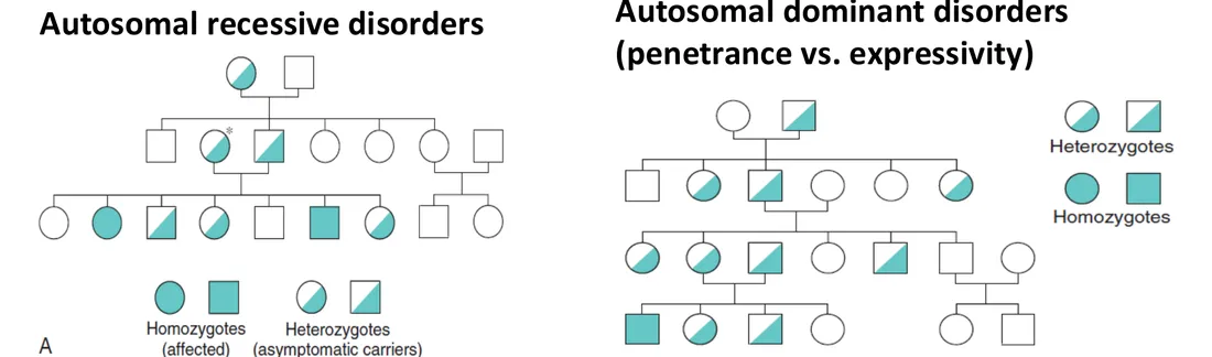
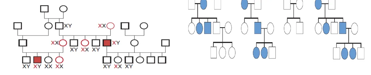
L5 p.5–6A pedigree is evidence. Each pattern leaves a signature that follows from two facts: autosomes come in pairs in both sexes, while a male has one X (from his mother) and passes his X to every daughter and his Y to every son. So the most powerful single observation is father-to-son transmission: it is possible only for autosomal (or Y-linked) traits and excludes anything carried on the X or in mitochondria.
Teaching schematic
Autosomal dominant
Every generation; affected parent → ½ of children; male-to-male transmission
Autosomal recessive
Unaffected carrier parents; ¼ of children affected; both sexes; often consanguinity
X-linked recessive
Mostly males, linked through carrier females; no male-to-male transmission
X-linked dominant
Affected father → all daughters, no sons; affected mother → ½ of either sex
Mitochondrial
Affected mother → all children (variable severity); affected father → none
- male
- female
- affected
- known carrier
- proband
Signatures and the lecture's disease examplesL5 p.5–6
| Pattern | Signature | Lecture examples |
|---|---|---|
| Autosomal dominant | Vertical: every generation; affected parent → each child 1/2; both sexes; male-to-male transmission possible | Marfan, osteogenesis imperfecta, familial hypercholesterolemia, von Willebrand disease, Huntington disease |
| Autosomal recessive | Horizontal: affected siblings of unaffected carrier parents; 1/4 per child; consanguinity raises risk | Cystic fibrosis, sickle cell disease, most metabolic disorders (PKU), lysosomal storage diseases (Tay–Sachs) |
| X-linked recessive | Mostly males; passed through carrier women (each son 1/2); affected father → all daughters carriers, no sons affected | Hemophilia A and B, chronic granulomatous disease |
| X-linked dominant | Affected father → all daughters, no sons; affected mother → 1/2 of children of either sex; more females affected | Hypophosphatemic (vitamin D–resistant) rickets, Alport syndrome, fragile X (lecture classification) |
Autosomal dominant
- Signature
- Vertical: every generation; affected parent → each child 1/2; both sexes; male-to-male transmission possible
- Lecture examples
- Marfan, osteogenesis imperfecta, familial hypercholesterolemia, von Willebrand disease, Huntington disease
Autosomal recessive
- Signature
- Horizontal: affected siblings of unaffected carrier parents; 1/4 per child; consanguinity raises risk
- Lecture examples
- Cystic fibrosis, sickle cell disease, most metabolic disorders (PKU), lysosomal storage diseases (Tay–Sachs)
X-linked recessive
- Signature
- Mostly males; passed through carrier women (each son 1/2); affected father → all daughters carriers, no sons affected
- Lecture examples
- Hemophilia A and B, chronic granulomatous disease
X-linked dominant
- Signature
- Affected father → all daughters, no sons; affected mother → 1/2 of children of either sex; more females affected
- Lecture examples
- Hypophosphatemic (vitamin D–resistant) rickets, Alport syndrome, fragile X (lecture classification)
What each example disease does (mechanism in one line)L5 p.5–6
| Disease | Gene / product | Why it causes disease |
|---|---|---|
| Marfan syndrome | FBN1 (fibrillin-1), chr 15 | Defective microfibrils and excess TGF-β signaling: tall stature, arachnodactyly, upward lens dislocation, aortic root dilation and dissection |
| Osteogenesis imperfecta | COL1A1/COL1A2 (type I collagen) | Abnormal collagen weakens bone, sclera, dentin: fractures, blue sclerae, hearing loss |
| Familial hypercholesterolemia | LDLR (LDL receptor) | Liver cannot clear LDL: very high LDL, xanthomas, early coronary disease; homozygotes far worse (gene dose) |
| von Willebrand disease | VWF | Poor platelet adhesion and less factor VIII carrier: mucosal bleeding |
| Cystic fibrosis | CFTR, chr 7 | Defective chloride channel: thick secretions in lungs, pancreas, vas deferens; salty sweat |
| Sickle cell disease | HBB Glu6Val | HbS polymerizes when deoxygenated: hemolysis and vaso-occlusion |
| Hemophilia A / B | F8 / F9 | Factor VIII / IX deficiency: deep tissue and joint bleeding |
| Chronic granulomatous disease | CYBB (NADPH oxidase, X-linked form) | No respiratory burst: infections by catalase-positive organisms, granulomas |
| Hypophosphatemic rickets | PHEX → ↑ FGF23 | Proximal tubule wastes phosphate: rickets despite vitamin D |
| Alport syndrome | COL4A5 (type IV collagen α5) | Defective basement membranes: hematuria → renal failure, sensorineural deafness, lens defects. (Goodpasture is autoimmune anti-GBM disease, not inherited.) |
Marfan syndrome
- Gene / product
- FBN1 (fibrillin-1), chr 15
- Why it causes disease
- Defective microfibrils and excess TGF-β signaling: tall stature, arachnodactyly, upward lens dislocation, aortic root dilation and dissection
Osteogenesis imperfecta
- Gene / product
- COL1A1/COL1A2 (type I collagen)
- Why it causes disease
- Abnormal collagen weakens bone, sclera, dentin: fractures, blue sclerae, hearing loss
Familial hypercholesterolemia
- Gene / product
- LDLR (LDL receptor)
- Why it causes disease
- Liver cannot clear LDL: very high LDL, xanthomas, early coronary disease; homozygotes far worse (gene dose)
von Willebrand disease
- Gene / product
- VWF
- Why it causes disease
- Poor platelet adhesion and less factor VIII carrier: mucosal bleeding
Cystic fibrosis
- Gene / product
- CFTR, chr 7
- Why it causes disease
- Defective chloride channel: thick secretions in lungs, pancreas, vas deferens; salty sweat
Sickle cell disease
- Gene / product
- HBB Glu6Val
- Why it causes disease
- HbS polymerizes when deoxygenated: hemolysis and vaso-occlusion
Hemophilia A / B
- Gene / product
- F8 / F9
- Why it causes disease
- Factor VIII / IX deficiency: deep tissue and joint bleeding
Chronic granulomatous disease
- Gene / product
- CYBB (NADPH oxidase, X-linked form)
- Why it causes disease
- No respiratory burst: infections by catalase-positive organisms, granulomas
Hypophosphatemic rickets
- Gene / product
- PHEX → ↑ FGF23
- Why it causes disease
- Proximal tubule wastes phosphate: rickets despite vitamin D
Alport syndrome
- Gene / product
- COL4A5 (type IV collagen α5)
- Why it causes disease
- Defective basement membranes: hematuria → renal failure, sensorineural deafness, lens defects. (Goodpasture is autoimmune anti-GBM disease, not inherited.)
Lecture slide
Lecture slide
Case image
Turning a pedigree into a probability
L5 p.5–6Every risk calculation has the same three steps: fix the genotypes that are certain (affected people and obligate carriers, whose children prove what they carry), list the equally likely outcomes of each mating, and then remove the outcomes the facts exclude. The last step is the one students skip, and it is the source of the famous 2/3.
Worked calculation · Healthy sibling of a child with an autosomal recessive disease
- Parents
- both carriers (+/m) — proven by the affected child
- Outcomes per child
- +/+, +/m, m/+, m/m (each 1/4)
- Known about the sibling
- healthy, so not m/m
Formula
P(carrier | healthy) = P(+/m) ÷ P(healthy) = (1/2) ÷ (3/4)
Ref. · Thompson & Thompson, risk calculationWorked calculation · Sister of a boy with an X-linked recessive disease
- Mother
- obligate carrier X⁺Xᵐ (affected son, healthy husband, affected brother)
- Father
- X⁺Y
- Question
- risk that the sister's next son is affected
Formula
P(sister carrier) × P(son receives Xᵐ)
Risks to each child, by patternL5 p.5–6, 21
| Mating | Sons | Daughters |
|---|---|---|
| AD: affected heterozygote × unaffected | 1/2 affected | 1/2 affected |
| AR: carrier × carrier | 1/4 affected, 1/2 carriers | 1/4 affected, 1/2 carriers |
| XLR: carrier mother × normal father | 1/2 affected | 1/2 carriers |
| XLR: affected father × normal mother | None affected | All carriers |
| XLD: affected father × normal mother | None affected | All affected |
| XLD: affected (heterozygous) mother × normal father | 1/2 affected | 1/2 affected |
| Mitochondrial: affected mother | All inherit (variable severity) | All inherit |
| Mitochondrial: affected father | None | None |
AD: affected heterozygote × unaffected
- Sons
- 1/2 affected
- Daughters
- 1/2 affected
AR: carrier × carrier
- Sons
- 1/4 affected, 1/2 carriers
- Daughters
- 1/4 affected, 1/2 carriers
XLR: carrier mother × normal father
- Sons
- 1/2 affected
- Daughters
- 1/2 carriers
XLR: affected father × normal mother
- Sons
- None affected
- Daughters
- All carriers
XLD: affected father × normal mother
- Sons
- None affected
- Daughters
- All affected
XLD: affected (heterozygous) mother × normal father
- Sons
- 1/2 affected
- Daughters
- 1/2 affected
Mitochondrial: affected mother
- Sons
- All inherit (variable severity)
- Daughters
- All inherit
Mitochondrial: affected father
- Sons
- None
- Daughters
- None
Pedigree Lab
L5 p.5–10, 21, 23Five families, one method. Pick the pattern and the clue that proves it, genotype the marked people by tapping them, then compute a risk. The second and third tabs carry the same logic into chromosomes: a 46-chromosome child with Down syndrome, and six children whose chromosome 15 tells a parent-of-origin story.
Centerpiece · Pedigree Lab
Pedigree Lab
A daughter with cystic fibrosis
III-1 has cystic fibrosis. Her parents and siblings are healthy, and so is the rest of the family. Her sister III-2 wants to know whether she could be a carrier.
- male
- female
- affected
- proband
Cystic fibrosis (CFTR) · dashed rings mark the people you will genotype in step 2.
Step 1
Inheritance pattern and the decisive clue
Step 2
Assign genotypes
Settle the pattern in step 1 first: genotypes depend on it.
Step 3
Compute the probability
Finish the genotypes first: the calculation starts from them.
Penetrance, expressivity and pleiotropy
L5 p.5Ch. V cases p.4Three words describe how a genotype shows up, and they answer different questions. Penetrance asks whether: the proportion of people with the genotype who show any sign of it. Most recessive and metabolic disorders are fully penetrant (the lecture's point); some dominant ones are not, so a carrier can look healthy and the disease seems to skip a generation. Expressivity asks how much: among affected people, how severe and in which organs. Pleiotropy asks how many places: one gene affecting several apparently unrelated systems.
| Term | Question it answers | Example |
|---|---|---|
| Penetrance | Does a person with the genotype show the trait at all? | Huntington: near-complete; BRCA1 or retinoblastoma: incomplete → apparent skipped generation |
| Expressivity | How severely, and with which features? | Marfan: one relative has only long fingers, another an aortic dissection; NF1 |
| Pleiotropy | How many organs does one gene affect? | Marfan (skeleton, eye, aorta); PKU (brain, pigment, odor); myotonic dystrophy (muscle, lens, heart, gonads) |
| Anticipation | Why is the next generation worse? | Repeat expansion: Huntington, myotonic dystrophy |
Penetrance
- Question it answers
- Does a person with the genotype show the trait at all?
- Example
- Huntington: near-complete; BRCA1 or retinoblastoma: incomplete → apparent skipped generation
Expressivity
- Question it answers
- How severely, and with which features?
- Example
- Marfan: one relative has only long fingers, another an aortic dissection; NF1
Pleiotropy
- Question it answers
- How many organs does one gene affect?
- Example
- Marfan (skeleton, eye, aorta); PKU (brain, pigment, odor); myotonic dystrophy (muscle, lens, heart, gonads)
Anticipation
- Question it answers
- Why is the next generation worse?
- Example
- Repeat expansion: Huntington, myotonic dystrophy
Case image
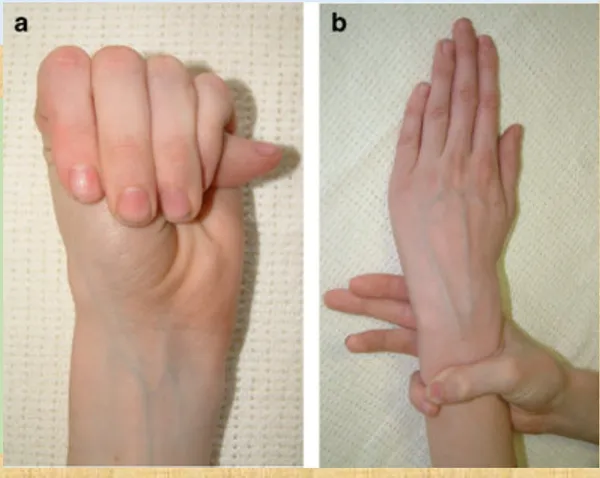
Marfan syndrome shows all three ideas. FBN1 encodes fibrillin-1, a scaffold for elastic fibers that also holds TGF-β inactive. Abnormal fibrillin weakens connective tissue and releases excess TGF-β: long bones overgrow (tall stature, arachnodactyly, pectus), the suspensory ligament of the lens fails (upward lens dislocation), the mitral valve becomes floppy, and the aortic media degenerates (cystic medial degeneration), so the aortic root dilates and can dissect. Penetrance is high, expressivity variable, and about a quarter of cases are new mutations.
Two recessive diseases from the cases: CF and PKU
Ch. V cases p.5–7L5 p.3, 5Cystic fibrosis (CFTR, chromosome 7) is the most common lethal autosomal recessive disease in people of European ancestry; about 1 in 25 are carriers. CFTR is an apical chloride (and bicarbonate) channel. Without it, epithelia secrete too little chloride and water, while airway cells absorb extra sodium, so secretions become thick. The sweat duct cannot reabsorb chloride, which is why sweat is salty and the sweat chloride test is diagnostic.
One channel, many organsCh. V cases p.5
| Organ | What thick secretions do | Clinical result |
|---|---|---|
| Pancreas (85–90%) | Ducts plug; acini atrophy and fibrose | No lipase reaches the gut → steatorrhea, failure to thrive, deficiency of vitamins A, D, E, K; later CF-related diabetes |
| Intestine | Inspissated meconium | Meconium ileus in newborns |
| Airways | Mucus is not cleared | Recurrent infection (S. aureus, P. aeruginosa), bronchiectasis, respiratory failure |
| Liver | Bile plugs ductules | Focal biliary cirrhosis |
| Male reproductive tract | Vas deferens fails to form or is obstructed | Infertility (azoospermia) |
| Sweat glands | Cl⁻ not reabsorbed | Salty sweat, salt depletion in heat |
Pancreas (85–90%)
- What thick secretions do
- Ducts plug; acini atrophy and fibrose
- Clinical result
- No lipase reaches the gut → steatorrhea, failure to thrive, deficiency of vitamins A, D, E, K; later CF-related diabetes
Intestine
- What thick secretions do
- Inspissated meconium
- Clinical result
- Meconium ileus in newborns
Airways
- What thick secretions do
- Mucus is not cleared
- Clinical result
- Recurrent infection (S. aureus, P. aeruginosa), bronchiectasis, respiratory failure
Liver
- What thick secretions do
- Bile plugs ductules
- Clinical result
- Focal biliary cirrhosis
Male reproductive tract
- What thick secretions do
- Vas deferens fails to form or is obstructed
- Clinical result
- Infertility (azoospermia)
Sweat glands
- What thick secretions do
- Cl⁻ not reabsorbed
- Clinical result
- Salty sweat, salt depletion in heat
Core activity · The Missing Arrow
The Missing Arrow
A 4-year-old with a positive sweat chloride test has greasy, foul-smelling stools (Ch. V Q4). Complete the chain from mutation to malabsorption.
- Both CFTR alleles carry ΔF508 (3-base, in-frame deletion: Phe508 lost)
- less Cl⁻ and HCO₃⁻ secreted, so less water follows into the duct
- Thick, dehydrated secretions in pancreatic ducts
Cards
Tap or drag a card. Placing a used card moves it.
Phenylketonuria (PKU) is an autosomal recessive deficiency of phenylalanine hydroxylase, which converts phenylalanine to tyrosine. The unmetabolized substrate accumulates: high phenylalanine impairs myelination and competes with other large neutral amino acids for transport into the brain, lowering serotonin and dopamine synthesis. Untreated infants look normal at birth, then develop intellectual disability, seizures and a musty (“mousy”) odor from phenylacetate. Less tyrosine means less melanin: fair skin, blond hair, blue eyes. Tyrosine becomes an essential amino acid.
Worked case · A boy adopted from an orphanage
A 10-month-old adopted from an orphanage is missing milestones. He is fair-skinned and blond with a mousy odor. Tests show an inborn error of amino acid metabolism (Ch. V Q5–Q6, one case with two questions).
Which amino acid must his diet restrict, and why is his brain injured?
Ch. V cases p.6–7Knowledge check · 5.1
Answer, read why, and try again whenever you like. Answers are kept in both study modes.
- Q1Marfan syndrome: autosomal dominantCh. V cases p.4L5 p.5
A 25-year-old man is 6 ft 5 in tall with the hand findings shown. A year later he suffers a dissecting aortic aneurysm (Ch. V Q3).
Case image
What type of inheritance is characteristic of his disease?
- Q2CF: pancreatic insufficiencyCh. V cases p.5
A 4-year-old boy is admitted with pneumonia. His stools are greasy and foul-smelling, and a sweat chloride test is positive (Ch. V Q4).
What mechanism most likely causes his steatorrhea?
- Q3PKU: phenylalanine restrictionCh. V cases p.6
A 10-month-old boy adopted from an orphanage is fair-skinned and blond, has a mousy odor and is missing milestones. An inborn error of amino acid metabolism is found (Ch. V Q5).
To prevent further intellectual disability, his diet should lack which essential amino acid?
- Q4PKU: substrate accumulationCh. V cases p.7
Same patient as the previous question (Ch. V Q6).
Which best describes the pathogenesis of his intellectual disability?
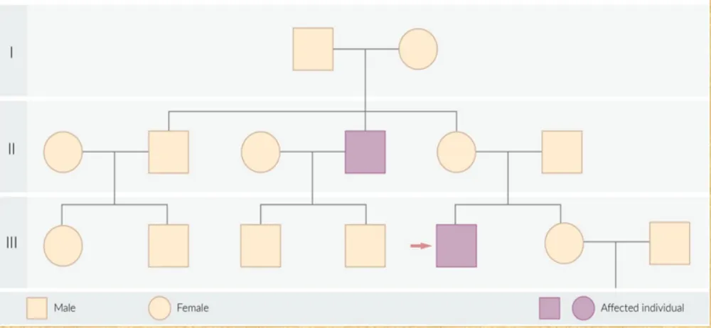
- Q5Pedigree: X-linked recessiveCh. V cases p.13L5 p.6
A 4-year-old boy cannot sort colored balls into matching baskets. His mother mentions an eye condition that runs in the family. The pedigree is shown; the proband is arrowed (Ch. V Q12).
- male
- female
- affected
- proband
Which mode of inheritance best explains his vision problem?
- Q6Anticipation (CTG expansion)Ch. V cases p.12L5 p.3
A 16-year-old with syncope, a pulse of 43/min, frontal balding, temporal wasting, testicular atrophy and distal weakness has myotonic dystrophy. His father had cataracts and frontal balding in his twenties only (Ch. V Q11).
The son's greater severity is best explained by which genetic property?
- Q7In-frame deletion vs frameshiftL5 p.3
The common CFTR allele deletes three nucleotides. What is the main consequence for the protein?
- Q82/3 carrier riskL5 p.5
A healthy 24-year-old woman's brother has cystic fibrosis; her parents are healthy. Her partner has no family history and has not been tested.
What is the probability that she is a carrier?