Who gets cancer, and which genes break
Cancer is common, rises with age and differs between populations. Underneath, every cancer is a set of mutations in four classes of genes: accelerators, brakes, apoptosis regulators and DNA repair.
Epidemiology: causes, age and populations
L7 p.13Cancer is the second most common cause of death in developed countries. The lecture sorts its causes into three groups: external factors (tobacco, the leading one; alcohol, for example squamous carcinoma of the esophagus; chemicals; radiation; microbes, for example Epstein–Barr virus and nasopharyngeal carcinoma), internal factors (hormones, immune status, inherited mutations) and geographic and ethnic factors.
Patterns the lecture highlightsL7 p.13
| Factor | Pattern | Why it makes sense |
|---|---|---|
| Age | Incidence rises after ~55 (colorectal, lung, prostate); some cancers peak earlier and then decline (melanoma) | Mutations accumulate with each division and each exposure |
| Ethnicity | Prostate cancer more common in Black than white Americans; low in Japanese men | Genetic background, hormones and diet |
| Skin pigment | Skin cancer more common in fair-skinned people | Melanin absorbs UV light |
| Geography | Breast cancer low in Japanese and other Asian women, high in North America and Europe | Reproductive patterns, diet and environment; risk shifts in migrants |
| Children (1–14 y) | Cancer is the leading disease-related cause of death; top three: leukemia (ALL), CNS tumors (cerebellar), neuroblastoma | Embryonal tumors arise from developing tissues |
Age
- Pattern
- Incidence rises after ~55 (colorectal, lung, prostate); some cancers peak earlier and then decline (melanoma)
- Why it makes sense
- Mutations accumulate with each division and each exposure
Ethnicity
- Pattern
- Prostate cancer more common in Black than white Americans; low in Japanese men
- Why it makes sense
- Genetic background, hormones and diet
Skin pigment
- Pattern
- Skin cancer more common in fair-skinned people
- Why it makes sense
- Melanin absorbs UV light
Geography
- Pattern
- Breast cancer low in Japanese and other Asian women, high in North America and Europe
- Why it makes sense
- Reproductive patterns, diet and environment; risk shifts in migrants
Children (1–14 y)
- Pattern
- Cancer is the leading disease-related cause of death; top three: leukemia (ALL), CNS tumors (cerebellar), neuroblastoma
- Why it makes sense
- Embryonal tumors arise from developing tissues
Screening: catching the clone early
L7 p.14Screening rests on three facts from the lecture. Cancer begins as one mutated cell. A cancer that causes symptoms only late has gone through more divisions and accumulated more mutations. Cancers detected late have a worse prognosis. Screening therefore looks for dysplasia before it becomes carcinoma, or for carcinoma before it causes symptoms or spreads.
Common screening methodsL7 p.14
| Test | Detects | Aim |
|---|---|---|
| Pap smear (with HPV testing) | Cervical dysplasia (CIN) | Treat CIN before it invades |
| Mammography | In situ carcinoma (DCIS, often as microcalcifications) or small invasive cancers | Find cancer before it is palpable |
| PSA and digital rectal exam | Prostate carcinoma | Find it before it spreads |
| Fecal occult blood and colonoscopy | Colonic adenomas and early carcinomas | Remove adenomas before they become carcinoma |
Pap smear (with HPV testing)
- Detects
- Cervical dysplasia (CIN)
- Aim
- Treat CIN before it invades
Mammography
- Detects
- In situ carcinoma (DCIS, often as microcalcifications) or small invasive cancers
- Aim
- Find cancer before it is palpable
PSA and digital rectal exam
- Detects
- Prostate carcinoma
- Aim
- Find it before it spreads
Fecal occult blood and colonoscopy
- Detects
- Colonic adenomas and early carcinomas
- Aim
- Remove adenomas before they become carcinoma
A screening test is not a diagnosis
Screening
- Applied to people without symptoms
- Cheap, safe, sensitive: Pap smear, mammogram, PSA, occult blood
- A positive result means 'look closer'
Tissue diagnosis
- Applied to a lesion that has been found
- Biopsy, excision or cytology read by a pathologist
- Names the tumor, its grade and whether it has invaded (7.6)
Four classes of cancer genes
L7 p.15, 18Cancer starts with DNA damage in a stem cell that overcomes repair but does not kill the cell. The damage can be a point mutation, a balanced translocation, insertion of a viral genome, a deletion, amplification of a gene or its overexpression. What matters is which genes are hit. The lecture names four classes:
Teaching schematic
Proto-oncogene → oncogene
Accelerator stuck down
One mutated allele is enough (gain of function)
RAS point mutation · ERBB2 amplification · BCR–ABL t(9;22) · MYC t(8;14)
Tumor suppressor
Brakes cut
Both alleles must be lost (two hits)
RB · TP53 · APC · NF1 · NF2 · VHL · WT1
Apoptosis regulator
Self-destruct disabled
Overexpression keeps damaged cells alive
BCL2 t(14;18) in follicular lymphoma
DNA repair gene
Proofreader asleep
Mutations in the other three classes accumulate faster
Mismatch repair (Lynch) · nucleotide excision repair (XP) · BRCA1/2
Proto-oncogenes are normal genes for growth factors, their receptors, signal transducers and nuclear transcription factors. A gain-of-function mutation turns one into an oncogene, and one mutated allele is enough because the abnormal protein acts regardless of the normal one. Tumor suppressor genes restrain growth; both alleles must be lost before the cell escapes (the two-hit hypothesis). Apoptosis regulators such as BCL2 keep damaged cells alive when overexpressed. DNA repair genes do not drive growth themselves; when they fail, mutations in the other three classes accumulate faster.
Five ways DNA damage changes a geneL7 p.15, 18
| Mechanism | What happens | Lecture example |
|---|---|---|
| Point mutation | One base change alters the protein | RAS locked in its active GTP-bound form; RET, KIT |
| Balanced translocation | A gene is moved next to a strong enhancer, or two genes fuse | t(9;22) BCR–ABL, t(8;14) MYC, t(14;18) BCL2, t(11;14) cyclin D1 |
| Amplification | Many copies of one gene | ERBB2 (HER2) in breast; MYCN in neuroblastoma; L-MYC in small cell carcinoma; CDK4 in melanoma |
| Deletion | A gene or chromosome segment is lost | Loss of the second RB1 or TP53 allele (the 'second hit') |
| Overexpression | Too much of a normal protein | PDGFB autocrine loop in astrocytoma; BCL2 |
Point mutation
- What happens
- One base change alters the protein
- Lecture example
- RAS locked in its active GTP-bound form; RET, KIT
Balanced translocation
- What happens
- A gene is moved next to a strong enhancer, or two genes fuse
- Lecture example
- t(9;22) BCR–ABL, t(8;14) MYC, t(14;18) BCL2, t(11;14) cyclin D1
Amplification
- What happens
- Many copies of one gene
- Lecture example
- ERBB2 (HER2) in breast; MYCN in neuroblastoma; L-MYC in small cell carcinoma; CDK4 in melanoma
Deletion
- What happens
- A gene or chromosome segment is lost
- Lecture example
- Loss of the second RB1 or TP53 allele (the 'second hit')
Overexpression
- What happens
- Too much of a normal protein
- Lecture example
- PDGFB autocrine loop in astrocytoma; BCL2
Genes organized by what their proteins doL7 p.15, 18–20
| What the protein does | Gene | Change in cancer | Tumor |
|---|---|---|---|
| Growth factor | PDGFB | Overexpression (autocrine loop) | Astrocytoma |
| Growth factor receptor | ERBB2 (HER2/neu) | Amplification | Subset of breast carcinomas |
| Growth factor receptor | RET | Point mutation (germline in MEN 2) | MEN 2A, 2B; medullary thyroid carcinoma |
| Growth factor receptor | KIT | Point mutation | Gastrointestinal stromal tumor |
| Signal transducer | RAS family | Point mutation | Carcinomas (pancreas, colon), melanoma, lymphoma |
| Signal transducer (tyrosine kinase) | ABL | t(9;22) BCR–ABL fusion | CML, some ALL |
| Nuclear transcription factor | c-MYC | t(8;14) beside IgH | Burkitt lymphoma |
| Nuclear transcription factor | MYCN / L-MYC | Amplification | Neuroblastoma / small cell lung carcinoma |
| Cell-cycle driver | Cyclin D1 (CCND1) | t(11;14) beside IgH | Mantle cell lymphoma |
| Cell-cycle driver | CDK4 | Amplification | Melanoma |
| Cell-cycle brake (G1 → S) | RB (holds E2F) | Loss of both alleles | Retinoblastoma, osteosarcoma; breast, lung, colon |
| Cell-cycle brake and DNA-damage sensor | TP53 (induces p21) | Loss of both alleles | Li–Fraumeni; most human cancers |
| Cell-cycle brake (growth inhibitory signal) | TGF-β pathway | Loss of signaling | Pancreatic and colorectal carcinoma |
| Brake on a growth pathway | APC (degrades β-catenin) | Loss → β-catenin reaches the nucleus | FAP; colon and stomach cancer |
| Brake on a growth pathway | NF1 (neurofibromin, a RAS GTPase activator) | Loss → RAS stays on | Neurofibromatosis 1 |
| Cytoskeletal stability | NF2 (merlin) | Loss | Schwannoma, meningioma (NF2) |
| Oxygen-sensing brake | VHL (degrades HIF) | Loss → HIF and VEGF ↑ | Renal cell carcinoma, hemangioblastoma |
| Nuclear transcription (kidney development) | WT1 | Loss | Wilms tumor |
| Apoptosis regulator | BCL2 | Overexpression, t(14;18) | Follicular lymphoma |
| DNA repair (double-strand breaks) | BRCA1 / BRCA2 | Loss | Breast, ovary (BRCA2: male breast) |
| DNA repair (base mismatches) | Mismatch repair genes | Loss → microsatellite instability | Lynch syndrome |
| DNA repair (UV pyrimidine dimers) | Nucleotide excision repair genes | Loss | Xeroderma pigmentosum |
Growth factor
- Gene
- PDGFB
- Change in cancer
- Overexpression (autocrine loop)
- Tumor
- Astrocytoma
Growth factor receptor
- Gene
- ERBB2 (HER2/neu)
- Change in cancer
- Amplification
- Tumor
- Subset of breast carcinomas
Growth factor receptor
- Gene
- RET
- Change in cancer
- Point mutation (germline in MEN 2)
- Tumor
- MEN 2A, 2B; medullary thyroid carcinoma
Growth factor receptor
- Gene
- KIT
- Change in cancer
- Point mutation
- Tumor
- Gastrointestinal stromal tumor
Signal transducer
- Gene
- RAS family
- Change in cancer
- Point mutation
- Tumor
- Carcinomas (pancreas, colon), melanoma, lymphoma
Signal transducer (tyrosine kinase)
- Gene
- ABL
- Change in cancer
- t(9;22) BCR–ABL fusion
- Tumor
- CML, some ALL
Nuclear transcription factor
- Gene
- c-MYC
- Change in cancer
- t(8;14) beside IgH
- Tumor
- Burkitt lymphoma
Nuclear transcription factor
- Gene
- MYCN / L-MYC
- Change in cancer
- Amplification
- Tumor
- Neuroblastoma / small cell lung carcinoma
Cell-cycle driver
- Gene
- Cyclin D1 (CCND1)
- Change in cancer
- t(11;14) beside IgH
- Tumor
- Mantle cell lymphoma
Cell-cycle driver
- Gene
- CDK4
- Change in cancer
- Amplification
- Tumor
- Melanoma
Cell-cycle brake (G1 → S)
- Gene
- RB (holds E2F)
- Change in cancer
- Loss of both alleles
- Tumor
- Retinoblastoma, osteosarcoma; breast, lung, colon
Cell-cycle brake and DNA-damage sensor
- Gene
- TP53 (induces p21)
- Change in cancer
- Loss of both alleles
- Tumor
- Li–Fraumeni; most human cancers
Cell-cycle brake (growth inhibitory signal)
- Gene
- TGF-β pathway
- Change in cancer
- Loss of signaling
- Tumor
- Pancreatic and colorectal carcinoma
Brake on a growth pathway
- Gene
- APC (degrades β-catenin)
- Change in cancer
- Loss → β-catenin reaches the nucleus
- Tumor
- FAP; colon and stomach cancer
Brake on a growth pathway
- Gene
- NF1 (neurofibromin, a RAS GTPase activator)
- Change in cancer
- Loss → RAS stays on
- Tumor
- Neurofibromatosis 1
Cytoskeletal stability
- Gene
- NF2 (merlin)
- Change in cancer
- Loss
- Tumor
- Schwannoma, meningioma (NF2)
Oxygen-sensing brake
- Gene
- VHL (degrades HIF)
- Change in cancer
- Loss → HIF and VEGF ↑
- Tumor
- Renal cell carcinoma, hemangioblastoma
Nuclear transcription (kidney development)
- Gene
- WT1
- Change in cancer
- Loss
- Tumor
- Wilms tumor
Apoptosis regulator
- Gene
- BCL2
- Change in cancer
- Overexpression, t(14;18)
- Tumor
- Follicular lymphoma
DNA repair (double-strand breaks)
- Gene
- BRCA1 / BRCA2
- Change in cancer
- Loss
- Tumor
- Breast, ovary (BRCA2: male breast)
DNA repair (base mismatches)
- Gene
- Mismatch repair genes
- Change in cancer
- Loss → microsatellite instability
- Tumor
- Lynch syndrome
DNA repair (UV pyrimidine dimers)
- Gene
- Nucleotide excision repair genes
- Change in cancer
- Loss
- Tumor
- Xeroderma pigmentosum
Read the table from left to right and the logic of cancer genes appears. Everything in the top half pushes a cell forward (growth signal → receptor → transducer → nucleus → cell cycle), and a gain of function at any step is enough. The middle rows are brakes placed along the same path; they must be lost on both alleles. The bottom rows do not touch growth directly: BCL2 keeps damaged cells alive and the repair genes decide how fast all the other mutations accumulate.
Translocations into the immunoglobulin heavy-chain locus on chromosome 14 are a recurring theme in B-cell tumors: the strongly active Ig enhancer drives whatever gene is moved next to it. In follicular lymphoma, t(14;18) places BCL2 (chromosome 18) beside IgH; B cells overexpress BCL2, cannot undergo apoptosis and accumulate.
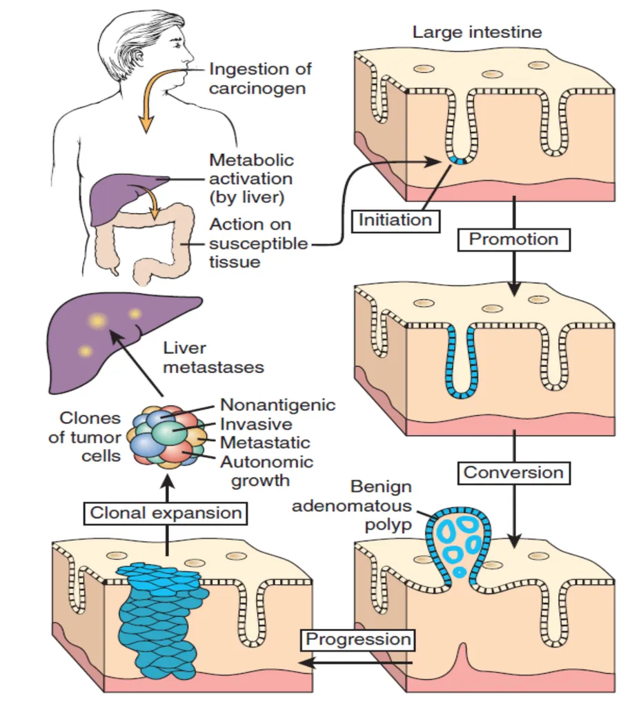
No single mutation makes a cancer. The lecture's colon diagram shows the steps: a carcinogen (activated by the liver) initiates a crypt cell by mutating its DNA; promotion makes the initiated cell proliferate; conversion produces a benign adenomatous polyp; further mutations bring progression to an invasive carcinoma, whose subclones become invasive, metastatic and nonantigenic. Each step adds a mutation from the table above (for colon: APC, then RAS, then TP53).
Lecture slide
The brakes: RB and p53
L7 p.19–20Both classic tumor suppressors guard the same door: the transition from G1 to S phase, the point after which a cell is committed to copying its DNA.
Teaching schematic
RB: the G1 gate
- Growth factor → cyclin D + CDK4
- Cyclin D–CDK4 phosphorylates RB
- E2F released from RB
- S-phase genes transcribed → G1 → S
RB lost (or cyclin D / CDK4 overactive) → E2F permanently free → the gate never closes.
p53: the damage sensor
- DNA damage → p53 stabilized
- p21 inhibits CDKs → RB stays active → G1 arrest
- DNA repair enzymes ↑
- Repair fails → BAX/BAK over BCL2 → cytochrome c → apoptosis
p53 lost → no arrest, no extra repair, no apoptosis: the cell divides carrying its damage.
RB is the gatekeeper. In its hypophosphorylated form it binds the transcription factor E2F, which is needed to transcribe S-phase genes. Growth factor signals induce cyclin D, which activates CDK4; cyclin D–CDK4 phosphorylates RB, E2F is released and the cell enters S phase. Losing RB (or overactivating cyclin D or CDK4, both in the lecture's oncogene table) leaves E2F permanently free.
p53 is the guardian that responds to DNA damage. It slows the cell cycle by inducing p21, a CDK inhibitor that keeps RB hypophosphorylated; it upregulates DNA repair; and if the damage cannot be repaired it induces apoptosis, activating BAX and BAK and counteracting BCL2 so the mitochondrion releases cytochrome c. A cell without p53 keeps dividing with damaged DNA, and every division adds mutations. TP53 is the most frequently mutated gene in human cancer.
DNA damage has three possible outcomes
- DNA damage (radiation, carcinogen, replication error)
- p53 stabilized → arrest in G1 (p21)
- Repair succeeds → the cell resumes dividing
- Repair fails → apoptosis (BAX/BAK, cytochrome c)
- p53 lost → none of these: the cell divides with the damage and the mutation is passed on
The two-hit hypothesis: retinoblastoma and Li–Fraumeni
L7 p.19Knudson studied retinoblastoma, a malignant tumor of the infant retina, and noticed two patterns: some children had tumors in both eyes, early, and often had affected relatives; others had a single tumor in one eye, later, with no family history. He proposed that the tumor needs two hits, one to each copy of the same gene (RB1, chromosome 13q14).
Teaching schematic
Familial (germline first hit)
Every cell at birth
One RB1 allele is already mutated, in all cells of the body.
Any retinal cell
A single somatic hit completes it. Tumors are bilateral, often multifocal, and early.
Later risk of **osteosarcoma**. Dominant in the pedigree, recessive in the cell.
Sporadic (both hits somatic)
Every cell at birth
Both RB1 alleles are normal.
One retinal cell
Two independent hits must strike the same cell: rare, so one tumor, one eye, later.
No inherited risk of second tumors.
Familial retinoblastoma
- Germline first hit in every cell; second hit somatic
- Bilateral, often multifocal; earlier onset
- Later osteosarcoma risk (and other sarcomas)
- Autosomal dominant in the pedigree
Sporadic retinoblastoma
- Both hits somatic, in the same retinal cell
- Unilateral, single tumor; later onset
- No inherited risk of second cancers
- No family history
Lecture image
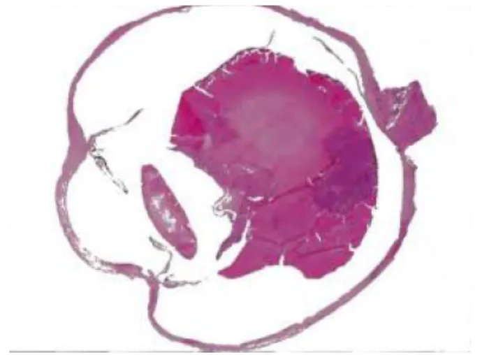
The same logic applies to TP53. A germline TP53 mutation causes Li–Fraumeni syndrome: one hit is inherited, the second is somatic, and carriers develop multiple carcinomas and sarcomas (breast carcinoma, brain tumors, leukemia, soft-tissue and bone sarcomas, adrenocortical carcinoma), often young and often more than one.
Core activity · The Missing Arrow
The Missing Arrow
A child is born to a parent with familial retinoblastoma. Complete the chain from the inherited mutation to a tumor.
- the remaining normal allele still makes enough RB protein
- Retinal cells behave normally; the child is healthy at birth
- That cell has no functional RB protein
- Uncontrolled proliferation: retinoblastoma, often in both eyes and early
- the same inherited first hit is present in osteoblast precursors
Cards
Tap or drag a card. Placing a used card moves it.
Inherited cancer syndromes
L7 p.15, 20–21Most inherited cancer syndromes are germline mutations of tumor suppressor or DNA repair genes. The family inherits one defective allele; each tumor arises when a cell loses the second. Recognize them by the pattern: young patients, multiple or bilateral tumors, the same cancer in relatives, and characteristic non-tumor signs.
Syndromes from the lectureL7 p.15, 20
| Syndrome | Gene and normal function | Tumors and clues |
|---|---|---|
| Familial adenomatous polyposis | APC: degrades β-catenin, a nuclear transcription activator | Hundreds of colonic adenomas → colorectal carcinoma almost certain without colectomy |
| Lynch syndrome (HNPCC) | Mismatch repair genes: correct base-pairing errors | Colorectal (often right-sided, younger), endometrial, ovarian and gastric carcinoma; microsatellite instability |
| Hereditary breast–ovarian cancer | BRCA1/BRCA2: repair of DNA double-strand breaks | Breast and ovarian carcinoma; BRCA2 also male breast, prostate, pancreas |
| Li–Fraumeni | TP53 | Sarcomas, breast, brain, leukemia, adrenocortical carcinoma |
| Familial retinoblastoma | RB1 | Bilateral retinoblastoma, osteosarcoma |
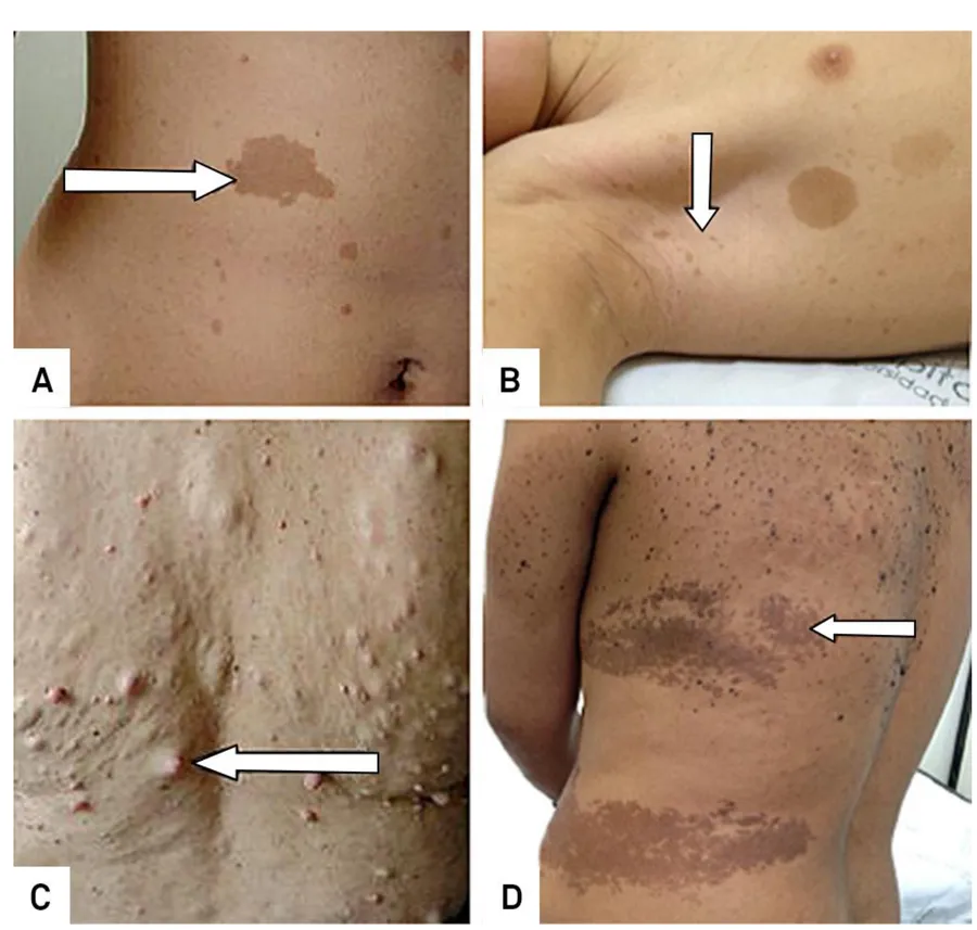
| Neurofibromatosis 1 | NF1 (neurofibromin): inhibits RAS signaling | Neurofibromas, café-au-lait macules, axillary freckling; pheochromocytoma; malignant nerve sheath tumors |
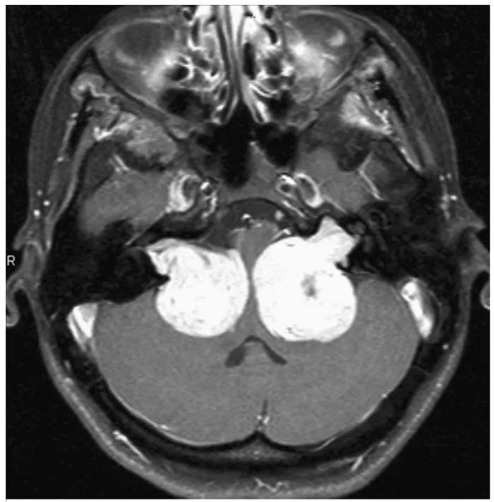
| Neurofibromatosis 2 | NF2 (merlin): cytoskeletal stability | Bilateral vestibular (acoustic) schwannomas, meningiomas |
| von Hippel–Lindau | VHL: targets HIF for degradation | Bilateral renal cell carcinoma, cerebellar hemangioblastoma, retinal angioma, pheochromocytoma |
| WT1 syndromes | WT1: nuclear transcription | Wilms tumor |
| Xeroderma pigmentosum | Nucleotide excision repair: removes UV pyrimidine dimers | Skin cancers (BCC, SCC, melanoma) in childhood on sun-exposed skin |
Familial adenomatous polyposis
- Gene and normal function
- APC: degrades β-catenin, a nuclear transcription activator
- Tumors and clues
- Hundreds of colonic adenomas → colorectal carcinoma almost certain without colectomy
Lynch syndrome (HNPCC)
- Gene and normal function
- Mismatch repair genes: correct base-pairing errors
- Tumors and clues
- Colorectal (often right-sided, younger), endometrial, ovarian and gastric carcinoma; microsatellite instability
Hereditary breast–ovarian cancer
- Gene and normal function
- BRCA1/BRCA2: repair of DNA double-strand breaks
- Tumors and clues
- Breast and ovarian carcinoma; BRCA2 also male breast, prostate, pancreas
Li–Fraumeni
- Gene and normal function
- TP53
- Tumors and clues
- Sarcomas, breast, brain, leukemia, adrenocortical carcinoma
Familial retinoblastoma
- Gene and normal function
- RB1
- Tumors and clues
- Bilateral retinoblastoma, osteosarcoma
Neurofibromatosis 1
- Gene and normal function
- NF1 (neurofibromin): inhibits RAS signaling
- Tumors and clues
- Neurofibromas, café-au-lait macules, axillary freckling; pheochromocytoma; malignant nerve sheath tumors
Neurofibromatosis 2
- Gene and normal function
- NF2 (merlin): cytoskeletal stability
- Tumors and clues
- Bilateral vestibular (acoustic) schwannomas, meningiomas
von Hippel–Lindau
- Gene and normal function
- VHL: targets HIF for degradation
- Tumors and clues
- Bilateral renal cell carcinoma, cerebellar hemangioblastoma, retinal angioma, pheochromocytoma
WT1 syndromes
- Gene and normal function
- WT1: nuclear transcription
- Tumors and clues
- Wilms tumor
Xeroderma pigmentosum
- Gene and normal function
- Nucleotide excision repair: removes UV pyrimidine dimers
- Tumors and clues
- Skin cancers (BCC, SCC, melanoma) in childhood on sun-exposed skin
Lecture image
Lecture image
Knowledge check · 7.4
Answer, read why, and try again whenever you like. Answers are kept in both study modes.
- Q1Two-hit hypothesisL7 p.19
An 8-month-old girl has a white pupillary reflex in both eyes. Examination shows several retinal tumors in each eye.
Which statement best explains her disease and her future risk?
- Q2BCL2 t(14;18)L7 p.15
A 58-year-old man has generalized painless lymphadenopathy. A node shows a follicular pattern of neoplastic B cells; cytogenetics shows t(14;18).
How does this translocation promote the tumor?
- Q3Lynch syndromeL7 p.15, 20
A 38-year-old woman has a carcinoma of the right colon. Her mother had endometrial carcinoma at 45 and her uncle colon cancer at 40. The tumor shows microsatellite instability.
Which gene class is most likely defective?
- Q4NF1 and RASL7 p.20–21
A 19-year-old man has the skin findings shown: light-brown macules on the trunk, freckling in the axilla and many soft cutaneous nodules.
Loss of which protein's function underlies these lesions, and what does that protein normally do?
- Q5Li–Fraumeni syndromeL7 p.19–20
A 29-year-old woman has breast carcinoma. She had a rhabdomyosarcoma at 6; her brother died of a brain tumor at 22; her mother had an adrenocortical carcinoma.
A germline mutation in which gene is most likely?
- Q6Screening: adenoma removalL7 p.9, 14
Why does removing colonic adenomas at colonoscopy reduce deaths from colorectal cancer?