What a tumor is and what it is called
A neoplasm is a clone of cells that keeps growing after the stimulus is gone. Its name records two things: the tissue it imitates and whether it behaves benignly or malignantly.
Neoplasia: one clone, growing without a reason
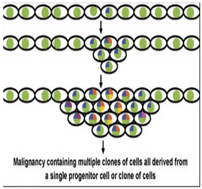
L7 p.2The lecture defines neoplasia as new tissue growth that is unregulated, irreversible and monoclonal. Each word separates it from the adaptations in Chapter 1. Hyperplasia and repair also add cells, but they answer a stimulus (a hormone, a wound) and stop when the stimulus stops, and they are built by many different cells dividing together: they are polyclonal. A neoplasm keeps growing when the stimulus is gone, because its cells carry mutations that switched growth on permanently, and all of its cells descend from one mother cell.
Teaching schematic
Normal tissue or hyperplasia
Each cell silenced one X at random, so both G6PD isoforms appear, about 1:1. Many cells are dividing: polyclonal.
Neoplasia
Every cell copies the X-inactivation of one mother cell, so only one isoform is found: monoclonal. The B-cell version is κ or λ light-chain restriction.
That is how clonality was first demonstrated. Glucose-6-phosphate dehydrogenase (G6PD) is encoded on the X chromosome. Early in female development each cell inactivates one X at random (lyonization), so in a woman who inherited two different G6PD isoforms, any normal or hyperplastic tissue contains both, roughly 1:1. A tumor from the same woman contains only one isoform, because all of its cells copy the X-inactivation pattern of their single ancestor. The androgen-receptor gene, also on the X, is used the same way today. For B cells there is a neater marker: each B cell commits to a κ or λ light chain. A reactive lymph node mixes both; a B-cell lymphoma shows only one (light-chain restriction).
Hyperplasia versus neoplasia
Hyperplasia (adaptation)
- Driven by a stimulus (hormone, growth factor, injury)
- Stops or regresses when the stimulus is removed
- Polyclonal: both G6PD isoforms, κ and λ B cells
- Normal maturation and architecture
Neoplasia
- Growth continues without the stimulus (unregulated)
- Irreversible: the change is in the cell's DNA
- Monoclonal: one isoform, one light chain
- Benign or malignant depending on how far the clone has escaped control
Every tumor has two parts. The parenchyma is the neoplastic clone itself; it decides the tumor's name and behavior. The stroma is the host's supporting tissue: connective tissue, vessels and inflammatory cells, recruited by the tumor. Some carcinomas provoke abundant dense collagen, called desmoplasia; such tumors are stony hard (scirrhous), which is why an invasive breast or pancreatic carcinoma feels firm and gritty when cut.
Lecture slide
Naming rules: -oma, carcinoma, sarcoma
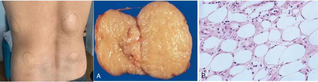
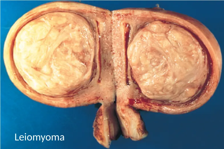
L7 p.3, 5–6A tumor's name answers two questions. Which tissue does it imitate? That is its lineage of differentiation. Benign or malignant? That is its behavior. A benign tumor usually takes the tissue name plus -oma (lipoma, adenoma, leiomyoma). A malignant tumor of epithelial origin is a carcinoma; a malignant tumor of mesenchymal (connective tissue, mesoderm) origin is a sarcoma. Carcinomas are named by the epithelium they imitate: squamous, glandular (adeno-) or transitional (urothelial).
Lineage plus behavior gives the nameL7 p.3, 5
| Tissue of origin | Benign | Malignant |
|---|---|---|
| Glandular epithelium | Adenoma | Adenocarcinoma |
| Surface epithelium in fronds | Papilloma | Papillary carcinoma |
| Squamous epithelium | Squamous papilloma | Squamous cell carcinoma |
| Urothelium (transitional epithelium) | Urothelial papilloma | Urothelial (transitional cell) carcinoma |
| Fat | Lipoma | Liposarcoma |
| Smooth muscle | Leiomyoma | Leiomyosarcoma |
| Skeletal muscle | Rhabdomyoma | Rhabdomyosarcoma |
| Bone | Osteoma | Osteosarcoma |
| Melanocytes | Nevus (mole) | Melanoma |
| Lymphocytes | No benign counterpart | Lymphoma / leukemia |
Glandular epithelium
- Benign
- Adenoma
- Malignant
- Adenocarcinoma
Surface epithelium in fronds
- Benign
- Papilloma
- Malignant
- Papillary carcinoma
Squamous epithelium
- Benign
- Squamous papilloma
- Malignant
- Squamous cell carcinoma
Urothelium (transitional epithelium)
- Benign
- Urothelial papilloma
- Malignant
- Urothelial (transitional cell) carcinoma
Fat
- Benign
- Lipoma
- Malignant
- Liposarcoma
Smooth muscle
- Benign
- Leiomyoma
- Malignant
- Leiomyosarcoma
Skeletal muscle
- Benign
- Rhabdomyoma
- Malignant
- Rhabdomyosarcoma
Bone
- Benign
- Osteoma
- Malignant
- Osteosarcoma
Melanocytes
- Benign
- Nevus (mole)
- Malignant
- Melanoma
Lymphocytes
- Benign
- No benign counterpart
- Malignant
- Lymphoma / leukemia
Lecture image
Lecture image
Lecture image
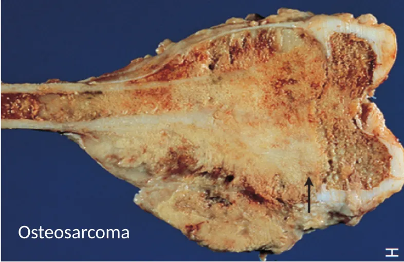
The lineage also predicts behavior. Carcinomas usually spread first through lymphatics to regional nodes; sarcomas usually spread through the blood (section 7.3). The lecture adds that about 40% of sarcomas arise in the lower extremity; osteosarcoma (bone) and rhabdomyosarcoma (skeletal muscle) are its examples.
Reading lineage under the microscope
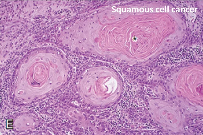
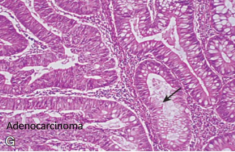
L7 p.3, 6A tumor betrays its lineage by what its cells try to make. Squamous cells make keratin; glandular cells make glands and mucin; mesenchymal cells make spindle-shaped cells and their matrix (osteoid in osteosarcoma, cartilage in chondrosarcoma). The better differentiated the tumor, the more clearly it still makes these products.
Lecture image
Lecture image
What the cells make tells you the lineageL7 p.3, 6
| Microscopic clue | Lineage | Example |
|---|---|---|
| Keratin pearls, intercellular bridges | Squamous epithelium | Squamous cell carcinoma of lung, cervix, esophagus, skin |
| Glands, mucin in the lumen | Glandular epithelium | Adenocarcinoma of colon, stomach, pancreas, lung |
| Spindle cells with osteoid | Mesenchyme (bone) | Osteosarcoma |
| Sheets of discohesive round cells | Lymphoid (or other small round cell tumors) | Lymphoma |
Keratin pearls, intercellular bridges
- Lineage
- Squamous epithelium
- Example
- Squamous cell carcinoma of lung, cervix, esophagus, skin
Glands, mucin in the lumen
- Lineage
- Glandular epithelium
- Example
- Adenocarcinoma of colon, stomach, pancreas, lung
Spindle cells with osteoid
- Lineage
- Mesenchyme (bone)
- Example
- Osteosarcoma
Sheets of discohesive round cells
- Lineage
- Lymphoid (or other small round cell tumors)
- Example
- Lymphoma
Tumor-like masses: hamartoma, choristoma, teratoma
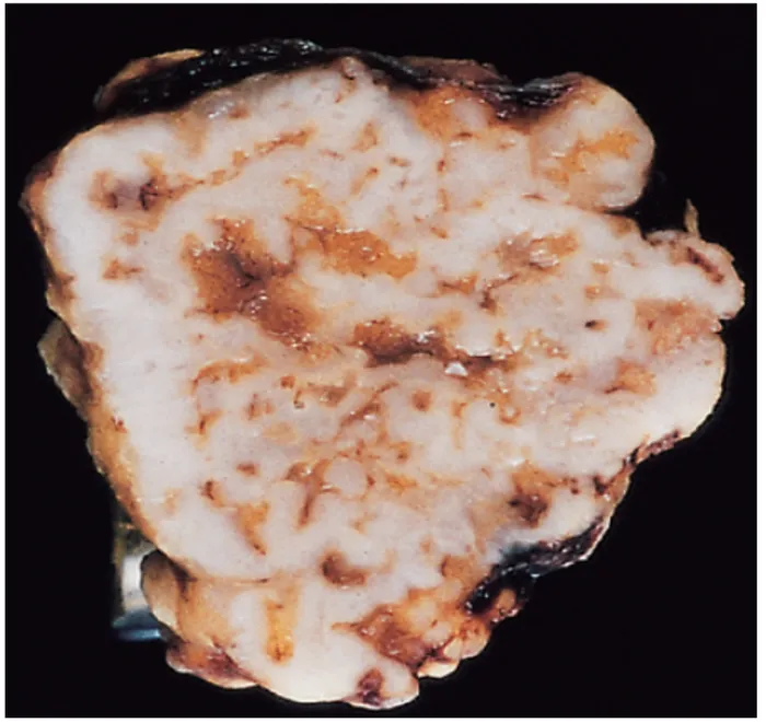
L7 p.7Not every lump is a neoplasm in the lecture's sense. Two tumor-like conditions are made of mature tissue that is not growing autonomously, and the question that separates them is simple: is the tissue native to that site?
Hamartoma
- Disorganized overgrowth of tissue that belongs at that site
- Pulmonary hamartoma: cartilage, fat, fibrous tissue and bronchial epithelium in a lung 'coin lesion'
- Peutz–Jeghers polyp: native glands and smooth muscle of the gut, arranged chaotically
Choristoma (heterotopic rest)
- Normal tissue in a foreign place
- Pancreatic tissue in the wall of the stomach
- Brain tissue in the nasal cavity; functioning thyroid tissue in the liver
Teratoma (a true neoplasm)
- Arises from totipotent germ cells
- Contains tissues of more than one germ layer (skin, hair, teeth, neural tissue, gut)
- Mature cystic teratoma of the ovary (dermoid) is benign; immature teratomas and most postpubertal testicular teratomas are malignant
Lecture image
Core activity · Almost Twins
Almost Twins
Look-alike A
Hamartoma
- Made of mature, cytologically normal tissue
- The tissue belongs at this site (cartilage, fat, bronchial glands in lung); only its arrangement is disorganized
- Forms a discrete, well-circumscribed mass
- Pulmonary 'coin lesion'; the polyps of Peutz–Jeghers syndrome
Look-alike A
Hamartoma
- Made of mature, cytologically normal tissue
- The tissue belongs at this site (cartilage, fat, bronchial glands in lung); only its arrangement is disorganized
- Forms a discrete, well-circumscribed mass
- Pulmonary 'coin lesion'; the polyps of Peutz–Jeghers syndrome
Look-alike B
Choristoma
- Made of mature, cytologically normal tissue
- The tissue does not belong at this site: it is normal tissue in a foreign place
- Forms a discrete nodule, often found incidentally
- Pancreatic rest in the stomach wall; thyroid tissue in the liver; brain tissue in the nose
Step 1 · What they share
What do they share?
Select every feature that both show.
Peutz–Jeghers syndrome
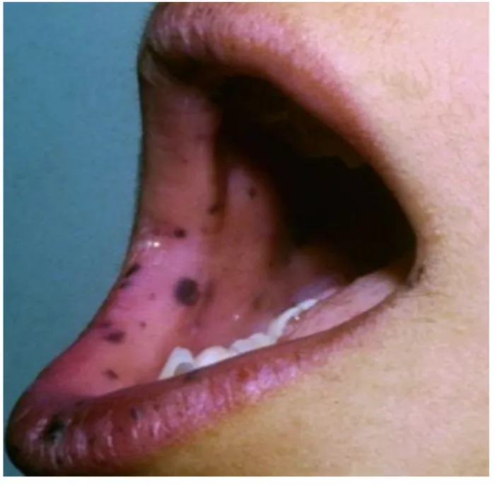
L7 p.7Peutz–Jeghers syndrome is an autosomal dominant disorder (germline mutation of the STK11/LKB1 tumor-suppressor gene) with two visible features: hamartomatous polyps throughout the gastrointestinal tract, most numerous in the small intestine, and hyperpigmented macules on the lips, buccal mucosa, hands and genitalia. The polyps themselves are not premalignant in the usual sense, but they can bleed or lead an intussusception, and patients carry a markedly increased risk of cancers of the colon, stomach, small bowel, pancreas and breast.
Lecture image
Knowledge check · 7.1
Answer, read why, and try again whenever you like. Answers are kept in both study modes.
- Q1Naming a benign mesenchymal tumorCh. VII cases p.8L7 p.4, 6
A 45-year-old woman has abdominal pain and heavy vaginal bleeding. A hysterectomy shows a benign tumor of the uterus derived from smooth muscle cells.
What is the appropriate diagnosis?
- Q2Lineage from morphology: glandsL7 p.3, 6
A 67-year-old man has a colonic mass that, on resection, has grown into the muscular wall of the bowel. A section of the tumor is shown.
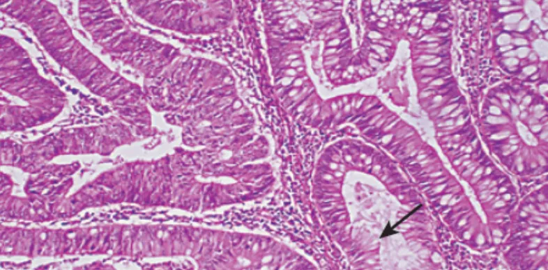
Which term fits this tumor?
- Q3Hamartoma vs choristomaL7 p.7
Endoscopy for dyspepsia in a 38-year-old woman finds a 1-cm smooth submucosal nodule in the gastric antrum. Biopsy shows normal pancreatic acini and ducts with no atypia.
What is the best term for this nodule?
- Q4Clonality by light-chain restrictionL7 p.2
A 59-year-old woman has painless, enlarging cervical lymph nodes. In an excised node, flow cytometry shows that 97% of the B cells express λ light chain and almost none express κ.
What does this result indicate?
- Q5Peutz–Jeghers syndromeL7 p.7
A 16-year-old boy has colicky abdominal pain from an intussusception. The lead point is a small-bowel polyp made of disorganized but normal-looking glands and branching smooth muscle. He has dark macules on his lips and buccal mucosa; his father had similar spots.
Which statement about his condition is correct?
- Q6Exceptions to the -oma ruleL7 p.3
Which of these tumors is malignant even though its name ends in -oma?