Benign or malignant: differentiation, dysplasia and invasion
Malignancy is decided by behavior: invasion and metastasis. Differentiation, growth rate and the basement membrane tell you how close a tumor is to that line, and a handful of acquired capabilities explain how it crosses it.
Four features that separate benign from malignant
L7 p.3–4, 22The lecture compares benign and malignant tumors on differentiation, rate of growth, local invasion and metastasis. The first three are tendencies; metastasis is the one feature that defines malignancy without exception, because benign tumors never metastasize.
Benign versus malignantL7 p.3–4, 22
| Feature | Benign | Malignant |
|---|---|---|
| Differentiation | Well differentiated; looks like the tissue of origin; nuclei of normal size and shape | Ranges from well differentiated to anaplastic; pleomorphic, hyperchromatic nuclei |
| Rate of growth | Usually slow, expansile | Usually fast; varies with differentiation |
| Local invasion | Pushes; often a fibrous capsule (uterine leiomyoma is circumscribed without one) | Infiltrates; no capsule; ragged borders; often necrosis |
| Metastasis | Never | Frequent; the decisive criterion |
| Clinical feel | Well circumscribed, mobile | Poorly circumscribed, fixed to surrounding structures |
Differentiation
- Benign
- Well differentiated; looks like the tissue of origin; nuclei of normal size and shape
- Malignant
- Ranges from well differentiated to anaplastic; pleomorphic, hyperchromatic nuclei
Rate of growth
- Benign
- Usually slow, expansile
- Malignant
- Usually fast; varies with differentiation
Local invasion
- Benign
- Pushes; often a fibrous capsule (uterine leiomyoma is circumscribed without one)
- Malignant
- Infiltrates; no capsule; ragged borders; often necrosis
Metastasis
- Benign
- Never
- Malignant
- Frequent; the decisive criterion
Clinical feel
- Benign
- Well circumscribed, mobile
- Malignant
- Poorly circumscribed, fixed to surrounding structures
Growth rate needs perspective. A tumor must go through about 30 population doublings to reach 1 g (about 10⁹ cells), the smallest mass that is usually detectable clinically; ten more doublings make 1 kg. By the time a cancer is found, most of its life history, and many of its mutations, lie behind it. Some tissues resist invasion well (mature cartilage, the elastic tissue of arteries), so the route a tumor takes is not random.
Worked calculation · How many doublings make a detectable tumor?
- Starting point
- 1 transformed cell
- Cells in 1 g of tumor
- ≈ 10⁹
- Cells in 1 kg of tumor
- ≈ 10¹²
Formula
cells after n doublings = 2ⁿ
L7 p.4Differentiation and anaplasia
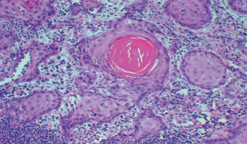
L7 p.3, 22Differentiation is how closely tumor cells resemble the normal cells they came from, in shape and in function. Anaplasia is the lack of differentiation, and it is a hallmark of malignancy. A well-differentiated squamous carcinoma still makes keratin pearls; an anaplastic one is a sheet of bizarre cells that no longer reveals its origin.
Teaching schematic
1. Well differentiated
Uniform cells and nuclei, low N:C ratio, orderly, few mitoses: resembles the tissue of origin.
2. Moderately differentiated
Nuclei larger and darker, some variation, more mitoses, order partly lost.
3. Poorly differentiated (anaplastic)
Pleomorphism, hyperchromasia, N:C ratio up to 1:1, atypical mitoses, loss of polarity and necrosis (pale patch).
What anaplasia looks like, and whyL7 p.4, 22
| Feature | What you see | What it reflects |
|---|---|---|
| Pleomorphism | Cells and nuclei vary in size and shape, giant cells | Unstable genome; no single program of maturation |
| Hyperchromasia | Dark, coarsely clumped chromatin | Extra DNA (aneuploidy) and active transcription |
| High N:C ratio | Nucleus nearly as large as the cell (up to 1:1) | Growth outpaces maturation |
| Atypical mitoses | Many mitoses, some tripolar or bizarre | Uncontrolled proliferation; defective checkpoints |
| Loss of polarity | Disorderly sheets instead of orderly layers | Cells no longer respond to positional cues |
| Necrosis | Pink, nuclear-debris-filled zones, often in the center of large nodules | Growth outruns the new, leaky blood supply |
Pleomorphism
- What you see
- Cells and nuclei vary in size and shape, giant cells
- What it reflects
- Unstable genome; no single program of maturation
Hyperchromasia
- What you see
- Dark, coarsely clumped chromatin
- What it reflects
- Extra DNA (aneuploidy) and active transcription
High N:C ratio
- What you see
- Nucleus nearly as large as the cell (up to 1:1)
- What it reflects
- Growth outpaces maturation
Atypical mitoses
- What you see
- Many mitoses, some tripolar or bizarre
- What it reflects
- Uncontrolled proliferation; defective checkpoints
Loss of polarity
- What you see
- Disorderly sheets instead of orderly layers
- What it reflects
- Cells no longer respond to positional cues
Necrosis
- What you see
- Pink, nuclear-debris-filled zones, often in the center of large nodules
- What it reflects
- Growth outruns the new, leaky blood supply
The lecture also describes the poorly differentiated cell from the inside: fewer mitochondria, less prominent rough ER, and loss of cadherins, the adhesion molecules that hold epithelial cells together. Losing cadherins lets cells slip away into the surrounding tissue, the first step of invasion (7.3).
Dysplasia → carcinoma in situ → invasion
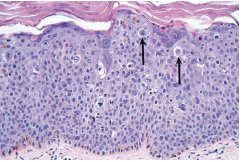
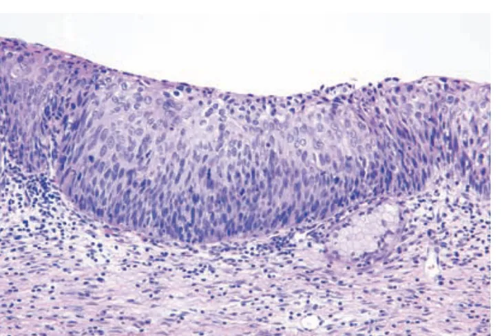
L7 p.8, 22L1 p.28In epithelium, malignancy usually announces itself before it invades. Dysplasia is disordered growth: the cells show atypia (enlarged, hyperchromatic nuclei, a high N:C ratio), lose their orderly maturation and divide above the basal layer. When these changes involve the full thickness of the epithelium, the lesion is carcinoma in situ (CIS): it has the cytology of cancer, but the basement membrane is intact, so it has not invaded and cannot yet metastasize.
Teaching schematic
1. Dysplasia
Atypia in the lower layers; maturation still visible at the surface. Basement membrane intact.
2. Carcinoma in situ
Atypia through the full thickness, no maturation. Basement membrane still intact: cannot metastasize.
3. Invasive carcinoma
Cells cross the basement membrane into stroma, where lymphatics and veins are.
The basement membrane is the line that matters because the epithelium has no lymphatics or blood vessels of its own. Above it, a clone can only grow; below it, it meets the vessels that carry it away. That is why carcinoma in situ, even when its cells look fully malignant, is cured by complete local excision, and why the first stromal invasion changes the prognosis.
Case image
Morphology comparison · Layers
Where are the cells?
Specimen A
Specimen B
Specimen C
Teaching schematics, not micrographs. Orange outline: lymphatic. Red outline: small vein.
0 of 3 answered correctly. Source: Lesson 7 Neoplasia, pp. 8 and 22; the same distinction is tested in Ch. VII Q2 and Midterm sim. Q5–Q6.
Core activity · Almost Twins
Almost Twins
Look-alike A
Dysplasia (low grade)
- Atypical nuclei: enlarged, hyperchromatic, high N:C ratio
- Atypia confined to the lower layers; cells still mature toward the surface
- Basement membrane intact
- Often regresses, especially after HPV clearance
Look-alike A
Dysplasia (low grade)
- Atypical nuclei: enlarged, hyperchromatic, high N:C ratio
- Atypia confined to the lower layers; cells still mature toward the surface
- Basement membrane intact
- Often regresses, especially after HPV clearance
Look-alike B
Carcinoma in situ (CIN 3)
- Atypical nuclei: enlarged, hyperchromatic, high N:C ratio
- Atypia through the full thickness; no maturation at all
- Basement membrane intact
- A true precursor: treated to prevent invasion
Step 1 · What they share
What do these two epithelia share?
Select every feature that both show.
Tumor Pathology Case Lab
Ch. VII cases p.3, 5, 6L7 p.8, 22–25Four specimens from the lecture and the Chapter VII cases, worked the way a pathologist works them: look at the slide before you read anything, decide whether the basement membrane has been crossed, choose the stain that answers the open question, sort the report into grade and stage, then take one more clue and say what it changes.
Centerpiece · Tumor Pathology Case Lab
Tumor Pathology Case Lab
Clinical history
A 60-year-old man has 4 months of weight loss, wheezing and breathlessness. He has smoked two packs a day for 40 years. A chest radiograph shows a 10-cm mass in the left lung, and bronchoscopy shows the left main bronchus obstructed. A biopsy is taken.
Before you answer, look for
- Are the cells arranged in nests, glands or sheets?
- Is anything being made by the cells: keratin, mucin, matrix?
- Are the nests confined to an epithelial layer, or sitting in stroma?
- 1. Look first
- 2. Has it invaded?
- 3. Choose a stain
- 4. Grade or stage
- 5. One more clue
Step 1 · Look first
What do you call this, and what is the lineage clue?
Decide benign or malignant first, then name what the cells are making.
Cervical dysplasia: the model sequence
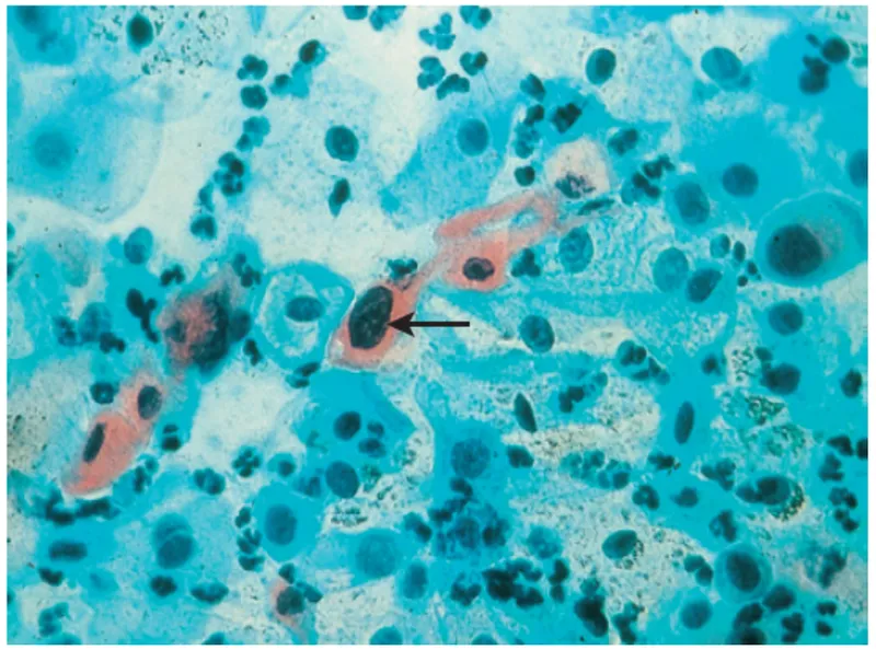
L7 p.14Midterm sim. p.2Ch. VII cases p.3The cervix shows the whole sequence in one organ. High-risk human papillomavirus (HPV 16 and 18) infects the immature metaplastic cells of the transformation zone. Its first visible sign is koilocytic atypia: squamous cells with a clear perinuclear halo and a wrinkled, dark nucleus, the cytopathic effect of the virus. When viral oncoproteins disable the cell's brakes (section 7.5), the epithelium becomes dysplastic, graded as CIN 1 (lower third), CIN 2 (up to two thirds) and CIN 3 (full thickness, the same as carcinoma in situ). Invasion through the basement membrane makes invasive squamous cell carcinoma.
Case image
Lecture image
Worked case · One specimen, two answers
A 25-year-old woman's Pap smear shows koilocytes; biopsy shows invasive squamous cell carcinoma, HPV positive. In the hysterectomy specimen, beside the invasive focus, dysplastic cells occupy the entire thickness of the epithelium with no maturation, and the basement membrane there is intact.
What is the lesion in the areas with an intact basement membrane, and why does the distinction matter in the same uterus?
Ch. VII cases p.3The hallmarks: what a clone must acquire
L7 p.3–4, 15, 19Every malignant tumor has acquired a small set of capabilities, each from a different kind of mutation or adaptation. The lecture names several of them; the others come from the gene classes in 7.4. Think of the list as the answer to 'what must change before a clone can invade and spread?'
Teaching schematic
Growth without a signal
Oncogenes: RAS, ERBB2
Ignoring the brakes
RB and p53 lost
Not dying
BCL2 overexpressed
Dividing forever
Telomerase reactivated
Building a blood supply
Hypoxia → HIF-1α → VEGF
Rewired metabolism
Aerobic glycolysis (Warburg) → FDG-PET
Hiding from the immune system
MHC I ↓, PD-L1, CTLA-4, DAF
Invading and spreading
E-cadherin ↓, MMPs (7.3)
Accumulating mutations faster
DNA repair genes lost
Limitless replication (telomerase). Every division shortens telomeres, and normal somatic cells eventually stop dividing (senescence) when telomeres get too short. Germ cells and stem cells express telomerase, which rebuilds telomeres. Most cancers reactivate it, so the clone can divide indefinitely. The lecture links telomerase to avoiding apoptosis; more precisely, it prevents the senescence and chromosomal crisis that short telomeres would cause.
Angiogenesis (VEGF). Oxygen diffuses only about 1–2 mm from a capillary, so a tumor cannot grow beyond that size without new vessels. Hypoxia in the tumor stabilizes HIF-1α, which switches on vascular endothelial growth factor (VEGF); tumor cells also release proteases that break down the vessel basement membrane so endothelial cells can migrate. The new vessels are leaky and disorganized, which is why large tumors still have areas of necrosis.
Altered metabolism (the Warburg effect). Cancer cells take up large amounts of glucose and run glycolysis even when oxygen is plentiful (aerobic glycolysis). Glycolysis yields little ATP, but its intermediates are the carbon skeletons for new lipids, nucleotides and amino acids, which a dividing cell needs. Clinically this is what PET sees: the radiolabeled glucose analogue FDG is taken up and trapped in the tumor, which is why PET is used for diagnosis, staging and monitoring.
Immune evasion. Cytotoxic T cells recognize tumor antigens on MHC class I, so tumors that downregulate MHC I escape them (natural killer cells, which kill cells lacking MHC I, are the backup). Many tumors also express PD-L1, which binds PD-1 on T cells and switches them off, and they exploit CTLA-4, a T-cell receptor that blocks costimulation. Antibodies that block PD-1, PD-L1 or CTLA-4 ('checkpoint inhibitors') release this brake. The lecture adds decay-accelerating factor (DAF): tumor cells limit C3 and C5 convertases, so complement cannot assemble the membrane attack complex on them.
Knowledge check · 7.2
Answer, read why, and try again whenever you like. Answers are kept in both study modes.
- Q1Carcinoma in situCh. VII cases p.3L7 p.22
A 25-year-old woman's Pap smear shows koilocytic atypia. Biopsy shows invasive squamous cell carcinoma, HPV positive, and she undergoes hysterectomy. Besides the invasive focus, the pathologist finds dysplastic squamous cells occupying the entire thickness of the cervical epithelium with no maturation; the basement membrane in these areas is intact.
Which term best describes these areas?
- Q2Telomerase in cancerCh. VII cases p.2L7 p.4
A 25-year-old man has a painless, enlarged left testis that does not transilluminate. Serum AFP and hCG are normal. Orchiectomy shows embryonal carcinoma.
Compared with normal adult somatic cells, this germ-cell tumor is most likely to express high levels of which protein?
- Q3Cervical dysplasiaMidterm sim. p.2L1 p.28
A 33-year-old woman has an abnormal Pap smear. On cervical biopsy the epithelium lacks normal polarity; individual cells have hyperchromatic nuclei and a high nucleus-to-cytoplasm ratio, and the tissue is disorderly. The lower border of the epithelium is smooth.
Which term best describes these changes?
- Q4Warburg effect and PETL7 p.3
A 61-year-old man with a lung mass has an FDG-PET scan. The mass and two mediastinal nodes take up the tracer intensely.
Why do the tumor cells accumulate FDG?
- Q5Immune evasion: PD-1/PD-L1L7 p.4Ref. · Robbins: immune evasion
A patient with metastatic melanoma responds dramatically to an antibody against PD-1. Biopsies before treatment showed tumor cells with strong PD-L1 expression surrounded by inactive T cells.
What did the antibody do?
- Q6AngiogenesisL7 p.4
A carcinoma nodule grows to about 1–2 mm and then stops enlarging for months. Which change most likely allows it to grow again?