Diagnosis, grade and stage
Tissue makes the diagnosis. Grade says how abnormal the cells look; stage says how far the tumor has gone. Stains and serum markers support an answer, they do not replace the histology or the clinical picture.
Getting the tissue: biopsy, FNA, frozen section, cytology
L7 p.22The lecture's rule is blunt: biopsy or excision is generally required before a tumor can be classified as benign or malignant with certainty, because some benign tumors grow in a malignant-looking way and some malignant tumors look circumscribed. Each sampling method answers a different question.
How tissue is obtainedL7 p.14, 22Ref. · Robbins: laboratory diagnosis
| Method | What it gives | Best for | Limits |
|---|---|---|---|
| Core or incisional biopsy | Intact tissue: architecture and the relation to the basement membrane | Deciding invasive versus in situ; grading; running stains | Sampling error if the needle misses the lesion |
| Fine-needle aspiration (FNA) | Cells only, no architecture | Quick assessment of a palpable mass: thyroid nodule, breast lump, lymph node | Cannot usually show invasion, because the basement membrane is not visible |
| Frozen section (during surgery) | A rapid diagnosis in minutes | Is the lesion malignant? Are the margins clear? Is the sentinel node involved? | Freezing distorts the cells; always confirmed on permanent sections |
| Exfoliative cytology (Pap smear, effusion fluid) | Shed cells | Screening (cervix) and detecting malignant cells in fluid | Shows atypia, not invasion |
| Excision | The whole lesion | Definitive diagnosis, grade, stage, margins | An operation |
Core or incisional biopsy
- What it gives
- Intact tissue: architecture and the relation to the basement membrane
- Best for
- Deciding invasive versus in situ; grading; running stains
- Limits
- Sampling error if the needle misses the lesion
Fine-needle aspiration (FNA)
- What it gives
- Cells only, no architecture
- Best for
- Quick assessment of a palpable mass: thyroid nodule, breast lump, lymph node
- Limits
- Cannot usually show invasion, because the basement membrane is not visible
Frozen section (during surgery)
- What it gives
- A rapid diagnosis in minutes
- Best for
- Is the lesion malignant? Are the margins clear? Is the sentinel node involved?
- Limits
- Freezing distorts the cells; always confirmed on permanent sections
Exfoliative cytology (Pap smear, effusion fluid)
- What it gives
- Shed cells
- Best for
- Screening (cervix) and detecting malignant cells in fluid
- Limits
- Shows atypia, not invasion
Excision
- What it gives
- The whole lesion
- Best for
- Definitive diagnosis, grade, stage, margins
- Limits
- An operation
Grade versus stage
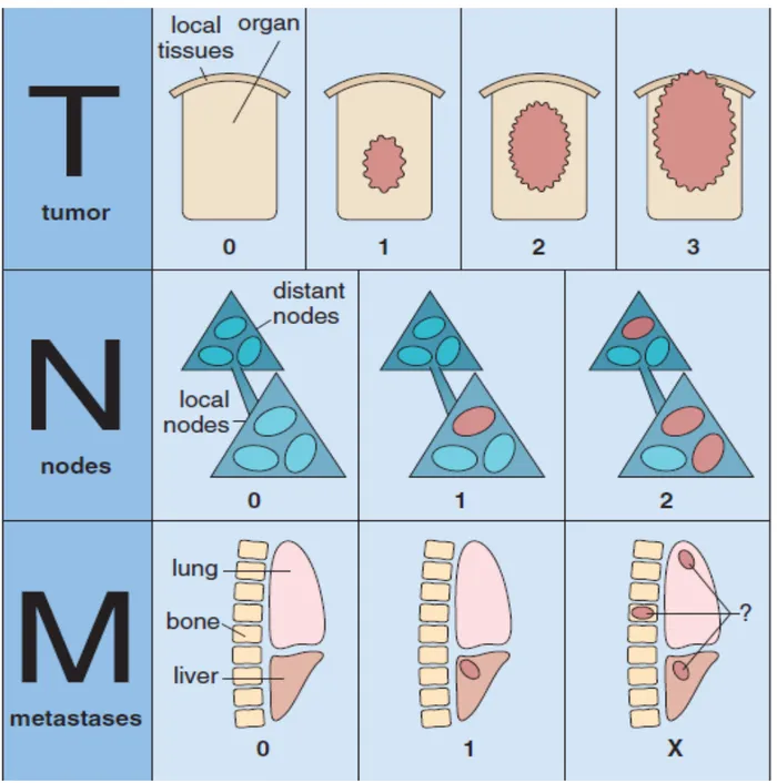
L7 p.24Ch. VII cases p.4Grade describes the tumor cells: how closely they still resemble the normal tissue of origin (differentiation), together with nuclear atypia and mitotic activity. Grades run low, intermediate, high (anaplastic). Stage describes the tumor's extent: how large or deep the primary is, whether regional nodes are involved, and whether there are distant metastases. Everything about the cells is grade; everything about distance travelled is stage.
Teaching schematic
Grade: what the cells look like
Differentiation, pleomorphism, mitoses. Low → high (anaplastic). Answered down the microscope.
Stage: how far it has gone
T size or depth, N nodes, M metastases. The strongest prognostic factor for survival.
Sorting facts into grade or stageL7 p.24
| Fact about a tumor | Grade or stage? | Why |
|---|---|---|
| Cells resemble the normal tissue of origin | Grade | Differentiation |
| Marked nuclear pleomorphism and 25 mitoses per high-power field | Grade | Cytologic abnormality |
| Tumor invades 8 mm into the bowel wall | Stage (T) | Extent of the primary |
| 3 of 14 axillary nodes contain tumor | Stage (N) | Regional spread |
| Nodules in both lungs | Stage (M) | Distant metastasis |
| Tumor extends through the prostatic capsule | Stage (T) | Local extent |
| Gleason pattern: fused, poorly formed glands | Grade | Architecture of the tumor cells themselves |
Cells resemble the normal tissue of origin
- Grade or stage?
- Grade
- Why
- Differentiation
Marked nuclear pleomorphism and 25 mitoses per high-power field
- Grade or stage?
- Grade
- Why
- Cytologic abnormality
Tumor invades 8 mm into the bowel wall
- Grade or stage?
- Stage (T)
- Why
- Extent of the primary
3 of 14 axillary nodes contain tumor
- Grade or stage?
- Stage (N)
- Why
- Regional spread
Nodules in both lungs
- Grade or stage?
- Stage (M)
- Why
- Distant metastasis
Tumor extends through the prostatic capsule
- Grade or stage?
- Stage (T)
- Why
- Local extent
Gleason pattern: fused, poorly formed glands
- Grade or stage?
- Grade
- Why
- Architecture of the tumor cells themselves
Lecture slide
Core activity · Almost Twins
Almost Twins
Look-alike A
Grade
Grade: what the cells look like
Differentiation, pleomorphism, mitoses. Low → high (anaplastic). Answered down the microscope.
- Carries prognostic information
- Describes the cells: differentiation, pleomorphism, mitoses, necrosis
- Determined on the slide alone
- Low, intermediate, high (anaplastic)
Look-alike A
Grade
Grade: what the cells look like
Differentiation, pleomorphism, mitoses. Low → high (anaplastic). Answered down the microscope.
- Carries prognostic information
- Describes the cells: differentiation, pleomorphism, mitoses, necrosis
- Determined on the slide alone
- Low, intermediate, high (anaplastic)
Look-alike B
Stage
Stage: how far it has gone
T size or depth, N nodes, M metastases. The strongest prognostic factor for survival.
- Carries prognostic information
- Describes the spread: size or depth (T), nodes (N), metastases (M)
- Needs imaging, surgery and examination of nodes
- TNM, combined into stage I–IV
Step 1 · What they share
What do grade and stage share?
Select every feature that both show.
Immunohistochemistry: asking the cell what it is
L7 p.23When a tumor is too poorly differentiated to name by morphology, immunohistochemistry answers with antibodies against proteins that each lineage still makes. Intermediate filaments are the backbone of the panel because every cell type keeps its own.
The lecture's panelL7 p.23
| Stain | Positive in | Use |
|---|---|---|
| Cytokeratin | Epithelium | Carcinoma (any epithelial tumor) |
| Vimentin | Mesenchyme | Sarcoma (and many other non-epithelial cells) |
| Desmin | Muscle | Leiomyosarcoma, rhabdomyosarcoma |
| GFAP | Neuroglia | Astrocytoma, ependymoma |
| Neurofilament | Neurons | Neuronal tumors, neuroblastoma |
| Chromogranin (and synaptophysin) | Neuroendocrine cells | Small cell carcinoma of lung, carcinoid |
| S-100 | Melanocytes, Schwann cells, Langerhans cells | Melanoma, schwannoma, Langerhans cell histiocytosis |
| PSA | Prostatic epithelium | Confirms a prostatic origin in a metastasis |
| ER (estrogen receptor) | Breast epithelium | Breast origin; predicts response to hormonal therapy |
| Thyroglobulin | Thyroid follicular cells | Follicular and papillary thyroid carcinoma |
Cytokeratin
- Positive in
- Epithelium
- Use
- Carcinoma (any epithelial tumor)
Vimentin
- Positive in
- Mesenchyme
- Use
- Sarcoma (and many other non-epithelial cells)
Desmin
- Positive in
- Muscle
- Use
- Leiomyosarcoma, rhabdomyosarcoma
GFAP
- Positive in
- Neuroglia
- Use
- Astrocytoma, ependymoma
Neurofilament
- Positive in
- Neurons
- Use
- Neuronal tumors, neuroblastoma
Chromogranin (and synaptophysin)
- Positive in
- Neuroendocrine cells
- Use
- Small cell carcinoma of lung, carcinoid
S-100
- Positive in
- Melanocytes, Schwann cells, Langerhans cells
- Use
- Melanoma, schwannoma, Langerhans cell histiocytosis
PSA
- Positive in
- Prostatic epithelium
- Use
- Confirms a prostatic origin in a metastasis
ER (estrogen receptor)
- Positive in
- Breast epithelium
- Use
- Breast origin; predicts response to hormonal therapy
Thyroglobulin
- Positive in
- Thyroid follicular cells
- Use
- Follicular and papillary thyroid carcinoma
Markers not on the lecture slide but used constantly: CD45 (leukocyte common antigen) for lymphoma, HMB-45 for melanoma (more specific than S-100), and TTF-1 for lung adenocarcinoma and thyroid.
Worked case · A panel in practice
A 62-year-old woman has a 4-cm mass in the neck. The biopsy shows sheets of discohesive large cells with no glands, no keratin and no pigment. Nothing can be named on H&E.
Which panel would you run, and how would you read it?
L7 p.23Serum tumor markers
L7 p.26Serum markers are substances a tumor releases into the blood. Their great strength is following a known cancer: a marker that falls after resection and then rises again signals recurrence long before imaging does. Their weakness is that benign conditions raise them too, so they are poor screening tests on their own.
Markers in the lectureL7 p.25–26
| Marker | Associated tumors | Notes |
|---|---|---|
| AFP | Hepatocellular carcinoma; yolk sac (endodermal sinus) tumor of ovary or testis | Also raised in pregnancy and cirrhosis |
| β-hCG | Choriocarcinoma, hydatidiform mole; testicular tumors with syncytiotrophoblast | Also the pregnancy test; can cause gynecomastia or hyperthyroidism |
| PSA | Prostate carcinoma | Also raised in benign prostatic hyperplasia and prostatitis |
| CEA | Colorectal and pancreatic carcinoma; also lung, stomach, breast | Mainly used to monitor for recurrence after colon cancer surgery |
| CA-125 | Surface-derived ovarian carcinoma (serous cystadenocarcinoma) | Helps separate benign from malignant adnexal masses; raised in endometriosis and peritonitis too |
| CA 19-9 | Pancreatic and colorectal carcinoma | Also raised in cholestasis |
| CA 15-3 | Breast cancer | Monitoring, not diagnosis |
| LDH | Malignant lymphoma (and many other tumors) | A measure of tumor burden and turnover; prognostic |
| Neuron-specific enolase | Small cell carcinoma of the lung (and neuroblastoma) | Neuroendocrine |
| Bence Jones protein (free light chains in urine) | Multiple myeloma, Waldenström macroglobulinemia | Monoclonal light chains, the clonality idea from 7.1 |
| Calcitonin | Medullary carcinoma of the thyroid | Used for diagnosis, monitoring and family screening in MEN 2 |
AFP
- Associated tumors
- Hepatocellular carcinoma; yolk sac (endodermal sinus) tumor of ovary or testis
- Notes
- Also raised in pregnancy and cirrhosis
β-hCG
- Associated tumors
- Choriocarcinoma, hydatidiform mole; testicular tumors with syncytiotrophoblast
- Notes
- Also the pregnancy test; can cause gynecomastia or hyperthyroidism
PSA
- Associated tumors
- Prostate carcinoma
- Notes
- Also raised in benign prostatic hyperplasia and prostatitis
CEA
- Associated tumors
- Colorectal and pancreatic carcinoma; also lung, stomach, breast
- Notes
- Mainly used to monitor for recurrence after colon cancer surgery
CA-125
- Associated tumors
- Surface-derived ovarian carcinoma (serous cystadenocarcinoma)
- Notes
- Helps separate benign from malignant adnexal masses; raised in endometriosis and peritonitis too
CA 19-9
- Associated tumors
- Pancreatic and colorectal carcinoma
- Notes
- Also raised in cholestasis
CA 15-3
- Associated tumors
- Breast cancer
- Notes
- Monitoring, not diagnosis
LDH
- Associated tumors
- Malignant lymphoma (and many other tumors)
- Notes
- A measure of tumor burden and turnover; prognostic
Neuron-specific enolase
- Associated tumors
- Small cell carcinoma of the lung (and neuroblastoma)
- Notes
- Neuroendocrine
Bence Jones protein (free light chains in urine)
- Associated tumors
- Multiple myeloma, Waldenström macroglobulinemia
- Notes
- Monoclonal light chains, the clonality idea from 7.1
Calcitonin
- Associated tumors
- Medullary carcinoma of the thyroid
- Notes
- Used for diagnosis, monitoring and family screening in MEN 2
Knowledge check · 7.6
Answer, read why, and try again whenever you like. Answers are kept in both study modes.
- Q1IHC: cytokeratin identifies carcinomaCh. VII cases p.5L7 p.23
A 60-year-old man who has smoked two packs a day for 40 years has 4 months of weight loss, wheezing and breathlessness. A chest radiograph shows a 10-cm left lung mass and bronchoscopy shows obstruction of the left main bronchus. The biopsy is shown.
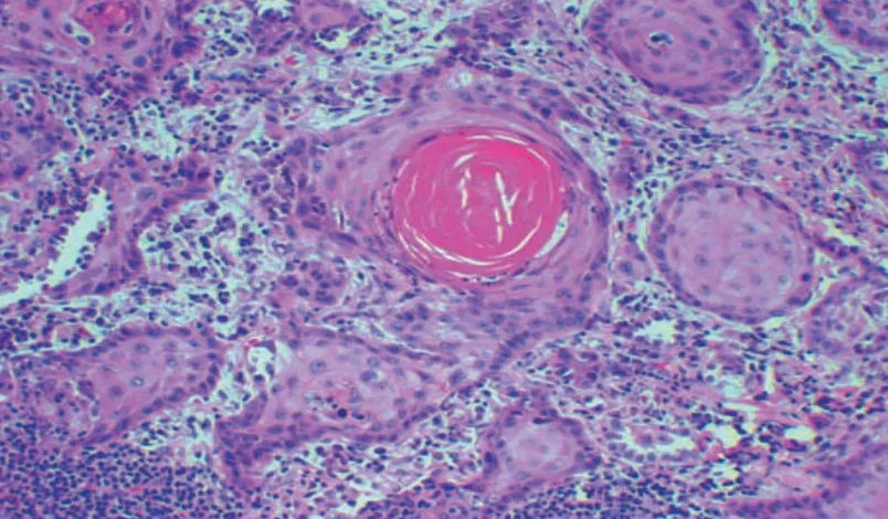
Immunohistochemistry of this biopsy would most likely show strong expression of which marker?
- Q2Grade versus stageCh. VII cases p.4L7 p.24
An 80-year-old man has lower abdominal pain, weakness and 7 kg of weight loss over 6 months. PSA is 8.5 ng/mL and the prostate is enlarged and nodular. Needle biopsy shows invasive prostatic adenocarcinoma.
Histologic grading of this carcinoma is based primarily on which criterion?
- Q3Stage outweighs gradeL7 p.24
Two women have invasive ductal carcinoma of the breast. Patient A has a high-grade (poorly differentiated) 1.2-cm tumor with no involved nodes and no metastases. Patient B has a low-grade (well-differentiated) 1.2-cm tumor with 9 involved axillary nodes and two liver metastases.
Who has the worse prognosis, and why?
- Q4Reading an IHC panelL7 p.23
A 58-year-old woman has a 3-cm axillary mass. Biopsy shows sheets of large, discohesive, poorly differentiated cells. Immunohistochemistry is negative for cytokeratin and for S-100, and strongly positive for CD45.
What is the most likely diagnosis?
- Q5Tumor markers monitor diseaseL7 p.26
After resection of a colonic adenocarcinoma, a patient's CEA falls from 42 to 3 ng/mL. Eight months later, with no symptoms, it is 29 ng/mL.
What is the best interpretation?