Interactive lab
Five activities
Every activity uses real chapter content and keeps your progress in both study modes. Each also appears inside the topic where it helps most.
Four specimens: look, decide about invasion, choose a stain, sort grade from stage, then take one more clue.
In context: 7.2 Dysplasia and invasion →Centerpiece · Tumor Pathology Case Lab
Tumor Pathology Case Lab
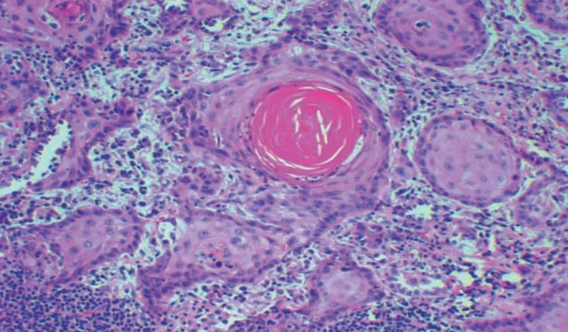
Clinical history
A 60-year-old man has 4 months of weight loss, wheezing and breathlessness. He has smoked two packs a day for 40 years. A chest radiograph shows a 10-cm mass in the left lung, and bronchoscopy shows the left main bronchus obstructed. A biopsy is taken.
Before you answer, look for
- Are the cells arranged in nests, glands or sheets?
- Is anything being made by the cells: keratin, mucin, matrix?
- Are the nests confined to an epithelial layer, or sitting in stroma?
- 1. Look first
- 2. Has it invaded?
- 3. Choose a stain
- 4. Grade or stage
- 5. One more clue
Step 1 · Look first
What do you call this, and what is the lineage clue?
Decide benign or malignant first, then name what the cells are making.
Three schematics of one epithelium with basement membrane, atypia and stroma overlays; decide which is invasive and what proves it.
In context: 7.2 Dysplasia and invasion →Morphology comparison · Layers
Where are the cells?
Specimen A
Specimen B
Specimen C
Teaching schematics, not micrographs. Orange outline: lymphatic. Red outline: small vein.
0 of 3 answered correctly. Source: Lesson 7 Neoplasia, pp. 8 and 22; the same distinction is tested in Ch. VII Q2 and Midterm sim. Q5–Q6.
HPV oncoproteins, the invasion cascade and the two-hit hypothesis; events versus mechanisms.
In context: 7.3, 7.4, 7.5 →Core activity · The Missing Arrow
The Missing Arrow
A cervical squamous cell at the transformation zone is infected by HPV 16, and the viral genome integrates into host DNA. Complete the chain to a dividing, mutating clone.
Two gaps ask for a mechanism, the link that explains how one event causes the next. Don't put an event there.
- High-risk HPV 16 integrates into the host genome
- integration disrupts viral E2, which normally restrains E6 and E7
- p53 protein is degraded
- no p53 means no p21, no DNA-repair induction and no apoptosis signal
- Continuous proliferation of cells carrying unrepaired DNA damage
- each division adds mutations the cell can no longer check
- Dysplasia → carcinoma in situ → invasive squamous cell carcinoma
Cards
Tap or drag a card. Placing a used card moves it.
Dysplasia vs in situ, in situ vs invasive, hamartoma vs choristoma, grade vs stage.
In context: 7.1, 7.2, 7.6 →Core activity · Almost Twins
Almost Twins
Look-alike A
Dysplasia (low grade)
- Atypical nuclei: enlarged, hyperchromatic, high N:C ratio
- Atypia confined to the lower layers; cells still mature toward the surface
- Basement membrane intact
- Often regresses, especially after HPV clearance
Look-alike A
Dysplasia (low grade)
- Atypical nuclei: enlarged, hyperchromatic, high N:C ratio
- Atypia confined to the lower layers; cells still mature toward the surface
- Basement membrane intact
- Often regresses, especially after HPV clearance
Look-alike B
Carcinoma in situ (CIN 3)
- Atypical nuclei: enlarged, hyperchromatic, high N:C ratio
- Atypia through the full thickness; no maturation at all
- Basement membrane intact
- A true precursor: treated to prevent invasion
Step 1 · What they share
What do these two epithelia share?
Select every feature that both show.
Change one laboratory value or one biopsy finding and decide whether the answer moves.
In context: 7.7 Paraneoplastic syndromes →Core activity · One Fact Changes
One Fact Changes
1 · The original case
A 64-year-old man who has smoked for 40 years has a central lung mass and weight loss. Serum calcium is 3.4 mmol/L (13.6 mg/dL), phosphate is low and intact PTH is undetectable; a bone scan shows no metastases. Biopsy of the mass is pending.
Which tumor and mediator best explain the laboratory findings?
Which fact decided it?