Invasion and metastasis
To spread, a carcinoma cell must let go of its neighbors, cut through the basement membrane, move through matrix and enter a vessel. Which vessel it enters decides where the metastases appear.
The invasion cascade
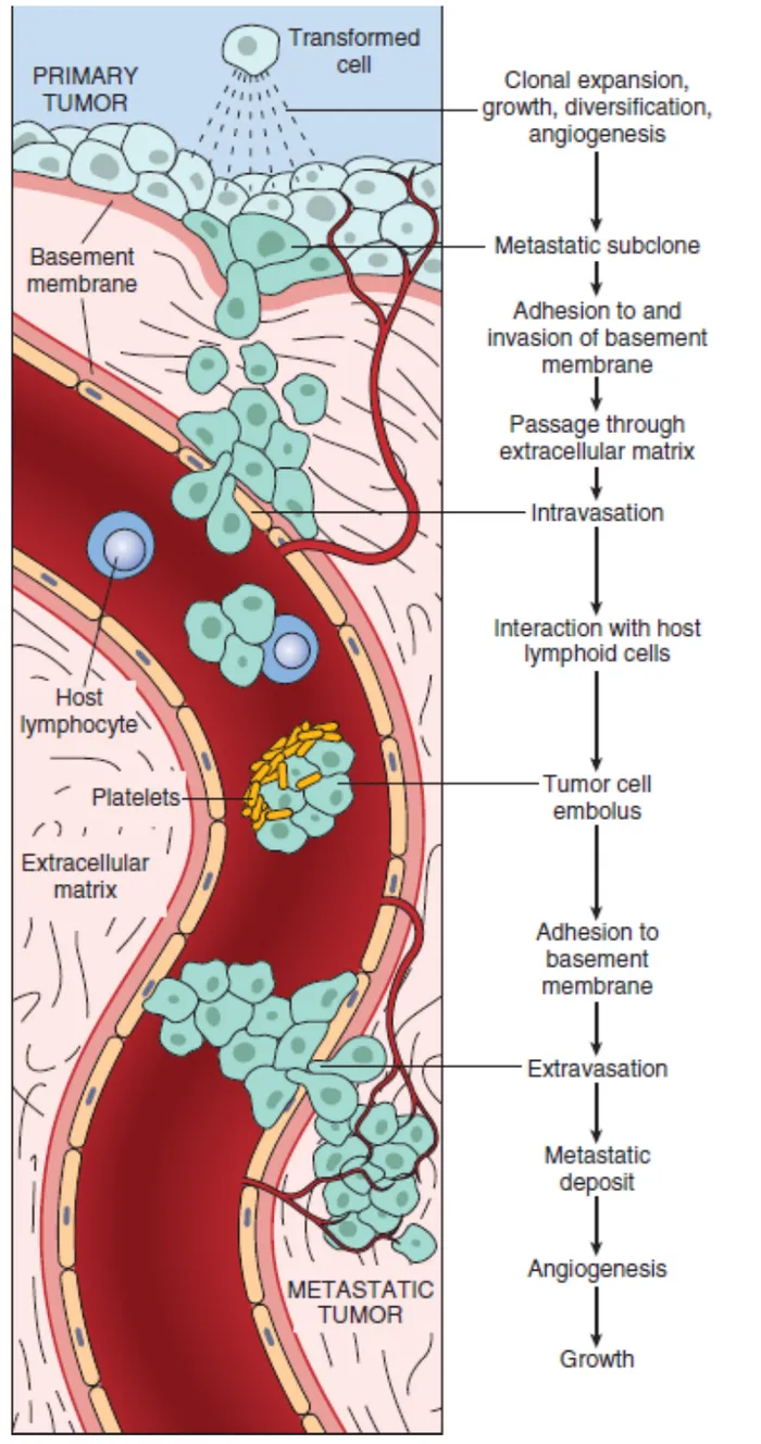
L7 p.8, 10Normal epithelial cells are held to each other by E-cadherin, a calcium-dependent adhesion molecule of the adherens junction, and anchored to the basement membrane by hemidesmosomes. Invasion reverses each of these attachments in turn. The lecture gives the sequence step by step:
- Detachment: tumor cells downregulate E-cadherin, so they dissociate from one another.
- Attachment to the basement membrane: cells bind laminin through laminin receptors.
- Degradation: they secrete proteases, above all matrix metalloproteinases (MMP-2 and MMP-9, the type IV collagenases), which dissolve the type IV collagen of the basement membrane.
- Locomotion: cells attach to fibronectin in the extracellular matrix through integrins and pull themselves through the stroma.
- Intravasation: they enter lymphatics or venules; in the blood they travel as emboli coated with platelets, which shield them from immune cells.
- Extravasation and colonization: they adhere to the endothelium of a distant capillary bed, cross it, and grow if they can recruit vessels (angiogenesis).
Teaching schematic
1. Detach
E-cadherin ↓: cells let go of each other
2. Attach
Bind laminin in the basement membrane
3. Degrade
MMPs digest type IV collagen
4. Migrate
Integrins grip fibronectin in the matrix
5. Enter a vessel
Intravasation into a lymphatic or venule
Lecture slide
Core activity · The Missing Arrow
The Missing Arrow
A colonic adenocarcinoma cell sits above an intact basement membrane. Complete the chain that takes it to the liver.
Each arrow is a mechanism; each box is an event. Keep them apart.
- Malignant cells above an intact basement membrane (carcinoma in situ)
- the cells bind laminin through laminin receptors
- Tumor cells attached to the basement membrane
- The basement membrane is breached: invasive carcinoma
- Cells migrate through the stroma and enter a portal venule
- the embolus, coated with platelets, travels in the portal vein
Cards
Tap or drag a card. Placing a used card moves it.
Three routes of spread
L7 p.10, 12Once cells reach a channel, the channel decides the destination. The lecture lists three routes plus perineural invasion.
Teaching schematic
Lymphatic
Most carcinomas
- Afferent lymphatic
- Sentinel (first draining) node
- Further nodes → thoracic duct → blood
Hematogenous
Sarcomas; RCC, HCC, follicular thyroid, choriocarcinoma
- Capillary or venule
- Portal vein → liver (gut, pancreas)
- Caval veins → lung (everything else)
Seeding
Ovarian surface carcinoma; medulloblastoma (CSF)
- Cells shed from a serosal surface
- Float in peritoneal, pleural fluid or CSF
- Implant: omental cake, ascites, drop metastases
Routes of metastasisL7 p.10
| Route | Typical tumors | Where it goes | Notes |
|---|---|---|---|
| Lymphatic | Most carcinomas | Regional nodes in the order of drainage; later the thoracic duct and blood | Cells arrive by afferent lymphatics in the subcapsular sinus. The first draining node is the sentinel node. |
| Hematogenous | Sarcomas; carcinoma exceptions: renal cell carcinoma, hepatocellular carcinoma, follicular thyroid carcinoma, choriocarcinoma | Portal vein → liver; caval veins → lungs | Veins (thin-walled) are entered far more often than arteries. RCC grows into the renal vein, HCC into the portal and hepatic veins. |
| Seeding (transcoelomic) | Ovarian surface carcinomas (serous cystadenocarcinoma); medulloblastoma | Peritoneum, omentum, pleura; CSF pathways | Malignant effusions (ascites); 'drop' metastases along the spinal cord |
| Perineural invasion | Prostate, pancreas, head and neck carcinomas | Along nerve sheaths | Painful; hard to excise completely |
Lymphatic
- Typical tumors
- Most carcinomas
- Where it goes
- Regional nodes in the order of drainage; later the thoracic duct and blood
- Notes
- Cells arrive by afferent lymphatics in the subcapsular sinus. The first draining node is the sentinel node.
Hematogenous
- Typical tumors
- Sarcomas; carcinoma exceptions: renal cell carcinoma, hepatocellular carcinoma, follicular thyroid carcinoma, choriocarcinoma
- Where it goes
- Portal vein → liver; caval veins → lungs
- Notes
- Veins (thin-walled) are entered far more often than arteries. RCC grows into the renal vein, HCC into the portal and hepatic veins.
Seeding (transcoelomic)
- Typical tumors
- Ovarian surface carcinomas (serous cystadenocarcinoma); medulloblastoma
- Where it goes
- Peritoneum, omentum, pleura; CSF pathways
- Notes
- Malignant effusions (ascites); 'drop' metastases along the spinal cord
Perineural invasion
- Typical tumors
- Prostate, pancreas, head and neck carcinomas
- Where it goes
- Along nerve sheaths
- Notes
- Painful; hard to excise completely
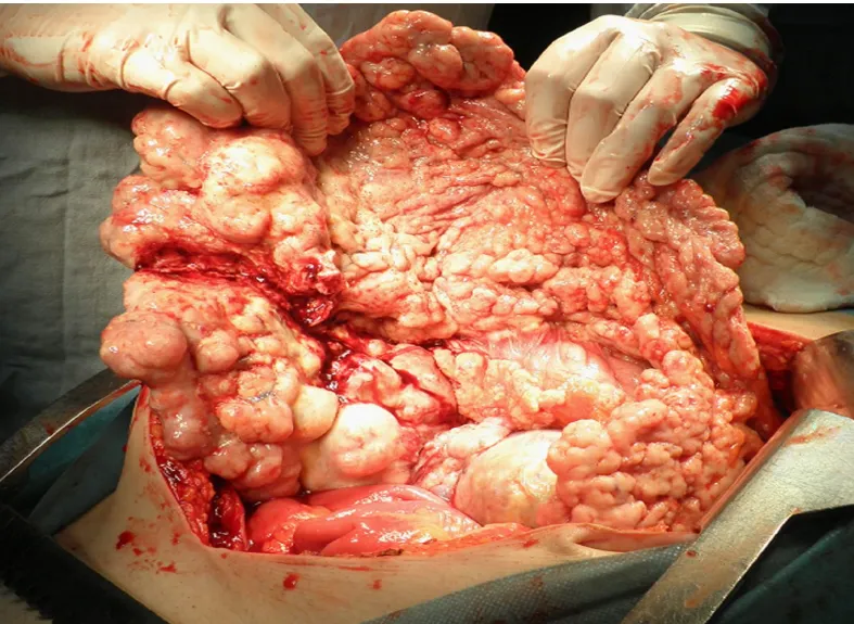
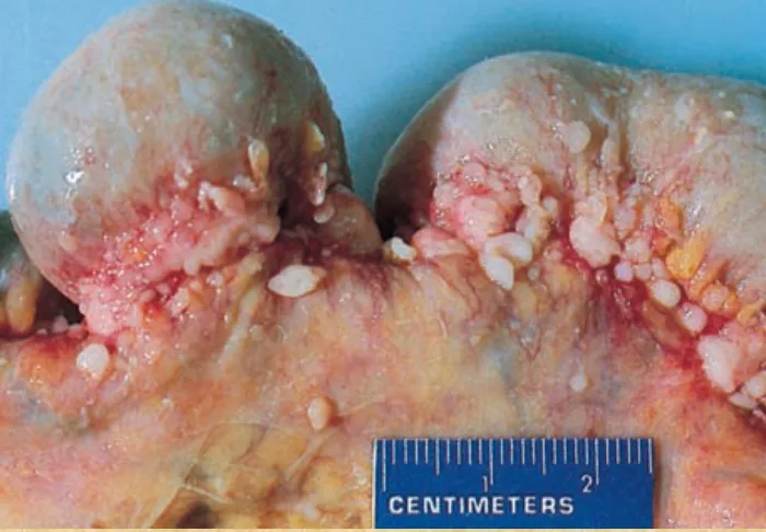
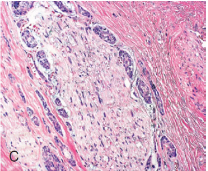
Two routes follow channels other than vessels. Cerebrospinal fluid works like a body cavity: medulloblastoma, a high-grade cerebellar tumor of children, sheds cells into the CSF that settle on the brainstem and spinal cord ('drop metastases'), which is why the whole neuraxis is imaged and treated. Perineural invasion tracks along the space around nerves; it causes pain and lets tumor reach well beyond the visible edge, so surgeons cannot trust a clean-looking margin. Neither route needs a blood vessel, and neither is stopped by a lymph node.
Lecture image
Case image
Lecture image
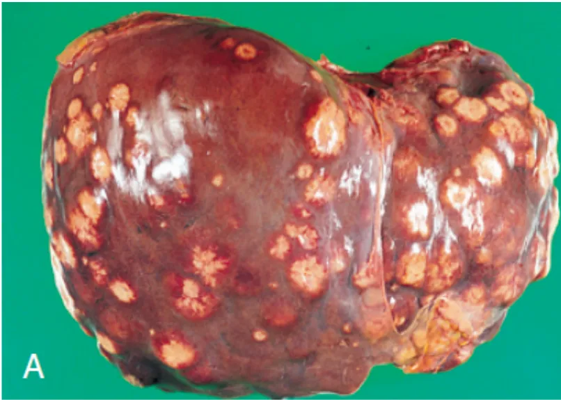
Why liver and lung: follow the venous drainage
L7 p.10, 12Ch. VII cases p.6Blood-borne tumor cells lodge in the first capillary bed downstream of the vein they entered. The stomach, intestine, pancreas and spleen drain through the portal vein, so their cancers metastasize first to the liver. Everything drained by the superior and inferior venae cavae (limbs, kidneys, most other organs) passes next through the lungs. That is why the liver and lungs are the organs most often involved by metastases, and why metastatic tumors in the liver far outnumber primary liver cancers.
Lecture image
Case image
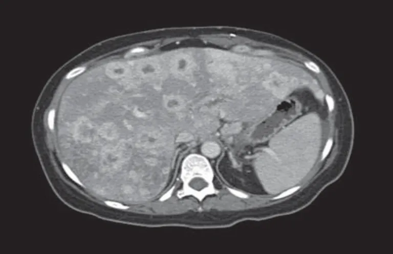
Worked case · Where did it start?
A 60-year-old man has weight loss, fatigue and hepatomegaly. CT shows multiple round nodules throughout the liver; a CT-guided biopsy shows mucin-secreting adenocarcinoma.
Which primary sites fit, and how would you narrow them?
Ch. VII cases p.6Bone metastases: blastic or lytic
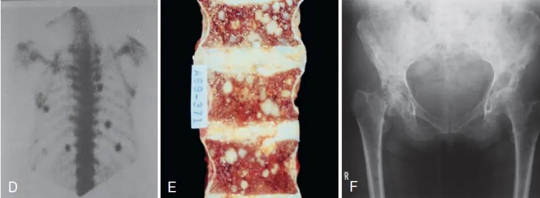
L7 p.11–12Metastases are the most common malignant tumors of bone. They favor the vertebral column (the most common site), pelvis, ribs and proximal long bones, where red marrow and a rich venous supply are; the valveless vertebral venous plexus offers a direct path from the pelvis to the spine. Breast cancer is the commonest cancer to metastasize to bone, prostate cancer the second. What the tumor cells secrete decides what the bone does.
Teaching schematic
Osteoblastic (blastic)
Tumor cytokines stimulate osteoblasts → reactive new bone. Prostate (and breast). Alkaline phosphatase ↑.
Osteolytic (lytic)
Tumor PGE2, IL-1 and PTHrP (via RANKL) activate osteoclasts → bone destroyed. Lung, renal, breast, myeloma. Fractures, hypercalcemia.
Osteoblastic (blastic)
- Tumor cytokines activate osteoblasts → reactive new bone
- Prostate carcinoma above all; breast can be blastic
- X-ray: radiodense (sclerotic) lesions
- Serum alkaline phosphatase ↑ (bone formation)
Osteolytic (lytic)
- Tumor products (PGE2, IL-1, local PTHrP → RANKL) activate osteoclasts
- Lung, renal cell, breast carcinoma (and myeloma, a primary bone marrow tumor)
- X-ray: radiolucent 'punched-out' lesions
- Pathologic fractures and hypercalcemia
Lecture image
Knowledge check · 7.3
Answer, read why, and try again whenever you like. Answers are kept in both study modes.
- Q1Seeding of body cavitiesCh. VII cases p.9L7 p.10
A 67-year-old woman was diagnosed with papillary serous cystadenocarcinoma of the ovary 3 years ago. She now has a massively swollen abdomen and dies a month later. At autopsy, the peritoneum is studded with small tumors and there are 4 L of ascites.
Which route of metastasis accounts for these findings?
- Q2Portal drainage → liver metastasesCh. VII cases p.6L7 p.10
A 60-year-old man has a 6-month history of weight loss and fatigue, and marked hepatomegaly. CT shows multiple round 'cannonball' nodules in the liver; CT-guided biopsy shows a mucin-secreting adenocarcinoma.
Where did this metastatic cancer most likely originate?
- Q3Invasion cascade: E-cadherinL7 p.8
In a colon adenocarcinoma, which change is the first step that allows tumor cells to leave the epithelial sheet?
- Q4Blastic bone metastasesL7 p.11–12
A 74-year-old man has low back pain. His PSA is 86 ng/mL. A bone scan shows many hot spots in the spine and pelvis.
Which pair of findings is most likely?
- Q5Hematogenous spread of sarcomasL7 p.10
A 15-year-old boy has an osteosarcoma of the distal femur. Staging CT shows three nodules in the lungs; regional lymph nodes are not enlarged.
How did the tumor most likely reach the lungs?