Interactive lab
Six activities
Every activity uses real unit content and keeps your progress in both study modes. Each also appears inside the topic where it helps most.
A named bone, a stated starting view, labels hidden: tap the bone on the specimen and read the cue that places it.
In context: 6 Practice on the specimen →Point to the bone
0 of 22All 22 bones, left and right asked separately. The prompt names one structure and states the starting view. Labels are hidden. Tap the structure on the specimen; you may rotate and zoom first. You get the recognition cue after a miss and can try again.
One side of a paired structure in a view that changes from prompt to prompt: work out where the patient's left is, then tap.
In context: 6 Practice on the specimen →Left or right?
0 of 10A paired bone is highlighted. Decide whether it is the patient's left or right in that view. The prompt names one structure and states the starting view. Labels are hidden. Choose the side. You get the recognition cue after a miss and can try again.
Processes, margins, sutures and large openings: find the anchor, confirm the neighbors, tap the landmark.
In context: 6 Practice on the specimen →Point to the landmark
0 of 50Surface landmarks, sutures and meeting points that this model shows clearly. The prompt names one structure and states the starting view. Labels are hidden. Tap the structure on the specimen; you may rotate and zoom first. You get the recognition cue after a miss and can try again.
One opening, its side and its view, for example the left foramen ovale from the internal cranial base; the feedback gives the cue that locates it.
In context: 5 The master table →Point to the opening
0 of 23Only openings that are individually recognisable on this model are asked. The prompt names one structure and states the starting view. Labels are hidden. Tap the structure on the specimen; you may rotate and zoom first. You get the recognition cue after a miss and can try again.
Five pairs that are easy to swap: rotundum and ovale, ovale and spinosum, internal acoustic meatus and stylomastoid foramen, optic canal and superior orbital fissure, jugular foramen and hypoglossal canal.
In context: 5 Three comparisons →Core activity · Almost Twins
Almost twins: openings
Look-alike A
Foramen rotundum
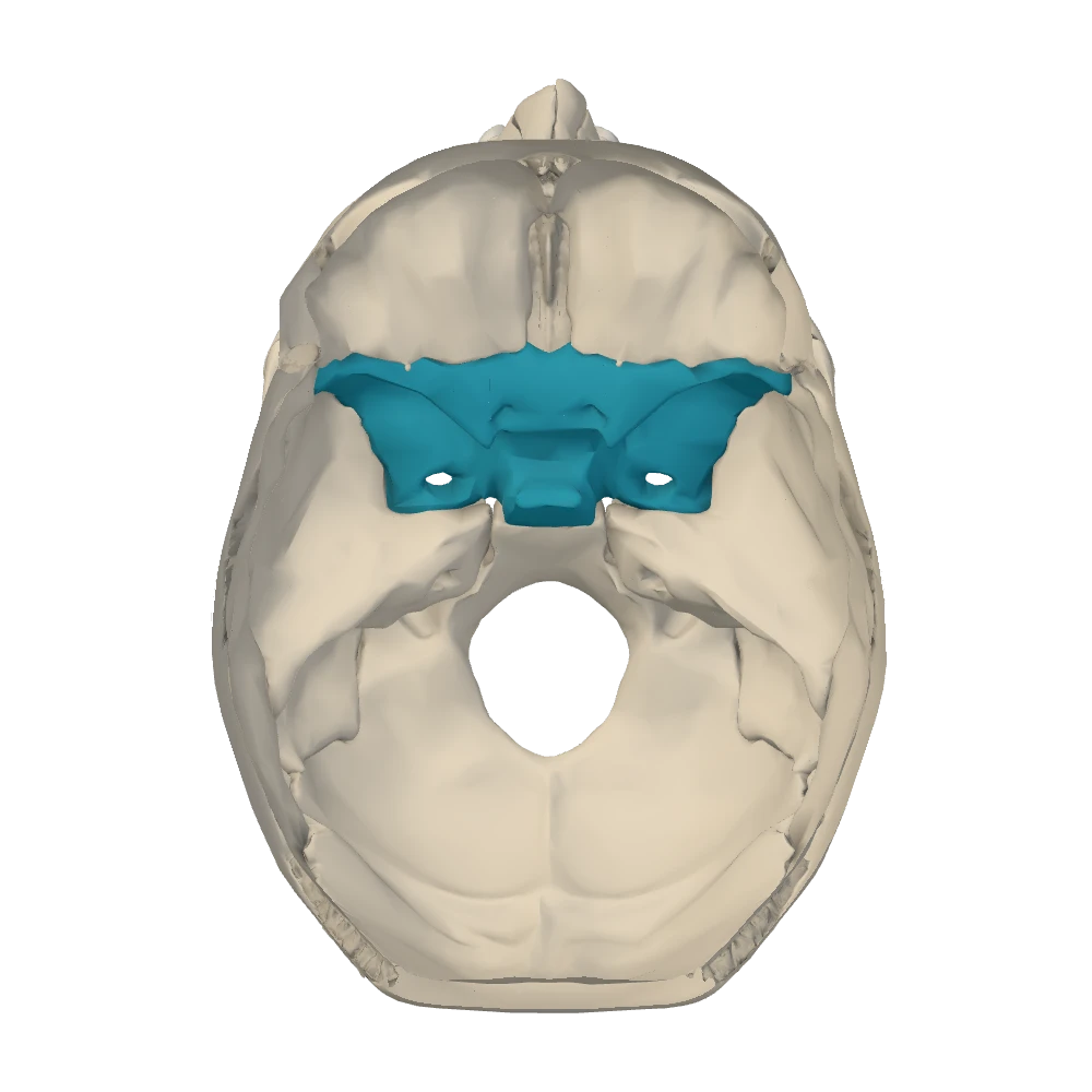
Internal cranial base view. Skull cap removed, looking down into the cranial cavity: the face is toward the top, the patient's right is on your right.
- Greater wing of the sphenoid, middle cranial fossa
- At the front, just below the superior orbital fissure; small, and it faces forward
- Leads to the pterygopalatine fossa; no mouth on the under-surface of the skull
- Maxillary nerve (V2)
Look-alike A
Foramen rotundum
Internal cranial base view. Skull cap removed, looking down into the cranial cavity: the face is toward the top, the patient's right is on your right.
- Greater wing of the sphenoid, middle cranial fossa
- At the front, just below the superior orbital fissure; small, and it faces forward
- Leads to the pterygopalatine fossa; no mouth on the under-surface of the skull
- Maxillary nerve (V2)
Look-alike B
Foramen ovale
Internal cranial base view. Skull cap removed, looking down into the cranial cavity: the face is toward the top, the patient's right is on your right.
- Greater wing of the sphenoid, middle cranial fossa
- Behind and lateral to the rotundum; the largest of the wing's openings, and it passes straight down
- Leads to the infratemporal fossa; found again in the inferior view
- Mandibular nerve (V3)
Step 1 · What they share
What do these two share?
Select every feature that both show.
Four routes with gaps: the facial nerve, a blow at the pterion, the olfactory filaments and the internal carotid artery.
In context: 5 Follow one structure through the bone →Core activity · The Missing Arrow
The missing arrow: pathways through the skull
The facial nerve (CN VII) starts inside the skull and ends on the face. On the way it crosses the temporal bone. Complete its route.
Events say where the nerve is. Links say which opening or tunnel takes it to the next place. Two gaps of each kind.
- CN VII and CN VIII lie side by side in the posterior cranial fossa
- the facial nerve goes on alone into its own bony tunnel
- The facial nerve is outside the skull and turns forward to the muscles of facial expression
Cards
Tap or drag a card. Placing a used card moves it.