Identify structures on the specimen
Naming a structure from a list is recall. Putting a fingertip on it is a different skill, and it is the one that a practical examination, a radiograph and an operating field all ask for. This section gives you one routine that finds any structure on the skull, the few look-alikes behind most wrong answers, and practice on the specimen with the labels switched off.
One routine for any structure: view, anchor, side, neighbors
OpenStaxNetter p.9–20Finding a structure on a skull is a procedure. People who do it reliably are not quicker at recognizing shapes. They ask the same four questions in the same order every time, so a difficult target is found by the same steps as an easy one. Learn the order once and use it for every prompt in this section.
- Choose the view. Ask which surface of the skull the structure is on, and open the preset that faces that surface. If a prompt states a starting view, begin there.
- Find an anchor. An anchor is a landmark too large to mistake, such as an orbit or the foramen magnum. Find the nearest one first and work outward from it. Do not hunt for a small structure directly.
- Fix the side. Say where the patient's left is in this view before you look at either half. Then look only at the half you need.
- Confirm with the neighbors. Name two things that must lie beside your answer and check that they do. If the neighbors are wrong, the answer is wrong.
Step 1: the view. Every structure is on a surface, and each preset faces one surface. The table repeats the views of section 1, sorted this time by what you are looking for.
Which view to openNetter p.9–20OpenStax
| You are looking for | Open | Why |
|---|---|---|
| A bone or an opening of the face or forehead | Anterior | The frontal bone, orbits, nasal and zygomatic bones, maxillae and mandible all face forward |
| Anything on the side wall: the temporal bone, the zygomatic arch, the pterion, the ramus of the mandible | Left or right lateral | The side facing you is the side the view is named for |
| The occipital bone, the lambdoid suture, the backs of the parietal bones | Posterior | It shows the back of the vault face on |
| The coronal and sagittal sutures, the bregma and the lambda | Superior | The vault from above |
| The palate, the pterygoid plates, the processes of the temporal bone, the occipital condyles, the outer mouth of an opening | Inferior | The outside of the base |
| A cranial fossa, the sella turcica, a petrous ridge, the inner mouth of an opening | Internal cranial base | The inside of the base, with the skullcap removed |
| The nasal septum, the sinus under the sella, the clivus, the palate in profile | Midsagittal section | Midline structures show in profile only on a cut surface |
A bone or an opening of the face or forehead
- Open
- Anterior
- Why
- The frontal bone, orbits, nasal and zygomatic bones, maxillae and mandible all face forward
Anything on the side wall: the temporal bone, the zygomatic arch, the pterion, the ramus of the mandible
- Open
- Left or right lateral
- Why
- The side facing you is the side the view is named for
The occipital bone, the lambdoid suture, the backs of the parietal bones
- Open
- Posterior
- Why
- It shows the back of the vault face on
The coronal and sagittal sutures, the bregma and the lambda
- Open
- Superior
- Why
- The vault from above
The palate, the pterygoid plates, the processes of the temporal bone, the occipital condyles, the outer mouth of an opening
- Open
- Inferior
- Why
- The outside of the base
A cranial fossa, the sella turcica, a petrous ridge, the inner mouth of an opening
- Open
- Internal cranial base
- Why
- The inside of the base, with the skullcap removed
The nasal septum, the sinus under the sella, the clivus, the palate in profile
- Open
- Midsagittal section
- Why
- Midline structures show in profile only on a cut surface
Step 2: the anchor. Six landmarks are large enough to find at a glance, and between them they fix every region of the skull. For each one, learn what lies next to it. The neighbors in this table are the routes you will use in practice.
Six anchors and where they leadNetter p.9–20OpenStax
| Anchor | Best seen in | From here you can find |
|---|---|---|
| Orbits | Anterior | Above: the supraorbital margin and the frontal bone. Below: the infraorbital margin, the maxilla and the infraorbital foramen. Lateral: the zygomatic bone. Medial: the nasal bones and the nasal opening. At the back: the apex, with the optic canal |
| Zygomatic arch | Lateral, inferior | Above it: the temple, the squamous part of the temporal bone and, about two finger-widths up, the pterion. Deep to it: the coronoid process. At its back end: the articular tubercle and the mandibular fossa |
| External acoustic meatus (the ear opening) | Lateral | Just in front: the head of the mandible in the mandibular fossa, the temporomandibular joint. Behind and below: the mastoid process. Below and deeper: the styloid process |
| Mastoid process | Lateral, posterior, inferior | In front and medial: the styloid process, with the stylomastoid foramen between the two. Behind and above: the occipital bone and the asterion |
| Foramen magnum | Inferior, internal cranial base | On each side: the occipital condyle, with the hypoglossal canal above it. In front: the basilar part of the occipital bone and the clivus. Lateral to the condyle: the jugular foramen |
| Sella turcica | Internal cranial base, midsagittal section | At its front outer corner: the optic canal, then the superior orbital fissure lateral to that. Lateral: the greater wing with the foramen rotundum, ovale and spinosum. Behind and lateral: the tip of the petrous part and the foramen lacerum. Below it: the sphenoidal sinus |
- Best seen in
- Anterior
- From here you can find
- Above: the supraorbital margin and the frontal bone. Below: the infraorbital margin, the maxilla and the infraorbital foramen. Lateral: the zygomatic bone. Medial: the nasal bones and the nasal opening. At the back: the apex, with the optic canal
- Best seen in
- Lateral, inferior
- From here you can find
- Above it: the temple, the squamous part of the temporal bone and, about two finger-widths up, the pterion. Deep to it: the coronoid process. At its back end: the articular tubercle and the mandibular fossa
External acoustic meatus (the ear opening)
- Best seen in
- Lateral
- From here you can find
- Just in front: the head of the mandible in the mandibular fossa, the temporomandibular joint. Behind and below: the mastoid process. Below and deeper: the styloid process
- Best seen in
- Lateral, posterior, inferior
- From here you can find
- In front and medial: the styloid process, with the stylomastoid foramen between the two. Behind and above: the occipital bone and the asterion
- Best seen in
- Inferior, internal cranial base
- From here you can find
- On each side: the occipital condyle, with the hypoglossal canal above it. In front: the basilar part of the occipital bone and the clivus. Lateral to the condyle: the jugular foramen
- Best seen in
- Internal cranial base, midsagittal section
- From here you can find
- At its front outer corner: the optic canal, then the superior orbital fissure lateral to that. Lateral: the greater wing with the foramen rotundum, ovale and spinosum. Behind and lateral: the tip of the petrous part and the foramen lacerum. Below it: the sphenoidal sinus
Specimen still
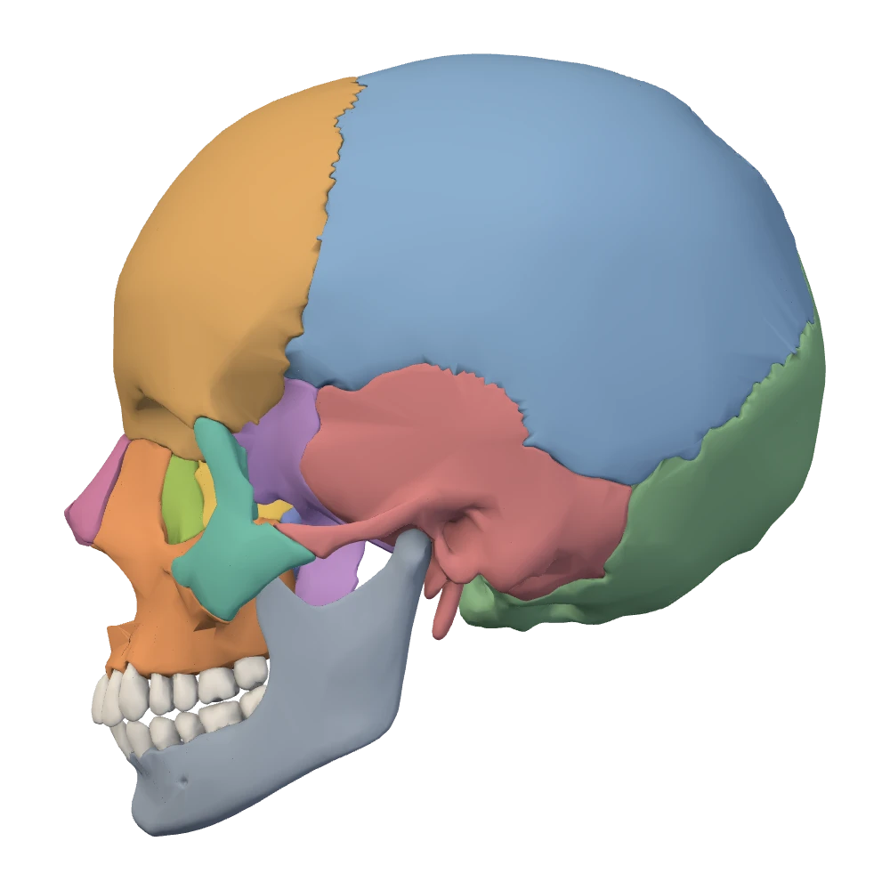
Left lateral view. The patient's left side: the face points to your left.
Step 3: the side. Left and right belong to the patient, never to the viewer (section 1). Two rules cover every preset. Seen from the front or from below, the picture is swapped: the patient's left is on your right. Seen from behind or from above, the picture agrees with you: the patient's left is on your left. The internal cranial base is a view from above, so with the face toward the top of the screen the patient's right is on your right. In a lateral view the side facing you is the side the view is named for, and the other side is hidden. In the midsagittal section of this specimen the left half has been removed, so you look at the cut surface of the right half with the face toward your left.
Step 4: the neighbors. A structure is identified by where it is, and a position is a statement about neighbors. Before you tap, finish a sentence of the form “it lies between … and …” or “it is just behind and lateral to …”. This is the step that separates look-alikes, and the next topic is built on it.
Worked example: “Select the left foramen ovale from the internal cranial base”
- View. Internal cranial base: the floor of the cranial cavity from above, with the face toward the top of the screen.
- Anchor. The sella turcica in the midline. The greater wing of the sphenoid spreads sideways from it as the floor of the middle cranial fossa.
- Side. This is a view from above, so the patient's left is on your left. Look only at the left half of the screen.
- Neighbors. Three openings cross the greater wing in a line that runs backward and outward: rotundum, ovale, spinosum. The foramen ovale is the middle one and the largest.
- Check. A much smaller opening, the foramen spinosum, lies just behind and lateral to it, and the ragged foramen lacerum lies medial to it at the tip of the petrous part. If both are where they should be, tap.
Specimen still
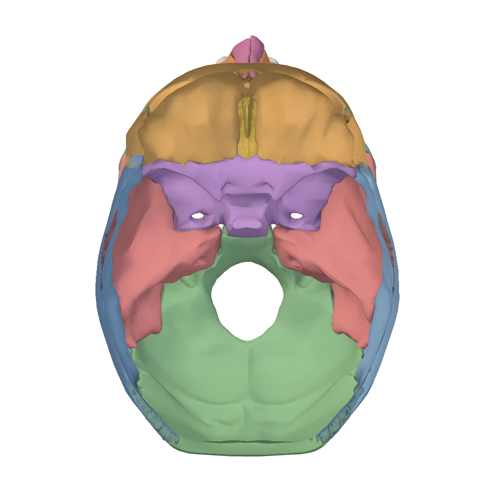
Internal cranial base view. Skull cap removed, looking down into the cranial cavity: the face is toward the top, the patient's right is on your right.
Look-alikes: five traps and the cue that separates each pair
Netter p.15–16, 19–22OpenStaxMost wrong answers in identification are not wild guesses. They are swaps between two structures that share a bone, a region, a name or a nerve. Each trap below has one cue that separates the pair, and in every case the cue is a position. Shape on its own is a poor guide, for a reason given at the end of this topic.
Five traps at a glanceNetter p.19–20Netter p.15–16, 21–22OpenStax · Middle cranial fossa; Temporal bone; The orbit; Nasal conchae; Mandible
| Look-alikes | Why they are confused | The cue that separates them | Confirm with |
|---|---|---|---|
| Rotundum, ovale, spinosum | Three small openings in the greater wing of the sphenoid, all in the middle cranial fossa | Order along the wing, from front and medial to back and lateral: rotundum, ovale, spinosum | Ovale is the largest. Spinosum is the smallest and lies beside the spine of the sphenoid. Only ovale and spinosum have a mouth on the under-surface |
| Internal acoustic meatus and stylomastoid foramen | Both are in the temporal bone and both carry the facial nerve | Inside or outside: the meatus is inside the skull, on the back slope of the petrous part; the foramen is outside, on the under-surface | The foramen lies between the styloid and mastoid processes. The meatus faces the posterior cranial fossa |
| Optic canal and superior orbital fissure | Side by side in the sphenoid, both leading from the middle cranial fossa to the back of the orbit | Round and medial, or slit and lateral: the canal is a round tunnel beside the sella; the fissure is a slit lateral to it | The canal opens at the apex of the orbit. The fissure has the lesser wing above it and the greater wing below |
| The three nasal conchae | Three similar curved shelves on the same wall | Which bone: the superior and middle conchae are parts of the ethmoid; the inferior nasal concha is a bone of its own | The inferior is the lowest and the largest. From the front only the inferior and the middle are in view |
| Coronoid and condylar processes | Two upward projections of the same ramus, with similar names | Front or back: coronoid in front, condylar behind, with the mandibular notch between them | The condylar process ends in a rounded head just in front of the ear opening. The coronoid is a thin point that passes under the zygomatic arch |
- Why they are confused
- Three small openings in the greater wing of the sphenoid, all in the middle cranial fossa
- The cue that separates them
- Order along the wing, from front and medial to back and lateral: rotundum, ovale, spinosum
- Confirm with
- Ovale is the largest. Spinosum is the smallest and lies beside the spine of the sphenoid. Only ovale and spinosum have a mouth on the under-surface
Internal acoustic meatus and stylomastoid foramen
- Why they are confused
- Both are in the temporal bone and both carry the facial nerve
- The cue that separates them
- Inside or outside: the meatus is inside the skull, on the back slope of the petrous part; the foramen is outside, on the under-surface
- Confirm with
- The foramen lies between the styloid and mastoid processes. The meatus faces the posterior cranial fossa
Optic canal and superior orbital fissure
- Why they are confused
- Side by side in the sphenoid, both leading from the middle cranial fossa to the back of the orbit
- The cue that separates them
- Round and medial, or slit and lateral: the canal is a round tunnel beside the sella; the fissure is a slit lateral to it
- Confirm with
- The canal opens at the apex of the orbit. The fissure has the lesser wing above it and the greater wing below
The three nasal conchae
- Why they are confused
- Three similar curved shelves on the same wall
- The cue that separates them
- Which bone: the superior and middle conchae are parts of the ethmoid; the inferior nasal concha is a bone of its own
- Confirm with
- The inferior is the lowest and the largest. From the front only the inferior and the middle are in view
Coronoid and condylar processes
- Why they are confused
- Two upward projections of the same ramus, with similar names
- The cue that separates them
- Front or back: coronoid in front, condylar behind, with the mandibular notch between them
- Confirm with
- The condylar process ends in a rounded head just in front of the ear opening. The coronoid is a thin point that passes under the zygomatic arch
Specimen still
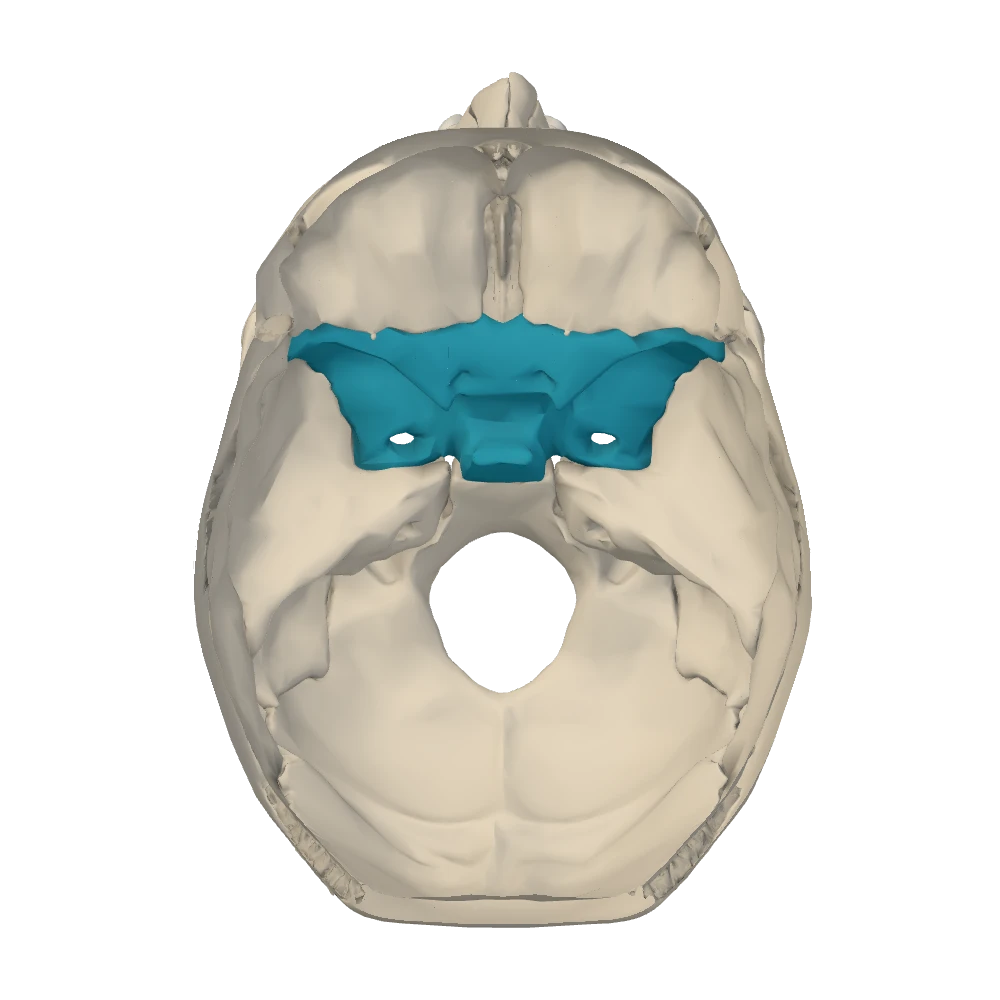
Internal cranial base view. Skull cap removed, looking down into the cranial cavity: the face is toward the top, the patient's right is on your right.
Rotundum, ovale, spinosum: count along the wing. Do not try to tell them apart by shape. “Round” and “oval” are hard to judge on an opening a few millimeters across. Start at the superior orbital fissure and move backward and outward along the greater wing. The first opening, just below the medial end of the fissure, is the foramen rotundum. It faces forward, toward the back of the upper jaw. The second, the foramen ovale, is the largest and passes straight down. The third, the foramen spinosum, is the smallest and lies just behind and lateral to the ovale. Turn the skull over and the count changes. On the under-surface only the ovale and the spinosum have a mouth, behind the root of the lateral pterygoid plate, and the spinosum is the one next to the pointed spine of the sphenoid. The rotundum cannot be found from below, because it opens forward into the pterygopalatine fossa.
Internal acoustic meatus and stylomastoid foramen: ask which surface. The temporal bone has three openings whose names are easy to cross. The external acoustic meatus is the ear opening on the side of the skull, seen in the lateral view. The internal acoustic meatus is inside the skull, on the back slope of the petrous part, and is found from the internal cranial base. The stylomastoid foramen is on the under-surface, between the styloid process and the mastoid process, and is found from the inferior view. The facial nerve links the last two. It goes in at the internal acoustic meatus together with the vestibulocochlear nerve and comes out, alone, at the stylomastoid foramen. When a prompt mentions the facial nerve, read on for enters or leaves, and check which view you are in.
Optic canal and superior orbital fissure: the words give the shape. A canal is a tunnel through bone and a fissure is a gap between two pieces of bone (section 5). At the back of the orbit the round tunnel is the optic canal. It lies medially, at the front outer corner of the sella turcica, and opens at the very apex of the orbit. The slit lateral to it is the superior orbital fissure, with the lesser wing above it and the greater wing below. Looking into the orbit from the front there is a second slit to keep apart from it. The inferior orbital fissure runs lower down, along the angle between the lateral wall and the floor, and does not lead to the cranial cavity.
The conchae: two belong to the ethmoid, one is a bone. Look into the nasal opening in the anterior view. On each side wall the lowest and largest shelf is the inferior nasal concha, one of the 14 facial bones. Above it is the middle nasal concha, and higher and further back, out of sight from the front, is the small superior nasal concha. Those two are not separate bones: they are scrolls of the ethmoid. The usual errors are to count all three as bones or to give all three to the ethmoid. A prompt for “the inferior nasal concha” asks for a bone. A prompt for “the middle nasal concha” asks for part of one.
Coronoid and condylar processes: the one near the ear makes the joint. In a lateral view the ramus of the mandible ends above in two projections separated by the mandibular notch. The front one, the coronoid process, is thin and pointed. A chewing muscle attaches to it, and it slips up under the zygomatic arch, so part of it is hidden in this view. The back one, the condylar process, has a narrow neck and a rounded head. The head sits in the mandibular fossa of the temporal bone, just in front of the ear opening, and forms the temporomandibular joint. If the names blur, use their roots: coronoid comes from the Greek for a crow, after its beak-like point, and condyle from the Greek for a knuckle.
More pairs, one line eachNetter p.11–12, 17–20Netter p.133–134OpenStax
| Pair | The cue |
|---|---|
| Styloid and mastoid processes | The mastoid process is the blunt mass behind the ear opening. The styloid process is the thin spike in front of and medial to it |
| Zygomatic process and temporal process | Each is named for the bone it reaches toward. The zygomatic process belongs to the temporal bone and the temporal process to the zygomatic bone. Together they are the zygomatic arch |
| Carotid canal and jugular foramen, from below | The carotid canal is the round opening in front, within the petrous part. The jugular foramen is the larger, irregular gap behind it, between the temporal and occipital bones |
| Jugular foramen and hypoglossal canal | The jugular foramen lies between two bones. The hypoglossal canal is a small tunnel in the occipital bone alone, above the condyle |
| Foramen lacerum and carotid canal | The foramen lacerum is a ragged gap between bones, closed by fibrocartilage in life. The carotid canal is a smooth tunnel, and the artery it carries only crosses the upper part of the lacerum |
| Bregma and lambda | Bregma in front, where the coronal suture meets the sagittal. Lambda behind, where the sagittal meets the lambdoid |
| Pterion and asterion | Pterion in the temple, in front of the ear: frontal, parietal, temporal, sphenoid. Asterion behind the ear: parietal, temporal, occipital |
| Lacrimal and nasal bones | The lacrimal bone is inside the orbit, at the front of its medial wall. The nasal bone is outside it, on the bridge of the nose |
| Medial and lateral pterygoid plates | From below, behind the hard palate: the medial plate borders the back opening of the nasal cavity; the lateral plate is broader and faces the infratemporal fossa |
Styloid and mastoid processes
- The cue
- The mastoid process is the blunt mass behind the ear opening. The styloid process is the thin spike in front of and medial to it
Zygomatic process and temporal process
- The cue
- Each is named for the bone it reaches toward. The zygomatic process belongs to the temporal bone and the temporal process to the zygomatic bone. Together they are the zygomatic arch
Carotid canal and jugular foramen, from below
- The cue
- The carotid canal is the round opening in front, within the petrous part. The jugular foramen is the larger, irregular gap behind it, between the temporal and occipital bones
Jugular foramen and hypoglossal canal
- The cue
- The jugular foramen lies between two bones. The hypoglossal canal is a small tunnel in the occipital bone alone, above the condyle
Foramen lacerum and carotid canal
- The cue
- The foramen lacerum is a ragged gap between bones, closed by fibrocartilage in life. The carotid canal is a smooth tunnel, and the artery it carries only crosses the upper part of the lacerum
Bregma and lambda
Pterion and asterion
Lacrimal and nasal bones
- The cue
- The lacrimal bone is inside the orbit, at the front of its medial wall. The nasal bone is outside it, on the bridge of the nose
Medial and lateral pterygoid plates
- The cue
- From below, behind the hard palate: the medial plate borders the back opening of the nasal cavity; the lateral plate is broader and faces the infratemporal fossa
Practice on the specimen
BodyParts3DThree short activities put the routine to work on the live specimen. The labels are off, nothing is selected for you, and you answer by tapping the structure itself. Each prompt is precise about what is wanted and states the view it starts in. You are free to rotate and zoom before you answer.
- Read the prompt. It names one structure and, where it matters, a side, for example “Select the left zygomatic bone.” The starting view is stated with it.
- Run the routine. View, anchor, side, neighbors. Say the neighbors to yourself before you tap.
- Tap the structure on the specimen.
- Read the feedback. It shows the correct structure and explains the cue that locates it, usually a neighbor or a landmark in that view.
- Retry. A missed prompt can be tried again. Afterward, find the same structure from a second view.
Point to the bone
0 of 22All 22 bones, left and right asked separately. The prompt names one structure and states the starting view. Labels are hidden. Tap the structure on the specimen; you may rotate and zoom first. You get the recognition cue after a miss and can try again.
Left or right?
0 of 10A paired bone is highlighted. Decide whether it is the patient's left or right in that view. The prompt names one structure and states the starting view. Labels are hidden. Choose the side. You get the recognition cue after a miss and can try again.
Point to the landmark
0 of 50Surface landmarks, sutures and meeting points that this model shows clearly. The prompt names one structure and states the starting view. Labels are hidden. Tap the structure on the specimen; you may rotate and zoom first. You get the recognition cue after a miss and can try again.
Read your misses
| What went wrong | Step that was skipped | What to do next time |
|---|---|---|
| Right structure, wrong side | Step 3, the side | Name the view and say where the patient's left is before you look at either half |
| The opening or process next door | Step 4, the neighbors | Count from the anchor and name two neighbors before you tap |
| Right region, wrong bone | Step 2, the anchor | Start again from the anchor and follow the sutures: a bone ends where its sutures are |
| Could not find it at all | Step 1, the view | Ask which surface it is on and change the view. Several structures show in one view only |
Right structure, wrong side
- Step that was skipped
- Step 3, the side
- What to do next time
- Name the view and say where the patient's left is before you look at either half
The opening or process next door
- Step that was skipped
- Step 4, the neighbors
- What to do next time
- Count from the anchor and name two neighbors before you tap
Right region, wrong bone
- Step that was skipped
- Step 2, the anchor
- What to do next time
- Start again from the anchor and follow the sutures: a bone ends where its sutures are
Could not find it at all
- Step that was skipped
- Step 1, the view
- What to do next time
- Ask which surface it is on and change the view. Several structures show in one view only
Knowledge check · 6
Answer, read why, and try again whenever you like. Answers are kept in both study modes.
- Q1Bone and side from an unlabeled anterior viewBodyParts3DOpenStax · Zygomatic bone
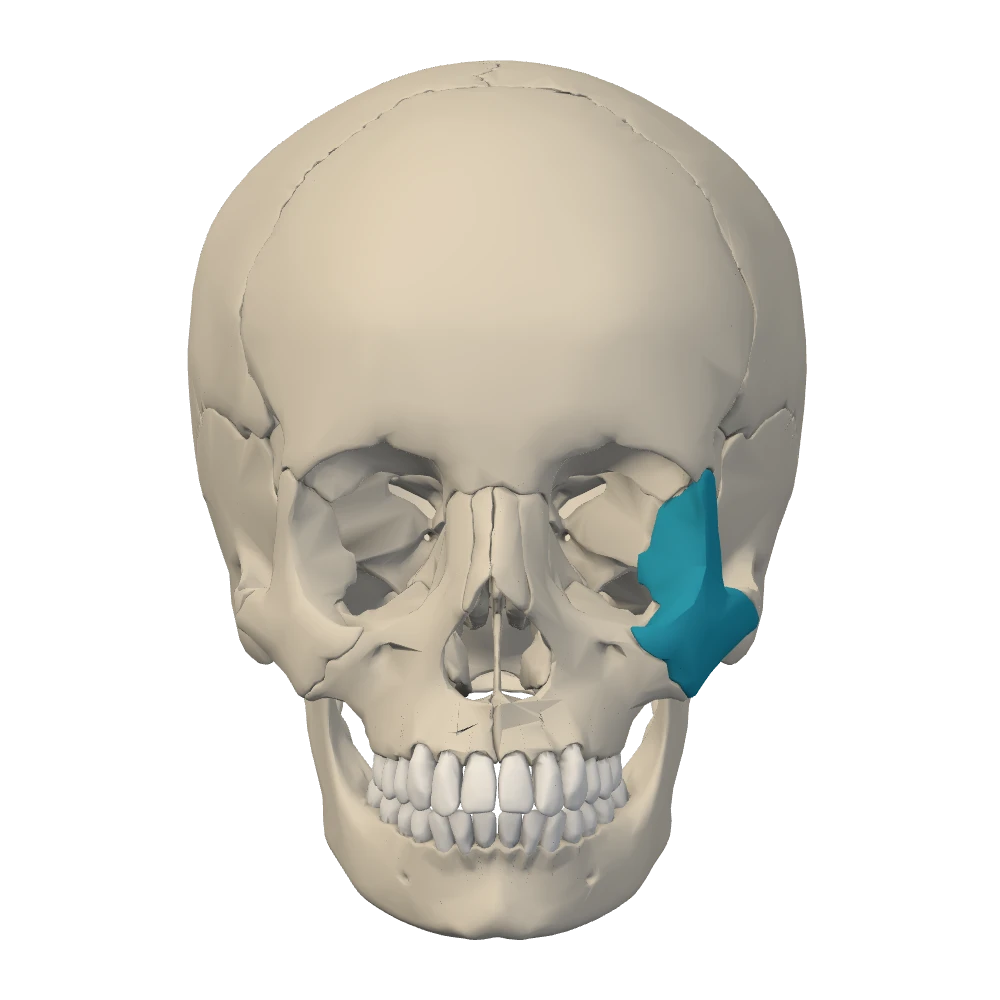
Anterior view. Facing the skull: the patient's left is on your right.
This is the anterior view with the labels off. One bone is highlighted, on the right-hand side of the picture. Which bone is it?
- Q2Opening and side from the internal cranial baseNetter p.19–20OpenStax · Middle cranial fossa
You are in the internal cranial base view with the face toward the top of the screen. On the right-hand side of the screen, in the greater wing of the sphenoid, you place the largest of three openings. A much smaller one lies just behind and lateral to it.
Which opening is it?
- Q3Choosing the view: internal acoustic meatus versus stylomastoid foramenNetter p.19–20OpenStax · Temporal bone
A prompt reads: “Select the right internal acoustic meatus.” Which plan finds it?
- Q4Conchae: the inferior is a bone, the superior and middle are ethmoidOpenStax · The nasal septum and nasal conchaeNetter p.15–16
Looking into the nasal opening in the anterior view, a student sees two curved shelves on each side wall and labels the lower, larger one “concha of the ethmoid bone”.
What is the correct identification of the lower shelf?
- Q5Coronoid versus condylar processNetter p.21–22OpenStax · Mandible
In the left lateral view the ramus of the mandible ends above in two projections with a notch between them.
Which statement identifies the posterior projection correctly?
- Q6Optic canal versus superior orbital fissure by position and shapeNetter p.19–20OpenStax · Middle cranial fossa
In the internal cranial base view you place a round opening at the front outer corner of the sella turcica. Just lateral to it, under the edge of the lesser wing of the sphenoid, is a slit.
Which pairing of opening and principal contents is correct?