Openings and what passes through them
Every nerve and vessel that enters or leaves the cranial cavity uses a named opening. Learn each one as a doorway: which bone it pierces, which two spaces it joins, what goes through it, and how its position separates it from the opening next door.
How to read an opening
Netter p.19–20OpenStax · Cranial fossaeAn opening in the skull is never just a hole. It is the place where a named structure crosses from one space to another, and its shape tells you something about what it carries. Four words cover almost every opening you will meet.
Four kinds of openingTA2 · terms
| Term | What it means | Example in this section |
|---|---|---|
| Foramen (plural foramina) | A hole through a plate of bone | Foramen ovale |
| Canal | A tunnel with some length | Optic canal, carotid canal |
| Fissure | A narrow slit, usually between two bones or two parts of one bone | Superior orbital fissure |
| Meatus | A passage that leads into a bone | Internal acoustic meatus |
Foramen (plural foramina)
- What it means
- A hole through a plate of bone
- Example in this section
- Foramen ovale
Canal
- What it means
- A tunnel with some length
- Example in this section
- Optic canal, carotid canal
Fissure
- What it means
- A narrow slit, usually between two bones or two parts of one bone
- Example in this section
- Superior orbital fissure
Meatus
- What it means
- A passage that leads into a bone
- Example in this section
- Internal acoustic meatus
A notch is an opening that the bone did not close; soft tissue completes it in life. The supraorbital notch is the usual example.
For every opening, answer the same four questions.
- Which bone, or between which bones?
- Which two spaces does it join?
- What passes through? Learn the principal structure first; add the others once that is secure.
- Which neighbor could I mistake it for, and what difference in position separates them?
The fourth question is the one students skip and examiners ask. On an unlabeled skull nothing tells you what runs through a hole; only its position does. So in this section position always comes first and contents second.
Twelve cranial nerves, front to back
- Anterior fossa: CN I (cribriform plate)
- Middle fossa: CN II (optic canal)
- CN III, IV, V1 and VI (superior orbital fissure)
- V2 (foramen rotundum), then V3 (foramen ovale)
- Posterior fossa: CN VII and VIII (internal acoustic meatus)
- CN IX, X and XI (jugular foramen)
- CN XII (hypoglossal canal)
Anterior cranial fossa: the cribriform plate
Netter p.19–20OpenStax · Ethmoid boneThe anterior fossa has one set of openings to learn, and it is a set, not a single hole. On each side of the crista galli the cribriform plate of the ethmoid forms a narrow gutter pierced by many small cribriform foramina (cribrum is Latin for a sieve). Treat the plate as a perforated region: you are not asked to find an individual hole.
Cribriform foraminaNetter p.19–20OpenStax · Ethmoid bone
| Question | Answer |
|---|---|
| Bone | Ethmoid (cribriform plate) |
| Where to look | Internal cranial base view, front of the midline: the sunken strip on each side of the crista galli, between the two orbital plates of the frontal bone |
| Connects | Anterior cranial fossa above with the roof of the nasal cavity below |
| Principal contents | Olfactory nerve filaments (CN I): many small bundles on each side |
| Do not confuse with | The optic canal, which is further back at the edge of the middle fossa and is a single round tunnel |
Bone
- Answer
- Ethmoid (cribriform plate)
Where to look
- Answer
- Internal cranial base view, front of the midline: the sunken strip on each side of the crista galli, between the two orbital plates of the frontal bone
Connects
- Answer
- Anterior cranial fossa above with the roof of the nasal cavity below
Principal contents
- Answer
- Olfactory nerve filaments (CN I): many small bundles on each side
Do not confuse with
- Answer
- The optic canal, which is further back at the edge of the middle fossa and is a single round tunnel
The direction of travel is upward. Receptor cells in the lining of the nasal roof send their fine axons up through the plate, and these end at once in the olfactory bulb, which rests on the upper surface of the plate. CN I is therefore not one trunk but a spray of filaments.
Middle cranial fossa: five openings in the sphenoid, and the carotid canal beside them
Netter p.19–20Netter p.133–134OpenStax · Middle cranial fossaMost of the openings of the cranial base are in the middle fossa, and five of them are in a single bone. Go to the internal cranial base view and find the sella turcica. On each side of it, the openings of the sphenoid form a curve that runs from the midline outward and backward: optic canal → superior orbital fissure → foramen rotundum → foramen ovale → foramen spinosum. Learn that curve as a sequence and each opening has an address.
Specimen still
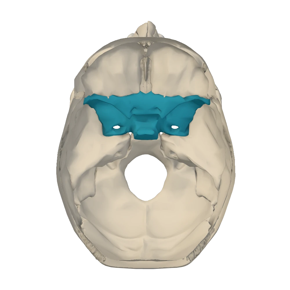
Internal cranial base view. Skull cap removed, looking down into the cranial cavity: the face is toward the top, the patient's right is on your right.
Optic canal. The optic canal is a short round tunnel through the root of the lesser wing, at the front outer corner of the sella turcica and just medial to the anterior clinoid process. It runs forward and a little outward into the apex of the orbit. It carries the optic nerve (CN II) and, with it, the ophthalmic artery.
Superior orbital fissure. Immediately lateral to the optic canal, and separated from it only by a thin bar of bone, is a long slit: the gap between the lesser wing above and the greater wing below. The superior orbital fissure is widest at its medial end and narrows as it runs upward and outward. It also opens into the orbit. Through it pass the three nerves that move the eye, the oculomotor (CN III), trochlear (CN IV) and abducens (CN VI) nerves; the three branches of the ophthalmic nerve (V1), named lacrimal, frontal and nasociliary; and the superior ophthalmic vein, which drains the orbit backward.
Foramen rotundum. Just below and behind the medial end of the fissure, in the greater wing, is a small round opening that faces forward more than downward. The foramen rotundum is really a short canal that runs forward into the pterygopalatine fossa. It carries the maxillary nerve (V2). Because it points forward, it has no mouth on the under-surface of the skull: from below you will not find it.
Foramen ovale. Further back and further out, still in the greater wing, is a larger oval hole that passes straight down through the floor. The foramen ovale opens into the infratemporal fossa and carries the mandibular nerve (V3). Other structures are listed for it but are variable: an accessory meningeal artery may enter here, and the lesser petrosal nerve occasionally leaves through it. Learn V3 as the answer and the others as possible companions.
Foramen spinosum. Behind and lateral to the foramen ovale, at the back corner of the greater wing, is the smallest opening of the set. The foramen spinosum is named for the spine of the sphenoid, a small downward point of bone right beside it on the external surface. It also opens into the infratemporal fossa, but its traffic runs the other way: the middle meningeal artery enters the skull here. From the foramen, branching grooves for the artery run up the inner surface of the side wall toward the pterion. A middle meningeal vein and a small meningeal branch of the mandibular nerve accompany the artery.
Foramen lacerum. Medial to the foramen ovale, at the side of the body of the sphenoid, the tip of the petrous part of the temporal bone stops short. The jagged gap left between it, the sphenoid and the basilar part of the occipital bone is the foramen lacerum (lacerum means torn). On a dry skull it can look like the largest opening in the region. In life it is filled with fibrocartilage, and no major structure passes down through it.
Carotid canal. The important structure here is the internal carotid artery, and it uses a different opening. The carotid canal begins as a round hole on the under-surface of the petrous part of the temporal bone, runs upward for a short distance, then bends forward and medially inside the bone. It ends at the petrous apex, in the upper part of the foramen lacerum. The artery emerges there, crosses the top of the cartilage, and turns upward beside the body of the sphenoid into the cavernous sinus next to the sella turcica. A plexus of sympathetic nerve fibers, the internal carotid plexus, travels on the artery through the canal.
Middle cranial fossa: the curve and its neighborsNetter p.19–20OpenStax · Middle cranial fossa
| Opening | Position cue | Leads to | Principal contents |
|---|---|---|---|
| Optic canal | Front outer corner of the sella; round; medial to the anterior clinoid process | Apex of the orbit | Optic nerve (CN II); ophthalmic artery |
| Superior orbital fissure | Slit lateral to the optic canal, between lesser and greater wings | Orbit | CN III, IV, VI; branches of V1; superior ophthalmic vein |
| Foramen rotundum | Greater wing, just below the medial end of the fissure; faces forward | Pterygopalatine fossa | Maxillary nerve (V2) |
| Foramen ovale | Greater wing, behind and lateral to rotundum; the largest of the three | Infratemporal fossa | Mandibular nerve (V3); variable companions |
| Foramen spinosum | Just behind and lateral to ovale, beside the spine of the sphenoid; the smallest | Infratemporal fossa | Middle meningeal artery |
| Foramen lacerum | Ragged gap at the petrous apex, medial to ovale | Closed by fibrocartilage in life | No major structure passes through; the internal carotid artery crosses above |
| Carotid canal | Inside the petrous part; its upper end opens at the petrous apex | From the neck to the middle cranial fossa | Internal carotid artery; internal carotid plexus |
- Position cue
- Front outer corner of the sella; round; medial to the anterior clinoid process
- Leads to
- Apex of the orbit
- Principal contents
- Optic nerve (CN II); ophthalmic artery
- Position cue
- Slit lateral to the optic canal, between lesser and greater wings
- Leads to
- Orbit
- Principal contents
- CN III, IV, VI; branches of V1; superior ophthalmic vein
- Position cue
- Greater wing, just below the medial end of the fissure; faces forward
- Leads to
- Pterygopalatine fossa
- Principal contents
- Maxillary nerve (V2)
- Position cue
- Greater wing, behind and lateral to rotundum; the largest of the three
- Leads to
- Infratemporal fossa
- Principal contents
- Mandibular nerve (V3); variable companions
- Position cue
- Just behind and lateral to ovale, beside the spine of the sphenoid; the smallest
- Leads to
- Infratemporal fossa
- Principal contents
- Middle meningeal artery
- Position cue
- Ragged gap at the petrous apex, medial to ovale
- Leads to
- Closed by fibrocartilage in life
- Principal contents
- No major structure passes through; the internal carotid artery crosses above
- Position cue
- Inside the petrous part; its upper end opens at the petrous apex
- Leads to
- From the neck to the middle cranial fossa
- Principal contents
- Internal carotid artery; internal carotid plexus
Deeper: why three neighboring openings carry one nerveMore detail
The trigeminal nerve (CN V) has a large sensory ganglion that rests in a shallow dent on the front slope of the petrous part, near its apex. Its three divisions leave the middle fossa through three neighboring openings, in order from front to back: V1 through the superior orbital fissure to the orbit and forehead, V2 through the foramen rotundum to the middle of the face, V3 through the foramen ovale to the lower jaw. Front to back on the skull matches top to bottom on the face.
Deeper: small nerves at the foramen lacerumMore detail
Sympathetic fibers leave the internal carotid plexus as the deep petrosal nerve. In the region of the foramen lacerum they join the greater petrosal nerve, a parasympathetic branch of the facial nerve, to form the nerve of the pterygoid canal, which runs forward through the root of the pterygoid process. You do not need this to identify the openings, but it explains why small nerves are described “at” the foramen lacerum even though nothing large passes through it.
Posterior cranial fossa: four openings around the brainstem
Netter p.19–20OpenStax · Posterior cranial fossaBehind the petrous ridge the openings are fewer and larger. Three are paired and one is the largest opening of the skull. Work from the petrous slope down to the midline.
Specimen still
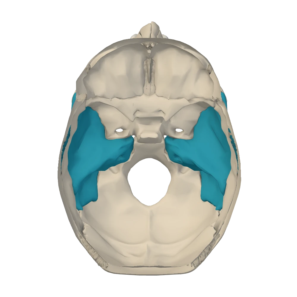
Internal cranial base view. Skull cap removed, looking down into the cranial cavity: the face is toward the top, the patient's right is on your right.
Internal acoustic meatus. About halfway along the back slope of the petrous part, facing the brainstem, is a round opening that leads sideways into the bone: the internal acoustic meatus. It is a short passage that ends blindly inside the bone. Two nerves enter it together: the facial nerve (CN VII) and the vestibulocochlear nerve (CN VIII), with the small labyrinthine artery to the inner ear. At the bottom of the meatus they separate. CN VIII ends in the inner ear, inside the petrous part, and never leaves the skull. CN VII continues into its own tunnel, the facial canal.
Jugular foramen. Directly below the internal acoustic meatus, in the seam between the petrous part of the temporal bone and the occipital bone, is a large, irregular opening: the jugular foramen. A broad curved groove on the inner wall of the fossa leads into it; this groove holds the sigmoid sinus, the last of the venous channels inside the skull. At the foramen the sigmoid sinus becomes the internal jugular vein. Three cranial nerves leave through the same opening: the glossopharyngeal (CN IX), vagus (CN X) and accessory (CN XI) nerves. A smaller venous channel, the inferior petrosal sinus, also drains through the foramen.
Hypoglossal canal. Lower and more medial still, just above the rim of the foramen magnum at its front outer edge, a short tunnel passes forward and outward through the occipital bone, above the occipital condyle. The hypoglossal canal carries the hypoglossal nerve (CN XII), the motor nerve of the tongue. From outside, its exit is hidden above the front of the occipital condyle.
Foramen magnum. The foramen magnum (“great opening”) is the oval hole in the floor of the fossa, entirely within the occipital bone, joining the cranial cavity to the vertebral canal. Here the medulla oblongata becomes the spinal cord, wrapped in the meninges. The two vertebral arteries come up through it to supply the brain. The spinal roots of the accessory nerve (CN XI) also ascend through it: they arise from the upper spinal cord, enter the skull through the foramen magnum, and then leave again through the jugular foramen.
Posterior cranial fossaNetter p.19–20OpenStax · Posterior cranial fossa
| Opening | Position cue | Leads to | Principal contents |
|---|---|---|---|
| Internal acoustic meatus | Back slope of the petrous part, about halfway along | Inside the petrous part: inner ear and facial canal | CN VII; CN VIII; labyrinthine artery |
| Jugular foramen | Below the meatus, between temporal and occipital bones, at the end of the sigmoid groove | Neck | CN IX, X, XI; sigmoid sinus → internal jugular vein; inferior petrosal sinus |
| Hypoglossal canal | Just above the front outer rim of the foramen magnum, above the condyle | Neck | CN XII |
| Foramen magnum | Floor of the fossa, midline, in the occipital bone | Vertebral canal | Medulla–spinal cord with meninges; vertebral arteries; spinal roots of CN XI |
- Position cue
- Back slope of the petrous part, about halfway along
- Leads to
- Inside the petrous part: inner ear and facial canal
- Principal contents
- CN VII; CN VIII; labyrinthine artery
- Position cue
- Below the meatus, between temporal and occipital bones, at the end of the sigmoid groove
- Leads to
- Neck
- Principal contents
- CN IX, X, XI; sigmoid sinus → internal jugular vein; inferior petrosal sinus
- Position cue
- Just above the front outer rim of the foramen magnum, above the condyle
- Leads to
- Neck
- Principal contents
- CN XII
- Position cue
- Floor of the fossa, midline, in the occipital bone
- Leads to
- Vertebral canal
- Principal contents
- Medulla–spinal cord with meninges; vertebral arteries; spinal roots of CN XI
From below: the stylomastoid foramen and the outer mouths
Netter p.17–18OpenStax · Temporal boneTurn to the inferior view. Most of what you find are the outer mouths of openings you already know, but one opening exists only on this surface.
Stylomastoid foramen. On the under-surface of the temporal bone, in the angle between the styloid process in front and the mastoid process behind, is a small round hole: the stylomastoid foramen. Its name is its address. It is the lower end of the facial canal, and the facial nerve (CN VII) leaves the skull here on its way to the muscles of facial expression.
Follow the facial nerve from one opening to the other
- Posterior cranial fossa: CN VII leaves the brainstem beside CN VIII
- Both enter the internal acoustic meatus on the back of the petrous part
- CN VIII stops at the inner ear; CN VII enters the facial canal
- The canal bends inside the temporal bone and runs down behind the middle ear
- CN VII exits at the stylomastoid foramen, between styloid and mastoid
- Muscles of facial expression
This route explains a clinical rule. A lesion at the internal acoustic meatus affects the facial nerve and hearing and balance, because CN VIII lies beside it there. A lesion at or beyond the stylomastoid foramen paralyzes the face alone.
The outer mouths. Medial to the styloid process find two openings, one in front of the other. The round, smooth-walled one in front is the lower opening of the carotid canal, where the internal carotid artery enters the bone. The larger, irregular one behind it is the jugular foramen, where the internal jugular vein begins. Artery in front, vein behind, with a ridge of bone between them. Further forward and medially, at the tip of the petrous part, is the ragged outer face of the foramen lacerum. On the under-surface of the greater wing, just behind the root of the lateral pterygoid plate, are the foramen ovale and, behind and lateral to it, the foramen spinosum next to the spine of the sphenoid.
Inside and outside: where to look for each openingOpenStax · Temporal bone; Cranial fossaeNetter p.17–20
| Opening | Internal cranial base view | Inferior view |
|---|---|---|
| Cribriform foramina | Beside the crista galli | No outer mouth on the base (they open into the nasal roof) |
| Optic canal | Front outer corner of the sella | No outer mouth on the base (its other end is at the apex of the orbit) |
| Superior orbital fissure | Slit under the lesser wing | No outer mouth on the base (opens into the orbit) |
| Foramen rotundum | Below the medial end of the fissure | No outer mouth on the base (opens forward into the pterygopalatine fossa) |
| Foramen ovale | Greater wing, behind and lateral to rotundum | Under-surface of the greater wing, behind the lateral pterygoid plate |
| Foramen spinosum | Just behind and lateral to ovale | Beside the spine of the sphenoid |
| Foramen lacerum | At the petrous apex, beside the body of the sphenoid | Ragged gap at the petrous apex |
| Carotid canal | Its upper end, at the petrous apex above the foramen lacerum | Round hole on the petrous part, in front of the jugular foramen |
| Internal acoustic meatus | Back slope of the petrous part | No outer mouth (ends blindly inside the bone) |
| Jugular foramen | Below the meatus, at the end of the sigmoid groove | Behind the carotid canal, medial to the styloid process |
| Hypoglossal canal | Just above the front outer rim of the foramen magnum | Above the front of the occipital condyle |
| Foramen magnum | Floor of the posterior fossa | Between the occipital condyles |
| Stylomastoid foramen | No inner mouth | Between the styloid and mastoid processes |
Cribriform foramina
- Internal cranial base view
- Beside the crista galli
- Inferior view
- No outer mouth on the base (they open into the nasal roof)
Optic canal
- Internal cranial base view
- Front outer corner of the sella
- Inferior view
- No outer mouth on the base (its other end is at the apex of the orbit)
Superior orbital fissure
- Internal cranial base view
- Slit under the lesser wing
- Inferior view
- No outer mouth on the base (opens into the orbit)
Foramen rotundum
- Internal cranial base view
- Below the medial end of the fissure
- Inferior view
- No outer mouth on the base (opens forward into the pterygopalatine fossa)
Foramen ovale
- Internal cranial base view
- Greater wing, behind and lateral to rotundum
- Inferior view
- Under-surface of the greater wing, behind the lateral pterygoid plate
Foramen spinosum
- Internal cranial base view
- Just behind and lateral to ovale
- Inferior view
- Beside the spine of the sphenoid
Foramen lacerum
- Internal cranial base view
- At the petrous apex, beside the body of the sphenoid
- Inferior view
- Ragged gap at the petrous apex
Carotid canal
- Internal cranial base view
- Its upper end, at the petrous apex above the foramen lacerum
- Inferior view
- Round hole on the petrous part, in front of the jugular foramen
Internal acoustic meatus
- Internal cranial base view
- Back slope of the petrous part
- Inferior view
- No outer mouth (ends blindly inside the bone)
Jugular foramen
- Internal cranial base view
- Below the meatus, at the end of the sigmoid groove
- Inferior view
- Behind the carotid canal, medial to the styloid process
Hypoglossal canal
- Internal cranial base view
- Just above the front outer rim of the foramen magnum
- Inferior view
- Above the front of the occipital condyle
Foramen magnum
- Internal cranial base view
- Floor of the posterior fossa
- Inferior view
- Between the occipital condyles
Stylomastoid foramen
- Internal cranial base view
- No inner mouth
- Inferior view
- Between the styloid and mastoid processes
This table says where each opening lies on a skull. Whether the model resolves a particular small opening is stated in that structure's panel.
Openings of the face, jaw and palate
Netter p.9–10, 21–24OpenStax · Anterior view of skull; Mandible; Maxillary boneThe remaining openings are on the facial skeleton. Three of them are the points where the skin nerves of the face come to the surface, one for each division of the trigeminal nerve, and they line up.
Three exits, one for each division of the trigeminal nerveNetter p.9–10, 21–22OpenStax · Anterior view of skull; Maxillary bone; Mandible
| Opening | Bone and position | Nerve and vessels | Skin supplied |
|---|---|---|---|
| Supraorbital notch (or foramen) | Frontal bone: on the supraorbital margin, toward its medial end | Supraorbital nerve (from V1) and vessels | Forehead |
| Infraorbital foramen | Maxilla: a short distance below the infraorbital margin | Infraorbital nerve (the continuation of V2) and vessels | Side of the nose, cheek, upper lip |
| Mental foramen | Mandible: outer surface of the body, below the premolar teeth | Mental nerve (the end branch of the inferior alveolar nerve, V3) and vessels | Chin and lower lip |
Supraorbital notch (or foramen)
- Bone and position
- Frontal bone: on the supraorbital margin, toward its medial end
- Nerve and vessels
- Supraorbital nerve (from V1) and vessels
- Skin supplied
- Forehead
- Bone and position
- Maxilla: a short distance below the infraorbital margin
- Nerve and vessels
- Infraorbital nerve (the continuation of V2) and vessels
- Skin supplied
- Side of the nose, cheek, upper lip
- Bone and position
- Mandible: outer surface of the body, below the premolar teeth
- Nerve and vessels
- Mental nerve (the end branch of the inferior alveolar nerve, V3) and vessels
- Skin supplied
- Chin and lower lip
In the anterior view these three openings lie close to one vertical line on each side of the face. From top to bottom they belong to V1, V2 and V3, the same order in which those divisions left the middle cranial fossa (superior orbital fissure, foramen rotundum, foramen ovale).
Mandibular foramen. Select the mandible and turn it to see the inner surface of the ramus. Near its center is the mandibular foramen, partly covered in front by a small tongue of bone, the lingula. It is the entrance to the mandibular canal, a tunnel that runs forward inside the body of the mandible beneath the roots of the teeth. The inferior alveolar nerve (a branch of V3) and its vessels enter here and supply the lower teeth. The nerve ends as the mental nerve, which comes back out to the surface at the mental foramen.
One nerve, two openings in the mandible
- Inferior alveolar nerve (V3) and vessels
- Enter the mandibular foramen (inner surface of the ramus, behind the lingula)
- Run forward in the mandibular canal under the teeth
- Mental nerve leaves at the mental foramen (outer surface of the body)
- Skin of the chin and lower lip
Openings of the palate. In the inferior view, three sets of openings mark the hard palate.
Palatal openingsNetter p.13–18
| Opening | Where to look | Connects | Contents |
|---|---|---|---|
| Incisive fossa and incisive canals | Midline pit just behind the upper central incisor teeth, in the maxillae | Floor of the nasal cavity, beside the septum, with the roof of the mouth | Nasopalatine nerve; the end branch of the greater palatine artery |
| Greater palatine foramen | Back outer corner of the hard palate on each side, medial to the last molar, where the horizontal plate of the palatine bone meets the maxilla | Pterygopalatine fossa, through the greater palatine canal, with the palate | Greater palatine nerve and vessels, which run forward to the hard palate |
| Lesser palatine foramina | One or more small holes just behind the greater palatine foramen, in the palatine bone | Pterygopalatine fossa with the palate | Lesser palatine nerves and vessels, which run backward to the soft palate |
Incisive fossa and incisive canals
- Where to look
- Midline pit just behind the upper central incisor teeth, in the maxillae
- Connects
- Floor of the nasal cavity, beside the septum, with the roof of the mouth
- Contents
- Nasopalatine nerve; the end branch of the greater palatine artery
- Where to look
- Back outer corner of the hard palate on each side, medial to the last molar, where the horizontal plate of the palatine bone meets the maxilla
- Connects
- Pterygopalatine fossa, through the greater palatine canal, with the palate
- Contents
- Greater palatine nerve and vessels, which run forward to the hard palate
- Where to look
- One or more small holes just behind the greater palatine foramen, in the palatine bone
- Connects
- Pterygopalatine fossa with the palate
- Contents
- Lesser palatine nerves and vessels, which run backward to the soft palate
The master table: every opening on one page
Netter p.19–20Netter p.9–10, 21–24OpenStax · Cranial fossae; MandibleEverything in this section on one page. Read each row as a sentence: this opening, in this bone, joins this space to that space and carries this. Cover the last column and test yourself, then cover the first.
Openings of the skull: bone, connection and principal contentsNetter p.19–20Netter p.9–10, 21–24OpenStax · Cranial fossae; Mandible
| Opening | Bone | Connects | Principal contents |
|---|---|---|---|
| Cribriform foramina | Ethmoid (cribriform plate) | Anterior cranial fossa ↔ nasal cavity | Olfactory nerve filaments (CN I) |
| Optic canal | Sphenoid (root of the lesser wing) | Middle cranial fossa ↔ apex of the orbit | Optic nerve (CN II); ophthalmic artery |
| Superior orbital fissure | Sphenoid (between lesser and greater wings) | Middle cranial fossa ↔ orbit | CN III, IV and VI; branches of V1 (lacrimal, frontal, nasociliary); superior ophthalmic vein |
| Foramen rotundum | Sphenoid (greater wing) | Middle cranial fossa → pterygopalatine fossa | Maxillary nerve (V2) |
| Foramen ovale | Sphenoid (greater wing) | Middle cranial fossa → infratemporal fossa | Mandibular nerve (V3). Variable: accessory meningeal artery; lesser petrosal nerve (occasionally) |
| Foramen spinosum | Sphenoid (greater wing, beside its spine) | Infratemporal fossa → middle cranial fossa | Middle meningeal artery (with its vein and a meningeal branch of V3) |
| Foramen lacerum | Between the sphenoid, the petrous apex of the temporal bone and the basilar part of the occipital bone | Closed by fibrocartilage in life | No major structure passes through; the internal carotid artery crosses its upper surface |
| Carotid canal | Temporal (petrous part) | Neck → middle cranial fossa, at the petrous apex | Internal carotid artery; internal carotid (sympathetic) plexus |
| Internal acoustic meatus | Temporal (back slope of the petrous part) | Posterior cranial fossa → inside the petrous part | Facial nerve (CN VII); vestibulocochlear nerve (CN VIII); labyrinthine artery |
| Stylomastoid foramen | Temporal (between the styloid and mastoid processes) | Facial canal → outside of the cranial base | Facial nerve (CN VII) |
| Jugular foramen | Between the temporal bone (petrous part) and the occipital bone | Posterior cranial fossa → neck | CN IX, X and XI; sigmoid sinus → internal jugular vein; inferior petrosal sinus |
| Hypoglossal canal | Occipital (above the condyle) | Posterior cranial fossa → neck | Hypoglossal nerve (CN XII) |
| Foramen magnum | Occipital | Posterior cranial fossa ↔ vertebral canal | Medulla oblongata continuing as the spinal cord, with the meninges; vertebral arteries; spinal roots of CN XI |
| Supraorbital notch or foramen | Frontal (supraorbital margin) | Orbit → forehead | Supraorbital nerve (V1) and vessels |
| Infraorbital foramen | Maxilla | Infraorbital canal in the orbital floor → face | Infraorbital nerve (V2) and vessels |
| Mental foramen | Mandible (body, outer surface) | Mandibular canal → face | Mental nerve (V3) and vessels |
| Mandibular foramen | Mandible (ramus, inner surface) | Infratemporal fossa → mandibular canal | Inferior alveolar nerve (V3) and vessels |
| Incisive fossa and canals | Maxillae (midline, behind the incisors) | Nasal cavity ↔ mouth (hard palate) | Nasopalatine nerve; end branch of the greater palatine artery |
| Greater palatine foramen | Palatine bone, at its junction with the maxilla | Pterygopalatine fossa → mouth (hard palate) | Greater palatine nerve and vessels |
| Lesser palatine foramina | Palatine bone | Pterygopalatine fossa → mouth (soft palate) | Lesser palatine nerves and vessels |
- Bone
- Ethmoid (cribriform plate)
- Connects
- Anterior cranial fossa ↔ nasal cavity
- Principal contents
- Olfactory nerve filaments (CN I)
- Bone
- Sphenoid (root of the lesser wing)
- Connects
- Middle cranial fossa ↔ apex of the orbit
- Principal contents
- Optic nerve (CN II); ophthalmic artery
- Bone
- Sphenoid (between lesser and greater wings)
- Connects
- Middle cranial fossa ↔ orbit
- Principal contents
- CN III, IV and VI; branches of V1 (lacrimal, frontal, nasociliary); superior ophthalmic vein
- Bone
- Sphenoid (greater wing)
- Connects
- Middle cranial fossa → pterygopalatine fossa
- Principal contents
- Maxillary nerve (V2)
- Bone
- Sphenoid (greater wing)
- Connects
- Middle cranial fossa → infratemporal fossa
- Principal contents
- Mandibular nerve (V3). Variable: accessory meningeal artery; lesser petrosal nerve (occasionally)
- Bone
- Sphenoid (greater wing, beside its spine)
- Connects
- Infratemporal fossa → middle cranial fossa
- Principal contents
- Middle meningeal artery (with its vein and a meningeal branch of V3)
- Bone
- Between the sphenoid, the petrous apex of the temporal bone and the basilar part of the occipital bone
- Connects
- Closed by fibrocartilage in life
- Principal contents
- No major structure passes through; the internal carotid artery crosses its upper surface
- Bone
- Temporal (petrous part)
- Connects
- Neck → middle cranial fossa, at the petrous apex
- Principal contents
- Internal carotid artery; internal carotid (sympathetic) plexus
- Bone
- Temporal (back slope of the petrous part)
- Connects
- Posterior cranial fossa → inside the petrous part
- Principal contents
- Facial nerve (CN VII); vestibulocochlear nerve (CN VIII); labyrinthine artery
- Bone
- Temporal (between the styloid and mastoid processes)
- Connects
- Facial canal → outside of the cranial base
- Principal contents
- Facial nerve (CN VII)
- Bone
- Between the temporal bone (petrous part) and the occipital bone
- Connects
- Posterior cranial fossa → neck
- Principal contents
- CN IX, X and XI; sigmoid sinus → internal jugular vein; inferior petrosal sinus
- Bone
- Occipital (above the condyle)
- Connects
- Posterior cranial fossa → neck
- Principal contents
- Hypoglossal nerve (CN XII)
- Bone
- Occipital
- Connects
- Posterior cranial fossa ↔ vertebral canal
- Principal contents
- Medulla oblongata continuing as the spinal cord, with the meninges; vertebral arteries; spinal roots of CN XI
- Bone
- Frontal (supraorbital margin)
- Connects
- Orbit → forehead
- Principal contents
- Supraorbital nerve (V1) and vessels
- Bone
- Maxilla
- Connects
- Infraorbital canal in the orbital floor → face
- Principal contents
- Infraorbital nerve (V2) and vessels
- Bone
- Mandible (body, outer surface)
- Connects
- Mandibular canal → face
- Principal contents
- Mental nerve (V3) and vessels
- Bone
- Mandible (ramus, inner surface)
- Connects
- Infratemporal fossa → mandibular canal
- Principal contents
- Inferior alveolar nerve (V3) and vessels
- Bone
- Maxillae (midline, behind the incisors)
- Connects
- Nasal cavity ↔ mouth (hard palate)
- Principal contents
- Nasopalatine nerve; end branch of the greater palatine artery
- Bone
- Palatine bone, at its junction with the maxilla
- Connects
- Pterygopalatine fossa → mouth (hard palate)
- Principal contents
- Greater palatine nerve and vessels
- Bone
- Palatine bone
- Connects
- Pterygopalatine fossa → mouth (soft palate)
- Principal contents
- Lesser palatine nerves and vessels
↔ marks a doorway used in both directions; → shows the direction of the main structure. The last three rows follow standard regional anatomy and are not listed on the supplied cards.
Point to the opening
0 of 23Only openings that are individually recognisable on this model are asked. The prompt names one structure and states the starting view. Labels are hidden. Tap the structure on the specimen; you may rotate and zoom first. You get the recognition cue after a miss and can try again.
Three comparisons: position first, contents second
Netter p.19–20OpenStax · Middle cranial fossa; Posterior cranial fossa; The orbitThree groups of openings are asked about together because they lie close together or carry the same nerve. In each, the contents are easy to memorize and easy to swap. What keeps them apart is where each opening is.
Foramen rotundum, foramen ovale and foramen spinosum
Rotundum
- Most anterior and medial of the three: just below the medial end of the superior orbital fissure
- Small and round; faces forward
- Leads to the pterygopalatine fossa; no mouth on the under-surface of the skull
- Maxillary nerve (V2)
Ovale
- In the middle: behind and lateral to rotundum, lateral to the foramen lacerum
- The largest of the three, oval; passes straight down
- Leads to the infratemporal fossa; found from above and from below
- Mandibular nerve (V3); variable accessory meningeal artery and lesser petrosal nerve
Spinosum
- Most posterior and lateral: just behind and lateral to ovale, beside the spine of the sphenoid
- The smallest, round; passes straight down
- Leads to the infratemporal fossa; found from above and from below
- Middle meningeal artery, entering the skull
A memory aid for the three divisions of the trigeminal nerve is “Standing Room Only”: Superior orbital fissure for V1, foramen Rotundum for V2, foramen Ovale for V3. The spinosum is not on that list because its principal content is an artery.
Internal acoustic meatus and stylomastoid foramen
Internal acoustic meatus
- Inside the skull: back slope of the petrous part, posterior cranial fossa
- Found only in the internal cranial base view
- The facial nerve enters the temporal bone here
- CN VII with CN VIII and the labyrinthine artery
- A lesion here: facial weakness plus hearing loss or vertigo
Stylomastoid foramen
- Outside the skull: under-surface of the temporal bone, between the styloid and mastoid processes
- Found only in the inferior view
- The facial nerve leaves the temporal bone here
- CN VII alone
- A lesion here: facial weakness only
Optic canal and superior orbital fissure
Optic canal
- Medial: at the front outer corner of the sella turcica, medial to the anterior clinoid process
- A round tunnel through the root of the lesser wing
- Opens at the apex of the orbit
- Optic nerve (CN II) and ophthalmic artery
- A lesion here: loss of vision in that eye
Superior orbital fissure
- Lateral to the optic canal, separated from it by a thin bar of bone
- A slit between the lesser wing (above) and the greater wing (below)
- Opens into the orbit just lateral to the apex
- CN III, IV, VI, the branches of V1, superior ophthalmic vein
- A lesion here: the eye cannot move normally and the forehead is numb, but the optic nerve is spared
Core activity · Almost Twins
Almost twins: openings
Look-alike A
Foramen rotundum
Internal cranial base view. Skull cap removed, looking down into the cranial cavity: the face is toward the top, the patient's right is on your right.
- Greater wing of the sphenoid, middle cranial fossa
- At the front, just below the superior orbital fissure; small, and it faces forward
- Leads to the pterygopalatine fossa; no mouth on the under-surface of the skull
- Maxillary nerve (V2)
Look-alike A
Foramen rotundum
Internal cranial base view. Skull cap removed, looking down into the cranial cavity: the face is toward the top, the patient's right is on your right.
- Greater wing of the sphenoid, middle cranial fossa
- At the front, just below the superior orbital fissure; small, and it faces forward
- Leads to the pterygopalatine fossa; no mouth on the under-surface of the skull
- Maxillary nerve (V2)
Look-alike B
Foramen ovale
Internal cranial base view. Skull cap removed, looking down into the cranial cavity: the face is toward the top, the patient's right is on your right.
- Greater wing of the sphenoid, middle cranial fossa
- Behind and lateral to the rotundum; the largest of the wing's openings, and it passes straight down
- Leads to the infratemporal fossa; found again in the inferior view
- Mandibular nerve (V3)
Step 1 · What they share
What do these two share?
Select every feature that both show.
Follow one structure through the bone
Netter p.11–12, 19–20Netter p.133–134OpenStax · Sutures of the skull; Middle cranial fossaOpenings make most sense when you follow one structure from where it starts to where it ends. The routes below are fixed by the anatomy, and each one ties two or more openings together.
Internal carotid artery: from the neck to the side of the sella
- Internal carotid artery in the neck
- Lower opening of the carotid canal on the petrous part, in front of the jugular foramen
- A short ascent, then a bend forward and medially inside the bone
- Emerges at the petrous apex, above the foramen lacerum
- Crosses the top of the cartilage (does not pass down through it)
- Turns up beside the body of the sphenoid into the cavernous sinus
- Curves back beneath the anterior clinoid process to join the arterial circle at the base of the brain
Middle meningeal artery: from the foramen spinosum to the pterion
- Middle meningeal artery in the infratemporal fossa
- Enters the middle cranial fossa through the foramen spinosum
- Runs outward and upward in grooves on the inner surface of the side wall
- A branch passes just deep to the pterion, where four bones meet and the wall is thin
- A fracture here can tear it
- Blood collects between bone and dura: an epidural (extradural) hematoma
Accessory nerve: in, then out
- Spinal roots arise from the upper spinal cord
- Ascend through the foramen magnum into the posterior cranial fossa
- Turn toward the jugular foramen
- Leave the skull there with CN IX and CN X
Maxillary nerve: three openings in a row
- V2 leaves the middle cranial fossa through the foramen rotundum
- Crosses the pterygopalatine fossa
- Enters the orbit through the inferior orbital fissure as the infraorbital nerve
- Runs in the groove and canal in the floor of the orbit
- Reaches the face at the infraorbital foramen
Worked case · Localize the fracture
After a road accident a patient has a fracture of the cranial base on CT. On examination the left side of the face does not move and hearing is reduced in the left ear. Eye movements, the pupils and the tongue are normal.
Which part of which bone is most likely broken, and which opening lies in the fracture line?
Netter p.17–20Core activity · The Missing Arrow
The missing arrow: pathways through the skull
The facial nerve (CN VII) starts inside the skull and ends on the face. On the way it crosses the temporal bone. Complete its route.
Events say where the nerve is. Links say which opening or tunnel takes it to the next place. Two gaps of each kind.
- CN VII and CN VIII lie side by side in the posterior cranial fossa
- the facial nerve goes on alone into its own bony tunnel
- The facial nerve is outside the skull and turns forward to the muscles of facial expression
Cards
Tap or drag a card. Placing a used card moves it.
Knowledge check · 5
Answer, read why, and try again whenever you like. Answers are kept in both study modes.
- Q1Opening → contents: foramen rotundum, V2Netter p.19–20
Which structure passes through the foramen rotundum?
- Q2Contents → opening: middle meningeal artery, foramen spinosumNetter p.19–20OpenStax · Middle cranial fossa
A surgeon needs to control the middle meningeal artery at the point where it enters the cranial cavity.
Through which opening does it enter?
- Q3Position comparison: rotundum, ovale, spinosumOpenStax · Middle cranial fossaNetter p.19–20
In the internal cranial base view you find three openings in the greater wing of the sphenoid.
Which description fits the foramen ovale?
- Q4Position comparison: optic canal versus superior orbital fissureNetter p.19–20OpenStax · Middle cranial fossa
A small tumor at the back of the orbit compresses the contents of one opening. The patient loses vision in that eye. Eye movements and sensation over the forehead are normal.
Which opening is involved, and where does it lie relative to the superior orbital fissure?
- Q5Foramen lacerum and the carotid canalNetter p.133–134OpenStax · Middle cranial fossa
Which statement about the foramen lacerum in a living person is correct?
- Q6Internal acoustic meatus versus stylomastoid foramenNetter p.19–20OpenStax · Temporal bone
A patient has complete paralysis of the muscles of facial expression on the right. Hearing and balance are normal.
A single lesion at which site best explains this, and where is that opening?
- Q7Contents → opening: CN IX, X, XI and the jugular foramenNetter p.19–20Netter p.133–134
A tumor at the base of the skull causes difficulty swallowing, a hoarse voice and a weak shoulder shrug, all on the left.
Through which opening do all the affected nerves pass?
- Q8Which bone: hypoglossal canal in the occipital boneNetter p.19–20OpenStax · Posterior cranial fossa
The hypoglossal canal carries CN XII. Where is it?
- Q9Foramen magnum and the accessory nerveNetter p.19–20
Which structure enters the skull through the foramen magnum and then leaves it through the jugular foramen?
- Q10Which fossa and bone: cribriform plate, CN INetter p.19–20OpenStax · Ethmoid bone
After a blow to the forehead a patient loses the sense of smell and has a clear watery discharge from the nose.
Which bone is most likely fractured, and which fossa does that part of it floor?
- Q11Mandibular foramen and mental foramenNetter p.21–24OpenStax · Mandible
A dentist places local anesthetic beside the inner surface of the ramus of the mandible. Soon the lower teeth, the chin and the lower lip on that side are numb.
Near which opening was the anesthetic placed, and through which opening does the nerve to the chin emerge?
- Q12Which view shows it: stylomastoid foramenOpenStax · Temporal bone
You are asked to find the stylomastoid foramen. Which view should you start from, and why?