Orientation: finding your way around a skull
Before you name a single bone, fix the frame of reference: how the head is held, whose left and right you mean, and what each standard view is for. Everything later in the unit is described in these terms.
Anatomical position, applied to the head
Anatomy is described as if every body were standing the same way. In the anatomical position the person stands upright, faces straight ahead with the eyes on the horizon, and holds the arms by the sides with the palms forward. Every term of direction in this unit assumes that posture, however the patient is really lying and however you have turned the skull on your screen. The top of the head is still superior when the patient is flat on a CT table.
A skull on its own has no body to tell you when it is level, so the rule is written on the bone. A skull is in the anatomical position when two landmarks sit at the same height on each side: the lowest point of the bony rim under the eye, the infraorbital margin, and the top of the opening of the ear canal, the external acoustic meatus. The horizontal plane through those points is the Frankfort horizontal plane. Open the left lateral view and test it: a line from the lower orbital rim to the top of the ear opening should run level across the screen.
Whose left? The patient's left and right
Left and right always belong to the patient, the person the skull came from, and never to the observer. The left parietal bone is the one above the patient's left ear, wherever it happens to be on your screen.
The difficulty is that a picture does not turn with the patient. Face someone across a table and their left hand is on your right. The anterior view of the skull works the same way: the patient's left is on the viewer's right. Walk around to the back and the two agree again: in the posterior view the patient's left is on the viewer's left.
Where the patient's left appears in each view
| View | You are looking from | The patient's left is |
|---|---|---|
| Anterior | The front, at the face | On the viewer's right |
| Posterior | Behind | On the viewer's left |
| Left lateral | The patient's left side | The side facing you. The face points to the viewer's left and the right side is hidden |
| Right lateral | The patient's right side | The hidden side. The face points to the viewer's right |
| Superior | Above, down onto the vault | On the viewer's left when the face is toward the top of the screen |
| Inferior | Below, up at the outside of the base | On the viewer's right when the face is toward the top of the screen |
| Internal cranial base | Above, with the skullcap removed | On the viewer's left when the face is toward the top of the screen, as in the superior view |
| Midsagittal section | The side, at a skull cut in the midline | Not laid out as a side: the picture runs from front to back along the cut |
Anterior
- You are looking from
- The front, at the face
- The patient's left is
- On the viewer's right
Posterior
- You are looking from
- Behind
- The patient's left is
- On the viewer's left
Left lateral
- You are looking from
- The patient's left side
- The patient's left is
- The side facing you. The face points to the viewer's left and the right side is hidden
Right lateral
- You are looking from
- The patient's right side
- The patient's left is
- The hidden side. The face points to the viewer's right
Superior
- You are looking from
- Above, down onto the vault
- The patient's left is
- On the viewer's left when the face is toward the top of the screen
Inferior
- You are looking from
- Below, up at the outside of the base
- The patient's left is
- On the viewer's right when the face is toward the top of the screen
Internal cranial base
- You are looking from
- Above, with the skullcap removed
- The patient's left is
- On the viewer's left when the face is toward the top of the screen, as in the superior view
Midsagittal section
- You are looking from
- The side, at a skull cut in the midline
- The patient's left is
- Not laid out as a side: the picture runs from front to back along the cut
Each atlas preset states where the patient's left is. Read it before you name a side.
The superior and inferior views are mirror images, because they look at the same skull from opposite directions. This is the trap at the cranial base: an opening that lies on the left of the screen from above lies on the right of the screen from below, and it has not changed sides.
In this unit a chip that names a side selects one bone and a chip that names a pair selects both. In the anterior view, tap left parietal bone and note which half of the screen it is on. Switch to the posterior view and tap it again. Then tap both parietal bones.
Directional terms on the skull
Directional terms come in opposite pairs, and each pair measures along one axis of the body in the anatomical position. Learn them by pointing: every row below names something you can tap on the specimen.
Directional terms, each with a skull example
| Term | Means | On the skull |
|---|---|---|
| Superior (cranial) | Toward the top of the head | The parietal bones are superior to the temporal bones |
| Inferior (caudal) | Toward the feet | The mandible is inferior to the maxillae |
| Anterior (ventral) | Toward the front, the face | The frontal bone is anterior to the parietal bones |
| Posterior (dorsal) | Toward the back | The occipital bone is the most posterior bone of the skull |
| Medial | Toward the median plane | The nasal bones are medial to the zygomatic bones |
| Lateral | Away from the median plane | The zygomatic arches are the most lateral parts of the face |
| Median | On the median plane itself | The vomer and the sagittal suture are median structures |
| External and internal | Toward the outer surface, or toward the cavity inside | The external surface of the occipital bone lies under the scalp; its internal surface faces the brain |
| Superficial and deep | Nearer to, or farther from, the surface of the body | The coronoid process of the mandible passes deep to the zygomatic arch |
| Ipsilateral and contralateral | On the same side, or on the opposite side | The left mastoid process is ipsilateral to the left ear opening and contralateral to the right one |
Superior (cranial)
- Means
- Toward the top of the head
- On the skull
- The parietal bones are superior to the temporal bones
Inferior (caudal)
- Means
- Toward the feet
Anterior (ventral)
- Means
- Toward the front, the face
- On the skull
- The frontal bone is anterior to the parietal bones
Posterior (dorsal)
- Means
- Toward the back
- On the skull
- The occipital bone is the most posterior bone of the skull
Medial
- Means
- Toward the median plane
- On the skull
- The nasal bones are medial to the zygomatic bones
Lateral
- Means
- Away from the median plane
- On the skull
- The zygomatic arches are the most lateral parts of the face
Median
- Means
- On the median plane itself
- On the skull
- The vomer and the sagittal suture are median structures
External and internal
- Means
- Toward the outer surface, or toward the cavity inside
- On the skull
- The external surface of the occipital bone lies under the scalp; its internal surface faces the brain
Superficial and deep
- Means
- Nearer to, or farther from, the surface of the body
- On the skull
- The coronoid process of the mandible passes deep to the zygomatic arch
Ipsilateral and contralateral
- Means
- On the same side, or on the opposite side
- On the skull
- The left mastoid process is ipsilateral to the left ear opening and contralateral to the right one
Three sets of planes cut the head at right angles to one another. The median plane is the single vertical plane that divides the skull into right and left halves. Planes parallel to it are sagittal, and a cut exactly in the median plane is called midsagittal. Coronal (frontal) planes are vertical and divide front from back. Axial (transverse, horizontal) planes divide top from bottom. Two sutures are named for the plane they lie in: the coronal suture crosses the vault from side to side, and the sagittal suture runs from front to back along the midline.
The standard views and what each is for
OpenStaxNetter p.9–18Anatomists look at the skull from a small set of agreed directions, so that “seen from below” means the same thing to everyone. The atlas beside this text has eight presets: six views of the outside and two that open the skull. Older books give the outside views Latin names beginning with norma; they are added here in case you meet them.
The standard viewsOpenStax
| View | What you see | Use it for |
|---|---|---|
| Anterior (frontal view, norma frontalis) | The forehead, both orbits, the nasal opening, the cheekbones and both jaws | The facial skeleton and the frontal bone; comparing right with left |
| Lateral, left or right (norma lateralis) | The side of the braincase, the temple, the ear opening, the zygomatic arch and the jaws in profile | The temporal bone, the sutures of the side wall, the pterion, the mandible and its joint |
| Posterior (norma occipitalis) | The back of the vault, with a mastoid process at each lower corner | The occipital bone, the backs of the parietal bones and the lambdoid suture |
| Superior (norma verticalis) | The oval dome of the vault from above | The frontal and parietal bones, the coronal and sagittal sutures and the point where they meet |
| Inferior (norma basalis) | The external surface of the base: the palate in front, the large opening for the spinal cord behind | The hard palate, the processes that hang from the base, and the outer ends of its openings |
| Internal cranial base | The floor of the cranial cavity from above, after the skullcap has been lifted off | The three cranial fossae and the inner ends of the openings (sections 4 and 5) |
| Midsagittal section | One half of a skull cut in the median plane, seen from the cut side | The nasal septum, the air sinuses, and the thickness and slope of the base |
Anterior (frontal view, norma frontalis)
- What you see
- The forehead, both orbits, the nasal opening, the cheekbones and both jaws
- Use it for
- The facial skeleton and the frontal bone; comparing right with left
Lateral, left or right (norma lateralis)
- What you see
- The side of the braincase, the temple, the ear opening, the zygomatic arch and the jaws in profile
- Use it for
- The temporal bone, the sutures of the side wall, the pterion, the mandible and its joint
Posterior (norma occipitalis)
- What you see
- The back of the vault, with a mastoid process at each lower corner
- Use it for
- The occipital bone, the backs of the parietal bones and the lambdoid suture
Superior (norma verticalis)
- What you see
- The oval dome of the vault from above
Inferior (norma basalis)
- What you see
- The external surface of the base: the palate in front, the large opening for the spinal cord behind
- Use it for
- The hard palate, the processes that hang from the base, and the outer ends of its openings
Internal cranial base
- What you see
- The floor of the cranial cavity from above, after the skullcap has been lifted off
- Use it for
- The three cranial fossae and the inner ends of the openings (sections 4 and 5)
Midsagittal section
- What you see
- One half of a skull cut in the median plane, seen from the cut side
- Use it for
- The nasal septum, the air sinuses, and the thickness and slope of the base
The lower jaw covers part of the base from below, so atlases usually show the inferior view with the mandible taken away.
No view shows everything, and each bone has one view in which it is easiest to recognize. Section 3 names that view for every bone. Start the habit now: find a structure in its best view, then rotate to a second view and find it again.
Calvaria versus cranial base
OpenStaxThe part of the skull that holds the brain has a roof and a floor. The roof is the calvaria (skullcap), the smooth dome you can feel through the scalp. It is made mainly by the frontal bone in front, the two parietal bones on top and at the sides, and the upper part of the occipital bone behind, with the thin upper plates of the temporal bones low on each side.
The floor is the cranial base (base of the skull). It is thicker and far more irregular, and it is pierced by the openings that let nerves and vessels in and out. Parts of five bones build it: the frontal, ethmoid, sphenoid, temporal and occipital bones. Like any floor it has two surfaces. The internal surface is stepped into three hollows, the anterior, middle and posterior cranial fossae (a fossa is a hollow), each lower than the one in front of it. The external surface faces down toward the neck and throat and carries the joints for the vertebral column and the lower jaw.
Roof and floor of the braincase
Calvaria
- Domed roof of the cranial cavity
- Broad curved plates of bone joined by long sutures
- Few openings
- Best views: superior, lateral, posterior
Cranial base
- Floor of the cranial cavity
- Thick ridges and thin plates, with three fossae inside
- Many openings for nerves and vessels
- Best views: inferior (outside) and internal base (inside)
The line between them is a convention, not a suture. Picture a horizontal ring around the head that passes just above the supraorbital margins (the upper rims of the orbits) in front and close to the external occipital protuberance (the midline bump at the back of the head) behind. What lies above the ring is calvaria. The internal cranial base view shows the skull with that cap removed.
Neurocranium versus viscerocranium at a glance
OpenStaxNetter p.9–10A second way to divide the skull is by what the bones are for. The neurocranium (braincase) is the box around the brain: the calvaria and the cranial base together, built from 8 cranial bones. The viscerocranium (facial skeleton) hangs from the front of the base and frames the mouth, the nose and most of each orbit; it is built from 14 facial bones. Section 2 lists all 22.
Specimen still
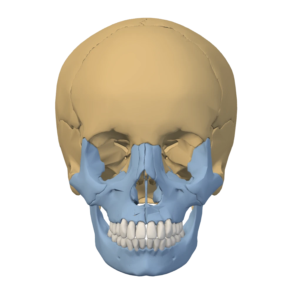
Anterior view. Facing the skull: the patient's left is on your right.
From the front, the boundary runs through the orbits. The forehead and the upper rim of each orbit are frontal bone, a cranial bone. The cheeks, the bridge of the nose and both jaws are facial bones. From the side, the braincase is the large rounded mass above and behind, and the face is the smaller block below and in front of it.
Specimen still
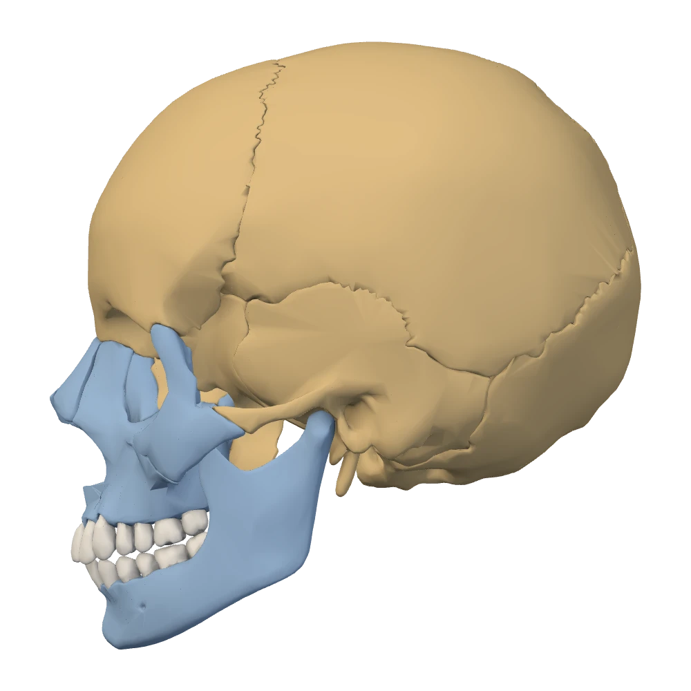
Left lateral view. The patient's left side: the face points to your left.
One landmark shows how closely the two parts are joined. The zygomatic arch, the bar of bone you can feel running from the cheek to just in front of the ear, is half facial and half cranial. Its front part belongs to the zygomatic bone and its back part to the temporal bone.
How to use the specimen beside the text
BodyParts3DThe skull beside this text is a three-dimensional model of one adult skull. All 22 bones are separate pieces, so every suture shows as the seam between two bones, and the teeth are separate from the jaws. Use it as you would a real skull on the bench: look, turn, name, and check.
- Tap a chip. A highlighted name in the text, such as frontal bone, selects that structure on the specimen. A chip for a pair, such as temporal bones, selects both sides. A chip that names a side, such as right temporal bone, selects one.
- Start from a preset. When the text names a view, switch to it before you look. Then rotate a little away from the preset and watch how the outline of the structure changes.
- Read the side. Every preset states where the patient's left is. Check it before you call anything left or right.
- Describe before you move on. Place the structure with two directional terms and one neighbor: “the mastoid process is posterior and inferior to the ear opening, on the temporal bone”.
- Find it a second way. Look for the same structure from another direction. A bone you can recognize in two views is a bone you know.
Knowledge check · 1
Answer, read why, and try again whenever you like. Answers are kept in both study modes.
- Q1Left and right in the anterior viewBodyParts3D
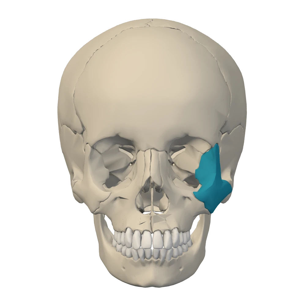
Anterior view. Facing the skull: the patient's left is on your right.
This is the anterior view. One cheekbone (zygomatic bone) is highlighted. On which side of the patient is it?
- Q2Choosing a view: the vault from aboveOpenStax
You want one picture that shows both parietal bones, the suture between them, and the suture that separates them from the frontal bone. Which preset is the best choice?
- Q3External versus internal views of the baseOpenStaxNetter p.17–18
A classmate wants to see the hard palate and the two occipital condyles together. Which view shows them?
- Q4Directional termsOpenStax
Which statement uses directional terms correctly?
- Q5Calvaria versus cranial baseOpenStax
Which of these belongs to the cranial base rather than to the calvaria?
- Q6Neurocranium versus viscerocranium: the ethmoidOpenStax
Which bone is counted with the cranial bones although most of it lies between the orbits and in the walls of the nasal cavity?
- Q7Left and right in the posterior viewBodyParts3DOpenStax
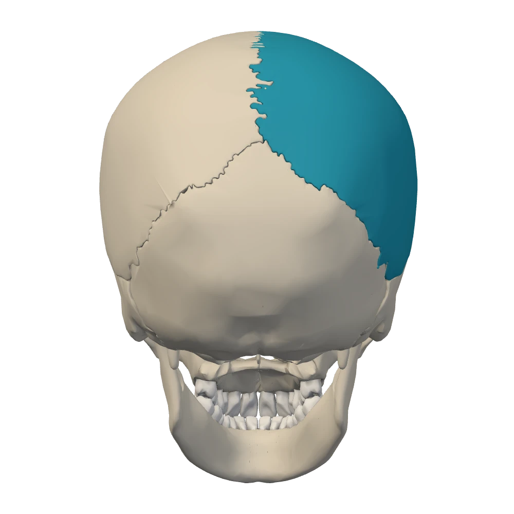
Posterior view. From behind: the patient's left is on your left.
This is the posterior view. Name the highlighted bone.
- Q8Zygomatic arch: one facial bone, one cranial boneOpenStax
The zygomatic arch joins the face to the braincase. Which two bones form it?