The bones one by one, and the sutures that join them
Thirteen short lessons take the skull apart and put it back together. Eleven go through the cranial and facial bones in the same order every time: where the bone is, its parts and landmarks, its neighbors, the best view, and one clinical relationship. The last two reassemble the skull: the mandible with its two movable joints, and the sutures that lock the other 21 bones into one piece.
Frontal bone
OpenStaxNetter p.9–10The frontal bone is the single bone of the forehead. It has two parts set at a right angle. The upright part is the frontal squama (a squama is a thin, scale-like plate of bone): it forms the forehead and the front of the calvaria. At the level of the eyebrows the bone turns backward into two thin horizontal shelves, the orbital plates, one above each eye. Between the two shelves there is a gap in the midline, the ethmoidal notch, which is filled by the ethmoid bone.
Run down the front of the bone in the anterior view. Above each orbit a low curved ridge lies under the eyebrow: the superciliary arch, or brow ridge. Between the two ridges, in the midline, is a smooth area called the glabella. Just below it the frontal bone meets the two nasal bones, and the midline point of that junction is the nasion, the dip at the root of the nose. The upper rim of each orbit is the supraorbital margin. About a third of the way along it from the nose, the rim is nicked by the supraorbital notch. In some skulls a bar of bone closes the notch into a supraorbital foramen, and the two sides of one skull may differ. The supraorbital nerve and vessels turn up onto the forehead here (Openings). At its outer end the margin runs down into the zygomatic process of the frontal bone, which meets the zygomatic bone on the lateral rim of the orbit.
Now the horizontal part. Each orbital plate does two jobs at once. Its lower surface is most of the roof of the orbit; its upper surface is most of the floor of the anterior cranial fossa, where the frontal lobe of the brain rests. One thin plate is all that separates the eye from the brain. Behind the brow ridges the bone is hollow: the two frontal sinuses are air spaces inside it, best understood from a midsagittal section.
Follow the orbital roof out to its edges to see what the plate meets. At the back it joins the lesser wing of the sphenoid, which completes the roof at the narrow back end of the orbit. On the medial side it joins the orbital plate of the ethmoid and, in front of that, the lacrimal bone. On the lateral side it joins the greater wing of the sphenoid and, at the rim, the zygomatic bone.
Neighbors (12 bones). Behind, the two parietal bones at the coronal suture. Between the orbits, the two nasal bones and the two maxillae. In and around each orbit, the lacrimal and zygomatic bones. In the base, the sphenoid and the ethmoid. Best view: anterior for the squama, glabella and supraorbital margins; internal cranial base for the two orbital plates with the ethmoid between them.
Parietal bones
OpenStaxNetter p.11–12The two parietal bones form most of the roof and the upper side walls of the calvaria. Each is a curved, four-sided plate, like a square sheet bent over the top of the head, so it has four borders and four corners. Learn the borders and you have learned three of the four named sutures of the vault.
The four borders of a parietal boneOpenStaxNetter p.11–12
| Border | Meets | Suture |
|---|---|---|
| Upper (sagittal) | The other parietal bone, in the midline | Sagittal suture |
| Front (frontal) | The frontal bone | Coronal suture |
| Back (occipital) | The occipital bone | Lambdoid suture |
| Lower (squamous) | From front to back: the greater wing of the sphenoid, the squamous part of the temporal bone, then its mastoid region | Mainly the squamous suture, where the temporal bone overlaps the parietal |
Upper (sagittal)
- Meets
- The other parietal bone, in the midline
- Suture
- Sagittal suture
Front (frontal)
- Meets
- The frontal bone
- Suture
- Coronal suture
Back (occipital)
- Meets
- The occipital bone
- Suture
- Lambdoid suture
Lower (squamous)
- Meets
- From front to back: the greater wing of the sphenoid, the squamous part of the temporal bone, then its mastoid region
- Suture
- Mainly the squamous suture, where the temporal bone overlaps the parietal
Specimen still
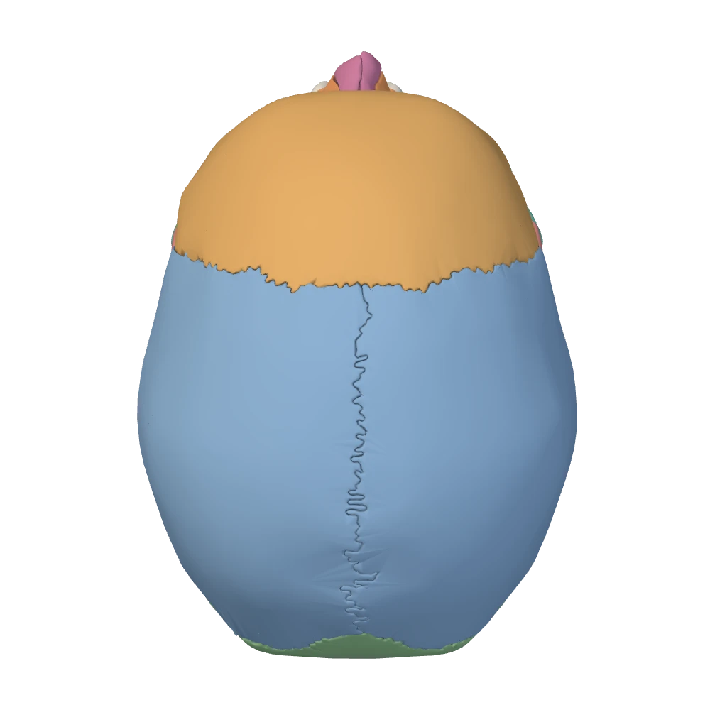
Superior view. From above: the face is toward the top, the patient's right is on your right.
Each corner lies at a named meeting point of sutures. The front upper corner is at the bregma and the back upper corner at the lambda, both on the midline. The front lower corner reaches down to the sphenoid at the pterion. The back lower corner meets the occipital and temporal bones at the asterion. These points are described with the sutures.
The outer surface is smooth and convex. Its most prominent point is the parietal eminence (parietal tuber), often the widest part of the head. Lower down, two faint curved ridges arch across the bone from front to back, one above the other: the superior temporal line and the inferior temporal line. They begin on the frontal bone behind the orbit, sweep back over the parietal bone, and turn down toward the temporal bone above the ear. The fascia that covers the temporalis muscle attaches to the upper line, and the muscle itself arises from the bone below the lower line. The lines are low, and often easier to feel on a real skull than to see: look for them as an arc across the side of the parietal bone, a little below the parietal eminence.
The lines mark a boundary. Below them is the temporal fossa, the shallow hollow of the temple, which is filled by the temporalis muscle. Above them the bone is covered only by the scalp. So the lower part of the parietal bone belongs to the temporal region, even though it is not part of the temporal bone.
Neighbors (5 bones): the other parietal, the frontal, the occipital, the temporal and the sphenoid. Best view: superior, for both bones with the sagittal suture between them; lateral, for one bone with all four borders.
Occipital bone
OpenStaxNetter p.17–18Netter p.11–12The occipital bone is the single bone at the back and underside of the braincase. It is built around a large oval opening, the foramen magnum (“great opening”), where the brainstem becomes the spinal cord. Its parts are named by where they lie around that opening: the broad curved squamous part behind and above it, the thick basilar part in front of it, and a lateral part on each side.
The squamous part, from behind. In the posterior view, find the midline bump roughly halfway up the bone, where the back of the head meets the neck: the external occipital protuberance. A ridge curves outward from it on each side toward the mastoid process: the superior nuchal line (nucha means the nape of the neck). The muscles of the back of the neck attach along and below this line; above it the bone is covered only by scalp. A second, fainter ridge, the inferior nuchal line, runs parallel to the first, about halfway between it and the foramen magnum. The smooth area above the superior nuchal lines belongs to the calvaria. The rougher area below them faces downward and belongs to the base.
The lateral parts, from below. In the inferior view, look on each side of the front half of the foramen magnum for a smooth oval knob: the occipital condyle. The two condyles sit in matching hollows on the first cervical vertebra, the atlas, and the head nods on these joints. The atlas itself belongs to the vertebral column unit; here, note only that the whole skull rests on the neck at these two small surfaces. Each lateral part is tunneled above its condyle by the hypoglossal canal, which carries the nerve to the muscles of the tongue. The canal begins inside the skull just above the rim of the foramen magnum and runs forward and outward to open above the front of the condyle. Lateral to the condyle the edge of the bone is notched, and with the temporal bone this notch completes the jugular foramen. What passes through these openings is followed in Openings.
The basilar part, from inside. In front of the foramen magnum the bone narrows to a thick bar that runs forward and upward to fuse with the body of the sphenoid. In a midsagittal section its upper surface is a smooth ramp, sloping down from the back of the sphenoid to the foramen magnum. This slope is the clivus (Latin for slope). The basilar part of the occipital bone forms its lower portion and the sphenoid its upper portion, and the brainstem lies against it.
The inner surface of the squamous part is divided by a cross-shaped ridge into four hollows. The lower two form the floor of the posterior cranial fossa and hold the cerebellum.
Neighbors (6 bones): the two parietal bones at the lambdoid suture, the two temporal bones at the sides, the sphenoid in front, and the atlas below. Best view: posterior for the squamous part; inferior for the foramen magnum and condyles; midsagittal for the clivus.
Temporal bones
OpenStaxNetter p.11–12Netter p.17–18Netter p.13–14Each temporal bone forms the lower part of the side wall of the braincase and a wedge of the base beneath it. It contains the organs of hearing and balance, and it carries the socket for the lower jaw. The easiest way to learn it is to stand at the ear opening, the external acoustic meatus (a meatus is a passage; older books say external auditory meatus), and name the part that lies in each direction.
The temporal bone, part by part, around the ear openingOpenStaxNetter p.11–12
| Part | Where it lies | What it carries |
|---|---|---|
| Squamous part | Above the ear opening: the temporal squama, a thin fan-shaped plate in the side wall of the vault | Its upper edge overlaps the parietal bone at the squamous suture. The zygomatic process springs from its lower part and runs forward |
| Tympanic part | Below and in front of the ear opening: the tympanic part, a small curved plate | It forms the front wall and floor of the external acoustic meatus and the back wall of the socket for the jaw |
| Mastoid region | Behind the ear opening | The mastoid process, the blunt cone you can feel behind the earlobe. It contains air cells that open into the middle ear |
| Petrous part | Medial to the ear opening: the petrous part, a dense pyramid of bone that points forward and inward into the base | The middle ear and inner ear, and the canal for the internal carotid artery. Its upper edge is the ridge between the middle and posterior cranial fossae |
| Styloid process | Below the ear opening: the styloid process, a slender spike pointing down and forward | Ligaments and small muscles of the tongue, throat and hyoid attach to it |
Squamous part
- Where it lies
- Above the ear opening: the temporal squama, a thin fan-shaped plate in the side wall of the vault
- What it carries
- Its upper edge overlaps the parietal bone at the squamous suture. The zygomatic process springs from its lower part and runs forward
Tympanic part
- Where it lies
- Below and in front of the ear opening: the tympanic part, a small curved plate
- What it carries
- It forms the front wall and floor of the external acoustic meatus and the back wall of the socket for the jaw
Mastoid region
- Where it lies
- Behind the ear opening
- What it carries
- The mastoid process, the blunt cone you can feel behind the earlobe. It contains air cells that open into the middle ear
Petrous part
- Where it lies
- Medial to the ear opening: the petrous part, a dense pyramid of bone that points forward and inward into the base
- What it carries
- The middle ear and inner ear, and the canal for the internal carotid artery. Its upper edge is the ridge between the middle and posterior cranial fossae
Styloid process
- Where it lies
- Below the ear opening: the styloid process, a slender spike pointing down and forward
- What it carries
- Ligaments and small muscles of the tongue, throat and hyoid attach to it
Books divide this bone differently. Some name three parts (squamous, tympanic, petrous) and include the mastoid process with the petrous part; others list a separate mastoid part or a combined petromastoid part. The bone is the same; only the grouping differs.
The zygomatic process is the handle-like bar that runs forward from the squamous part to meet the zygomatic bone; together they make the zygomatic arch. Turn to the inferior view and look under the root of the process. The smooth hollow there is the mandibular fossa, which receives the head of the mandible, and the rounded ridge just in front of it is the articular tubercle. Together they are the temporal half of the jaw joint, described under the temporomandibular joint.
Four openings belong to this bone, and each is found from a different direction. Their contents are followed in Openings; here, learn where each one is.
Openings of the temporal bone and where to look for themOpenStaxNetter p.19–20
| Opening | Where to look | View |
|---|---|---|
| External acoustic meatus | On the side of the skull, below the root of the zygomatic process and behind the mandibular fossa | Lateral |
| Internal acoustic meatus | On the back surface of the petrous part, facing the posterior cranial fossa | Internal cranial base |
| Stylomastoid foramen | On the underside, between the styloid process and the mastoid process | Inferior |
| Carotid canal | Its outer opening is on the underside of the petrous part, in front of the jugular foramen and medial to the styloid process | Inferior |
External acoustic meatus
- Where to look
- On the side of the skull, below the root of the zygomatic process and behind the mandibular fossa
- View
- Lateral
- Where to look
- On the back surface of the petrous part, facing the posterior cranial fossa
- View
- Internal cranial base
- Where to look
- On the underside, between the styloid process and the mastoid process
- View
- Inferior
- Where to look
- Its outer opening is on the underside of the petrous part, in front of the jugular foramen and medial to the styloid process
- View
- Inferior
A fifth opening, the jugular foramen, is not in the temporal bone but beside it: it is the gap between the petrous part and the occipital bone.
Neighbors (5 bones): the parietal bone above, the occipital bone behind, the sphenoid in front, the zygomatic bone at the arch, and the mandible at the jaw joint, the only movable one. Best view: lateral for the squamous part, zygomatic process, ear opening and mastoid process; inferior for the fossa, the styloid process and the openings of the underside; internal cranial base for the petrous part.
Sphenoid bone
OpenStaxNetter p.13–14Netter p.15–16Netter p.17–18Netter p.19–20The sphenoid bone is a single bone wedged into the middle of the cranial base, and it touches every other cranial bone. Its shape is easiest to hold as a bat with its wings spread: a central body, two pairs of wings reaching sideways, and two legs hanging down. The small upper wings are the lesser wings, the large lower wings are the greater wings, and the legs are the pterygoid processes. Because the bone is buried in the center of the skull, no single view shows all of it. Build it up from four views.
From inside (internal cranial base): the body and the wings. The body is a block of bone in the midline, hollowed by the two sphenoidal air sinuses. Its upper surface is shaped like a saddle and is called the sella turcica (“Turkish saddle”). The seat of the saddle is a rounded hollow, the hypophyseal fossa, which holds the pituitary gland (the hypophysis). In front of the seat is a low ridge. Behind it stands a square plate of bone, the back of the saddle (dorsum sellae), which slopes away behind into the clivus.
The lesser wings are two thin, pointed plates that spread sideways from the top front of the body. Their sharp back edge is the step between the anterior and middle cranial fossae. Each lesser wing is attached to the body by two small roots, and the optic canal passes between them: to find the canal from inside, look just in front of the sella, on either side of the midline. The greater wings arise lower down, from the sides of the body, and curve outward and upward to form the front part of the floor of the middle cranial fossa.
The openings, in order. The gap between the lesser wing above and the greater wing below is a slanting slit, the superior orbital fissure, which opens forward into the orbit. Behind it, three holes pierce the root of each greater wing in a curved line running backward and outward: first the foramen rotundum (round), then the larger foramen ovale (oval), then the small foramen spinosum at the back corner of the wing. The three do not point the same way. The foramen rotundum runs forward, so its far end cannot be seen on the underside of the skull. The foramen ovale and the foramen spinosum run downward and are found again in the inferior view, behind the root of the lateral pterygoid plate. What each opening transmits is the subject of Openings.
From the side (lateral view): the greater wing in the temple. The outer surface of the greater wing reaches the side of the skull as a patch of bone in the temple. It lies behind the frontal and zygomatic bones and in front of the temporal squama, and its upper end touches the parietal bone at the pterion.
From the front (anterior view, into the orbit): the back of the orbit. The greater wing forms the back part of the lateral wall of the orbit, and the lesser wing forms the back of its roof. The optic canal and the superior orbital fissure open at the apex, the narrow back end of the orbit.
From below (inferior view): the pterygoid processes. Each pterygoid process hangs down from the junction of the body and a greater wing, just behind the upper jaw. It divides into two plates. The medial pterygoid plate is narrow, forms the back end of the side wall of the nasal cavity, and ends below in a small hook. The lateral pterygoid plate is broader and faces outward; two of the muscles that move the jaw, the lateral and medial pterygoid muscles, arise from its two surfaces.
Neighbors (12 bones): all seven other cranial bones (the frontal, both parietal, both temporal, the occipital and the ethmoid) and five facial bones (both zygomatic, both palatine and the vomer). Best view: internal cranial base for the body, sella turcica and wings; then lateral, anterior and inferior for the parts described above.
Ethmoid bone
OpenStaxNetter p.13–14Netter p.15–16The ethmoid bone is a single, light, fragile bone hidden in the middle of the face, between the two orbits. It forms the roof and the upper side walls of the nasal cavity. It is counted as a cranial bone because its top surface is part of the floor of the anterior cranial fossa. Its name means sieve-like, after the perforated plate on top.
Its shape is simple once you see the plan. A horizontal plate lies across the top. A vertical plate crosses it in the midline, standing up above it and hanging down below it. And from each side edge of the horizontal plate hangs a box of thin-walled air cells.
- The horizontal plate is the cribriform plate: a narrow strip in the midline of the anterior cranial fossa, set into the gap between the two orbital plates of the frontal bone. It is pierced by many tiny holes for the filaments of the olfactory nerve, which pass from the nose to the brain. The holes are too small to pick out one by one: learn them as a perforated region on each side of the midline. The same plate is the roof of the nasal cavity.
- The vertical plate above is the crista galli (“cock's comb”): a midline crest that stands up from the cribriform plate into the cranial cavity. A fold of dura, the falx cerebri, is anchored to it.
- The vertical plate below is the perpendicular plate: it hangs down into the nose and forms the upper part of the bony nasal septum, the partition between the right and left halves of the nasal cavity.
- The two boxes are the ethmoidal labyrinths. Each lies between the nasal cavity and an orbit and is full of small air spaces, the ethmoidal cells. Its outer wall is a smooth, paper-thin sheet, the orbital plate, which forms most of the medial wall of the orbit. Its inner wall faces the nasal cavity and carries two curled shelves of bone: the small superior nasal concha high at the back, and the larger middle nasal concha below it.
Neighbors (13 bones): two cranial bones, the frontal above and the sphenoid behind, and eleven facial bones: both nasal, both maxillae, both lacrimal, both palatine, both inferior nasal conchae, and the vomer. Best view: internal cranial base for the cribriform plate and crista galli; anterior, looking into the orbit for the orbital plate and into the nasal opening for the perpendicular plate and middle concha; midsagittal for the perpendicular plate in the septum.
Maxillae
OpenStaxNetter p.9–10Netter p.15–16Netter p.17–18The two maxillae (singular maxilla) form the upper jaw and the center of the face. Between them they reach the orbits, the nose and the mouth, and each one touches every other facial bone except the mandible. A maxilla is a hollow body with four processes growing out of it.
The body is the block of bone beside the nose and below the orbit. It is hollow: almost all of it is occupied by an air space, the maxillary sinus. Each of its surfaces faces a different region. The front surface lies under the cheek. The upper surface is most of the floor of the orbit. The inner surface is part of the side wall of the nasal cavity. In the anterior view, the lower rim of the orbit is the infraorbital margin; the maxilla forms its inner half and the zygomatic bone its outer half. About a finger's breadth below the margin, on the front of the body, is the infraorbital foramen, where the infraorbital nerve and vessels come out onto the face (Openings).
- Frontal process. It climbs beside the nose to reach the frontal bone, with the nasal bone in front of it and the lacrimal bone behind. It forms the inner rim of the orbit.
- Zygomatic process. A short, thick buttress that runs outward to meet the zygomatic bone.
- Alveolar process. The alveolar process is the curved ridge that hangs down from the body and carries the sockets for the upper teeth, eight on each side in a full adult set.
- Palatine process. The palatine process is a horizontal shelf that runs inward to meet its partner in the midline. The two shelves form the larger, front part of the hard palate, which is both the roof of the mouth and the floor of the nasal cavity. The palatine bones complete it behind. In the midline just behind the incisor teeth is the incisive foramen.
Where the two maxillae meet below the nasal opening, a small sharp spur of bone points forward in the midline: the anterior nasal spine. The pear-shaped nasal opening of the skull is framed by the two nasal bones above and by the two maxillae at the sides and below.
The maxillary sinus cannot be seen on an intact skull, so learn it by its walls. Its roof is the floor of the orbit. Its floor is the alveolar process, where the roots of the upper back teeth lie just beneath it. Its inner wall is the side wall of the nose, and its only opening is high on that wall, well above the floor of the sinus. It is the largest of the air sinuses around the nose.
Neighbors (9 bones): the frontal and ethmoid bones above; the nasal, lacrimal and zygomatic bones around the nose and orbit; the palatine bone, the inferior nasal concha and the vomer inside the nose and palate; and the other maxilla. Best view: anterior for the body, the processes and the infraorbital margin; inferior for the palatine processes in the hard palate.
Fracture lines of the midface (Le Fort)More detail
Severe blows to the face break the upper jaw along three typical levels, each higher than the last. They are worth knowing here because each line is defined by the bones it crosses.
- Le Fort I is horizontal. It separates the tooth-bearing part of the upper jaw from the rest of the maxillae along the floor of the nose.
- Le Fort II is shaped like a pyramid. It passes through both maxillae, the nasal bones, the infraorbital margins and the floors of the orbits.
- Le Fort III runs higher still, across the root of the nose and through the orbits to the zygomatic bones, so that the whole facial skeleton comes away from the cranial base.
Zygomatic bones and the zygomatic arch
OpenStaxNetter p.9–10Netter p.11–12Netter p.17–18Each zygomatic bone is a cheekbone (older name: malar bone). It makes the prominence of the cheek, the outer and lower rim of the orbit, and the front part of the orbit's lateral wall. It is a small, strong, four-cornered bone that braces the upper jaw against the side of the skull.
Think of it as a diamond with an arm reaching toward each neighbor. The rule for the names is worth learning once, because it applies all over the skeleton: a process is named for the bone it reaches toward.
- The frontal process runs up the outer rim of the orbit to meet the frontal bone. You can feel the junction as a slight dip at the outer end of the eyebrow.
- The temporal process runs backward to meet the zygomatic process of the temporal bone. They join at a slanting suture, and together they make the zygomatic arch.
- Medially, a broad edge joins the zygomatic process of the maxilla. Here the zygomatic bone forms the outer half of the infraorbital margin.
- Inside the orbit, a plate of the bone turns backward to form the front of the lateral wall and a little of the floor, and meets the greater wing of the sphenoid.
Specimen still
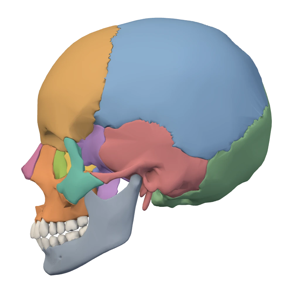
Left lateral view. The patient's left side: the face points to your left.
The zygomatic arch is the bar you can feel from the cheek to just in front of the ear. Its shorter front part is zygomatic bone and its longer back part is temporal bone. It stands away from the side of the skull, leaving a gap. The temporalis muscle passes down through that gap to reach the coronoid process of the mandible, and the masseter muscle hangs from the lower edge of the arch. The hollow above the arch is the temporal fossa; the space below and deep to it is the infratemporal fossa.
Neighbors (4 bones): frontal, sphenoid, temporal and maxilla. Best view: anterior for the cheek prominence and the orbital rim; lateral for the temporal process and the arch in profile; inferior to see how far the arch stands out from the skull.
Nasal and lacrimal bones
OpenStaxNetter p.9–10Netter p.11–12Netter p.15–16This lesson covers four small bones in two pairs. They are easy to tell apart by position: the nasal bones are on the outside of the nose, and the lacrimal bones are just inside the orbits.
Nasal bones. The two nasal bones are small oblong plates that lie side by side and form the bony bridge of the nose. Each one meets its partner in the midline, the frontal bone above at the nasion, and the frontal process of the maxilla at its outer edge. Their lower edges are free and form the top of the nasal opening of the skull. The rest of the external nose is cartilage, which is why a dry skull has a large pear-shaped hole where the nose was. Each nasal bone has four bony neighbors: the other nasal bone, the frontal bone, the maxilla, and the ethmoid, which supports it from behind.
Lacrimal bones. Each lacrimal bone is a thin plate about the size of a small fingernail, the smallest and most fragile bone of the face. It lies at the front of the medial wall of the orbit. To find it, start at the inner rim of the orbit, which is the frontal process of the maxilla, and step just behind it: the first small plate you meet is the lacrimal bone. Behind it is the orbital plate of the ethmoid, above it the frontal bone, and below it the maxilla in the floor of the orbit. Its four bony neighbors are the frontal bone, the ethmoid, the maxilla and the inferior nasal concha.
The lacrimal groove. A vertical ridge runs down the lacrimal bone, and in front of the ridge the bone is hollowed into the lacrimal groove. The back edge of the frontal process of the maxilla carries a matching groove. Put together, the two grooves make a hollow at the inner corner of the orbit that holds the lacrimal sac, the small reservoir that collects tears. From the bottom of the hollow a bony canal, the nasolacrimal canal, leads down into the nasal cavity and opens beneath the inferior nasal concha. This is the path tears take from the eye to the nose, and it is why the nose runs when you cry.
Best view: anterior. The nasal bones are at the bridge of the nose. For each lacrimal bone, look into the orbit at the front of its medial wall. A lateral view shows the nasal bone in profile and the lacrimal bone through the opening of the orbit.
Palatine bones
OpenStaxNetter p.13–14Netter p.15–16Netter p.17–18Each palatine bone is a small L-shaped bone at the back of the nasal cavity, wedged between the maxilla in front and the pterygoid process of the sphenoid behind. The foot of the L lies flat in the palate. The upright of the L stands in the side wall of the nose. Most of the bone is hidden; on an assembled skull only the foot is easy to see.
- The foot is the horizontal plate. It meets its partner in the midline and the palatine process of the maxilla in front, and the two horizontal plates form the back part of the hard palate. Their back edge is free: it is the back border of the hard palate, where the soft palate is attached in life.
- The upright is the perpendicular plate. It is a thin sheet that rises from the outer edge of the horizontal plate and forms the back part of the side wall of the nasal cavity, between the maxilla in front and the medial pterygoid plate behind.
- At the top of the perpendicular plate are two small processes. One of them, the orbital process, tilts forward and outward to reach the very back of the floor of the orbit. It is the smallest of the seven bony contributions to the orbit.
Specimen still
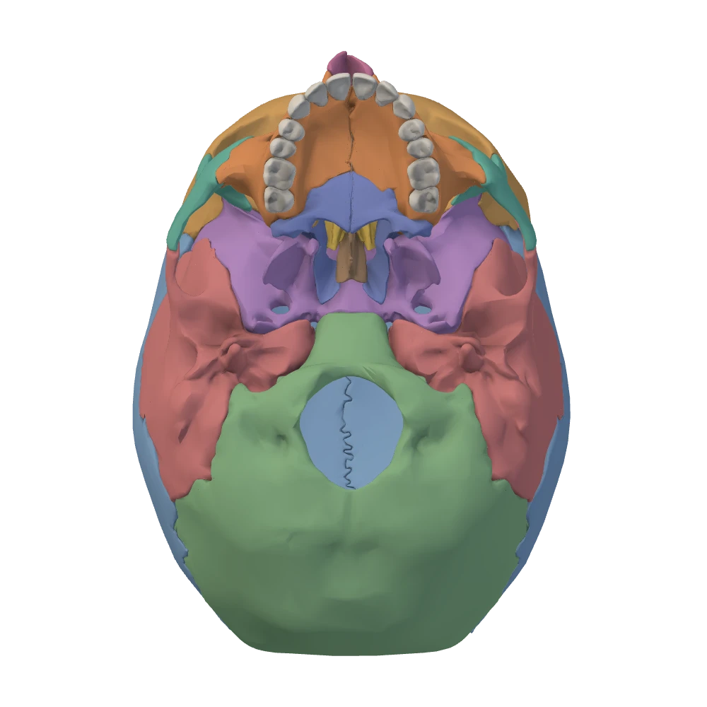
Inferior (external base) view. From below, mandible removed: the face is toward the top, the patient's left is on your right.
The greater palatine region. In the inferior view, look at the back outer corner of the hard palate, beside the last molar tooth. Here, where the horizontal plate meets the maxilla, is the greater palatine foramen. Just behind it are the smaller lesser palatine foramina. Nerves and vessels reach the palate through them: those of the greater foramen run forward to the hard palate, and those of the lesser foramina turn back to the soft palate (Openings).
Neighbors (6 bones): the other palatine bone, the maxilla, the sphenoid, the ethmoid, the inferior nasal concha and the vomer. Best view: inferior for the horizontal plates at the back of the palate. The perpendicular plate is seen only on the side wall of the nasal cavity, in a skull cut in the midline with the septum removed.
Inferior nasal conchae and vomer
OpenStaxNetter p.13–14Netter p.15–16Netter p.17–18Two kinds of bone remain inside the nasal cavity, and they complete the 22: a pair of inferior nasal conchae on the side walls, and the single vomer in the midline.
Inferior nasal conchae. Each inferior nasal concha is a separate bone: a thin, curled shelf attached along the lower part of the side wall of the nasal cavity, with its free edge rolling downward and inward like a scroll (concha means shell). It is the largest of the three conchae on each side and the only one that is a bone in its own right; the middle and superior conchae above it are parts of the ethmoid. Covered with its lining in life, it is what a clinician calls the inferior turbinate. The conchae enlarge the surface of the nasal lining and stir the air that passes over it, which warms, moistens and cleans the air on its way to the lungs. The channel beneath each concha is called a meatus, and the tear passage opens into the one beneath the inferior concha.
Each inferior concha has four neighbors: the maxilla and the palatine bone, to which it is attached, and the lacrimal bone and the ethmoid, which it touches above.
Vomer. The vomer is a single thin, flat plate standing upright in the midline. It forms the lower and back part of the bony nasal septum. Take its four edges in turn. The upper edge is thick and grips the underside of the body of the sphenoid. The lower edge stands on the midline ridge of the hard palate, on the maxillae and the palatine bones. The back edge is free: it is the sharp partition between the two back openings of the nasal cavity, and it is the part you see in the inferior view. The front edge slopes downward and forward. Its upper half is joined to the perpendicular plate of the ethmoid, and its lower half is grooved to hold the septal cartilage.
So the septum has three main pieces. The perpendicular plate of the ethmoid is the upper part, the vomer is the lower back part, and the septal cartilage fills in the front. Cartilage does not survive in a dry skull, which is why the bony septum seen through the nasal opening looks as if a piece is missing in front.
The vomer has six neighbors: the sphenoid, the ethmoid, both maxillae and both palatine bones. Best view: for the inferior conchae, anterior, looking into the nasal opening at the lower part of each side wall. For the vomer, inferior, where its back edge divides the two openings behind the hard palate; a midsagittal section shows the whole plate.
Mandible and the temporomandibular joint
OpenStaxNetter p.21–22Netter p.23–24Netter p.25–26The mandible is the lower jaw. It is the largest and strongest bone of the face and the only bone of the skull that moves. It is also the only one of the 22 that is not held to its neighbors by sutures: it hangs from the two temporal bones at a pair of movable joints. It begins as a right and a left half, which fuse at the chin during the first year of life. Seen from above, the adult bone is a horseshoe. Seen from the side, each half is an L: a horizontal body that carries the teeth and an upright ramus that reaches up to the skull.
The body. The body is the curved horizontal bar. Its upper edge is the alveolar part, also called the alveolar process, which holds the sockets for the lower teeth, 16 in a full adult set. Its lower edge is the thick rounded border you can feel along your jawline. In the midline at the front, where the two halves fused, the adult has no joint, but the place keeps the name mandibular symphysis and may be marked by a faint vertical ridge. Below the ridge the bone swells forward as the mental protuberance, the point of the chin. Mental here comes from the Latin mentum, chin; it has nothing to do with the mind.
On the outer surface of the body, on each side, is the mental foramen. Look below the premolar teeth, usually below the second one, about halfway between the upper and lower edges of the bone. The mental nerve and vessels come out here to supply the chin and the lower lip.
The angle and the ramus. Follow the lower edge of the body backward until it turns upward. The corner is the angle of the mandible, which you can feel below and a little in front of your earlobe. The broad flat plate that rises from it is the ramus (Latin for branch; plural rami). Two strong chewing muscles grip the ramus and the angle between them: the masseter on the outer surface and the medial pterygoid on the inner surface. The upper edge of the ramus carries two processes with a notch between them.
- Coronoid process. The front one is the coronoid process: thin, flat and pointed. It forms no joint. The temporalis muscle comes down deep to the zygomatic arch and attaches to it, so with the jaw closed its tip is tucked in under the arch.
- Condylar process. The back one is the condylar process. It has a narrow neck and, on top, the head of the mandible (the condyle): a smooth knob like a short roller set crosswise, wider from side to side than from front to back. The head is the mandible's half of the jaw joint.
- Mandibular notch. The curved dip between the two processes is the mandibular notch.
Specimen still
Left lateral view. The patient's left side: the face points to your left.
The inner surface and the mandibular canal. The mandible has a second pair of openings, on the opposite face of the bone. Near the middle of the inner (medial) surface of each ramus is the mandibular foramen, with a small tongue of bone, the lingula, overlapping its front edge. This surface faces the midline, so the anterior and lateral views do not show it: look between the two rami from behind and below. The foramen is the mouth of the mandibular canal, a tunnel inside the bone. The canal runs down the ramus and then forward through the body, just beneath the roots of the teeth, toward the mental foramen. Because it is buried in the bone it cannot be seen on the specimen. Picture it as the line that joins the two openings: in on the inside of the ramus, out on the outside of the body.
What runs through the canal
- The mandibular nerve (CN V3) leaves the cranial cavity through the foramen ovale and gives off the inferior alveolar nerve.
- The inferior alveolar nerve, with an artery and a vein of the same name, enters the mandibular foramen.
- In the mandibular canal they supply every lower tooth of that side.
- Near the premolar teeth the nerve divides. A small branch continues forward inside the bone to the front teeth.
- The other branch, the mental nerve, comes out through the mental foramen to the skin of the chin and the lower lip.
This path explains a routine of dentistry. Local anesthetic placed beside the mandibular foramen, just before the nerve enters the bone, numbs all the lower teeth of that side with one injection, and the chin and lower lip of that side go numb with them. What passes through each opening of the skull is followed in Openings.
The temporomandibular joints. Each condylar process ends at a temporomandibular joint (TMJ), just in front of the ear opening. These two joints are the only movable joints between the 22 bones of the skull. You can feel one at work: put a fingertip on your face just in front of the opening of your ear and open your mouth wide. The head of the mandible slides forward from under your finger and leaves a hollow behind it.
The parts of the temporomandibular jointOpenStaxNetter p.25–26
| Part | Belongs to | What to know |
|---|---|---|
| Head of the mandible | The condylar process of the mandible | The lower joint surface: a roller set crosswise |
| Mandibular fossa | The temporal bone, under the root of its zygomatic process | The hollow that holds the head when the mouth is closed |
| Articular tubercle | The temporal bone, immediately in front of the fossa | A rounded ridge. In wide opening the head slides forward out of the fossa and down the slope of the ridge, as far as its summit |
| Articular disc | Neither bone: a plate of tough fibrocartilage between them, attached around its edge to the joint capsule | It divides the joint into an upper and a lower compartment and travels with the head of the mandible |
| Capsule and ligaments | The soft tissue around the joint | A loose capsule, thickened on its outer side as the lateral ligament. The sphenomandibular and stylomandibular ligaments run from the base of the skull to the mandible |
Head of the mandible
- Belongs to
- The condylar process of the mandible
- What to know
- The lower joint surface: a roller set crosswise
- Belongs to
- The temporal bone, under the root of its zygomatic process
- What to know
- The hollow that holds the head when the mouth is closed
- Belongs to
- The temporal bone, immediately in front of the fossa
- What to know
- A rounded ridge. In wide opening the head slides forward out of the fossa and down the slope of the ridge, as far as its summit
Articular disc
- Belongs to
- Neither bone: a plate of tough fibrocartilage between them, attached around its edge to the joint capsule
- What to know
- It divides the joint into an upper and a lower compartment and travels with the head of the mandible
Capsule and ligaments
- Belongs to
- The soft tissue around the joint
- What to know
- A loose capsule, thickened on its outer side as the lateral ligament. The sphenomandibular and stylomandibular ligaments run from the base of the skull to the mandible
Only the three bony parts can be found on a skull. The disc, the capsule and the ligaments are soft tissue and are described here in words.
Two compartments, two movements. The disc turns one joint into two. In the lower compartment, between the head of the mandible and the disc, the head rotates like a hinge. In the upper compartment, between the disc and the temporal bone, the disc and the head glide together, forward and backward along the temporal bone. A small opening of the mouth is mostly hinge movement. A wide opening needs both. Pushing the chin forward and drawing it back are gliding alone. In chewing the two sides take turns: one head glides forward while the other stays back and pivots, which swings the chin toward the side that stayed back.
Opening the mouth wide, step by step
- Mouth closed: each head rests in its mandibular fossa, with the disc between head and bone.
- Opening begins: each head rotates like a hinge beneath its disc (lower compartment).
- Opening continues: disc and head glide forward together out of the fossa (upper compartment), while the head goes on rotating.
- Mouth wide open: disc and head lie beneath the summit of the articular tubercle, and the fossa behind them is empty.
- Closing: the same steps in reverse. Disc and head glide back, and the head rotates up into the fossa.
The mandible is one bone with a joint at each end, so the right and left joints can never move separately: every movement of the jaw uses both. The specimen shows the bones in one position only, which is why the movements are described here in words and not animated.
Specimen still
Inferior (external base) view. From below, mandible removed: the face is toward the top, the patient's left is on your right.
Neighbors (2 bones): the right and left temporal bones, at the two temporomandibular joints. The mandible touches no other bone. Its teeth meet the upper teeth, but that is tooth against tooth, not bone against bone. Best view: lateral for the body, angle, ramus, both processes and the notch; anterior for the mental protuberance and the two mental foramina; inferior, with the lower jaw removed as in the still above, for the two fossae and tubercles on the temporal bones.
More landmarks on the inner surfaceMore detail
- Lingula. The small tongue of bone at the front edge of the mandibular foramen. A ligament from the sphenoid, the sphenomandibular ligament, attaches to it.
- Mylohyoid line. A slanting ridge along the inner surface of the body. The mylohyoid muscle, which forms the floor of the mouth, arises from the line on each side.
- Sublingual and submandibular fossae. Two shallow hollows, one above the front of the mylohyoid line and one below the back of it, where the sublingual and submandibular salivary glands lie against the bone.
- Mental spines. Small points in the midline behind the chin, for muscles of the tongue and the floor of the mouth.
Fractures of the mandibleMore detail
- The mandible is the second most often fractured bone of the face, after the nasal bones.
- It is a rigid arch fixed to the skull at both ends, so a single blow often breaks it in two places. One fracture line is a reason to look for a second.
- Common sites are the region of the canine tooth and the region just in front of the third molar. The slender neck of the condylar process is another weak point.
Sutures, bregma, lambda and the pterion
OpenStaxNetter p.11–12BodyParts3DEvery bone has now had its turn. This lesson is about the lines between them. A suture is a kind of joint found only in the skull: two bones meet edge to edge and are bound together by a thin layer of dense fibrous tissue. There is no joint cavity and, in the adult, no movement. That makes a suture a fibrous joint, the opposite of the temporomandibular joint in the lesson before, which has a cavity and moves freely.
In the vault the meeting edges are not straight. They are cut into small teeth that interlock like the two sides of a zipper, so the bones cannot slide or be pulled apart. In a child the sutures are also where the vault grows: new bone is laid down along the edges as the brain enlarges. From early adult life the fibrous tissue slowly turns to bone, and in an old skull some sutures can no longer be traced.
The four named sutures of the vaultOpenStaxNetter p.11–12
| Suture | Bones it joins | Where it runs | The name |
|---|---|---|---|
| Coronal suture | The frontal bone and the two parietal bones | From side to side over the top of the skull, from one temple to the other | Corona, a crown or garland: it crosses the head where a headband sits |
| Sagittal suture | The right and left parietal bones | Along the midline of the vault, from front to back | Sagitta, an arrow |
| Lambdoid suture | The two parietal bones and the occipital bone | An inverted V across the back of the skull | The Greek capital letter lambda, Λ |
| Squamous suture, one on each side | The squamous part of the temporal bone and the parietal bone above it | An arch on the side of the skull, above the ear | Squama, a scale: the thin edge of the temporal bone overlaps the parietal bone like a scale |
- Bones it joins
- The frontal bone and the two parietal bones
- Where it runs
- From side to side over the top of the skull, from one temple to the other
- The name
- Corona, a crown or garland: it crosses the head where a headband sits
- Bones it joins
- The right and left parietal bones
- Where it runs
- Along the midline of the vault, from front to back
- The name
- Sagitta, an arrow
- Bones it joins
- The two parietal bones and the occipital bone
- Where it runs
- An inverted V across the back of the skull
- The name
- The Greek capital letter lambda, Λ
Squamous suture, one on each side
- Bones it joins
- The squamous part of the temporal bone and the parietal bone above it
- Where it runs
- An arch on the side of the skull, above the ear
- The name
- Squama, a scale: the thin edge of the temporal bone overlaps the parietal bone like a scale
The coronal suture lies in a coronal plane and the sagittal suture in the midsagittal plane. The planes and the sutures share their names.
Every other suture is named for its two bones. Only these four have names of their own. The rest take the names of the bones on either side: the frontonasal suture between the frontal and nasal bones, the sphenoparietal suture between the sphenoid and the parietal bone, the occipitomastoid suture between the occipital bone and the mastoid region of the temporal bone. So the test for any suture is always the same: name the bone on each side of the line.
Where sutures meet. Four meeting places have names. They are the four corners of the parietal bone. Two are single points on the midline. The bregma is on top of the head, where the sagittal suture meets the coronal suture: three bones meet there, the frontal and both parietals. The lambda is at the back, where the sagittal suture meets the lambdoid suture: three bones again, both parietals and the occipital. The other two are paired, one on each side of the head. The pterion is in the temple, at the lower end of the coronal suture. The asterion is behind the ear, at the lower end of the lambdoid suture, where the parietal, occipital and temporal bones meet.
Specimen still
Superior view. From above: the face is toward the top, the patient's right is on your right.
Tracing the sutures on the specimen. The bones of the specimen are separate pieces, so every suture is a seam you can see: the line where one piece ends and the next begins. With each bone in its own color, a suture is the boundary between two colors. Take them one at a time.
- Coronal suture. Superior view. Find the back edge of the frontal bone and follow it from the bregma down each side to the temple. It ends where the frontal and parietal bones give way to the sphenoid, at the pterion. The lateral views show its lower half.
- Sagittal suture. Superior view. The seam between the two parietal bones, from the bregma straight back to the lambda. On this specimen it is deeply toothed and wanders a millimeter or two from the exact midline, which is normal: a suture is rarely ruler-straight.
- Lambdoid suture. Posterior view. The upper edge of the occipital bone, an inverted V. Its tip is the lambda. Each limb runs downward and outward to the asterion, above and behind the mastoid process.
- Squamous suture. Lateral view, one on each side. The curved upper edge of the fan-shaped plate of the temporal bone, with the parietal bone above it. Toward the back the arch flattens into a short seam between the parietal bone and the mastoid region, which ends at the asterion.
- Bregma and lambda. Find the two ends of the sagittal suture. On this specimen the three pieces do not quite touch at the bregma and leave a small gap. The gap belongs to the model: an adult skull has no opening there.
The pterion. In the temple four bones come together in an area about the size of a fingertip: the frontal bone in front and above, the parietal bone behind and above, the greater wing of the sphenoid in front and below, and the squamous part of the temporal bone behind and below. The pterion is this meeting region, not a single point. Its sutures usually draw a rough letter H. The crossbar of the H is the short sphenoparietal suture, with the parietal bone above it and the greater wing below it. The frontal bone lies in front of the parietal and the temporal squama behind the greater wing, so three bones meet at each end of the crossbar. To place the pterion on a head, measure about two finger-breadths above the zygomatic arch and a thumb's breadth behind the outer rim of the orbit, which is the frontal process of the zygomatic bone.
That is the usual pattern, not the only one. In some skulls the frontal and temporal bones meet and keep the sphenoid away from the parietal bone, and in others a small separate sutural bone fills the spot. On this specimen the greater wing reaches the parietal bone on both sides, so the H can be traced; its crossbar is a little longer on the left than on the right.
Specimen still
Left lateral view. The patient's left side: the face points to your left.
What lies deep to the pterion. The bone here is thin, the weakest part of the side wall of the skull. Against its inner surface runs the middle meningeal artery, the main artery of the dura and of the bones of the vault. The artery enters the cranial cavity through the foramen spinosum, crosses the floor of the middle cranial fossa and divides. Its front branch climbs in a groove, sometimes a short tunnel of bone, just inside the pterion, and its branches then fan out under the parietal bone, where their grooves were described with the parietal bones. The artery is not on the specimen. Learn it as a relationship: outside, the pterion; inside, the artery.
A suture that closes too early, and one that stays openMore detail
- Too early. If a suture turns to bone in infancy (craniosynostosis), the vault can no longer widen across that suture and grows more in the other directions instead. Early closure of the sagittal suture, the most common form, gives a long, narrow head.
- Still open. The frontal bone forms as a right and a left half joined by a midline frontal suture, also called the metopic suture, which normally disappears in childhood. In a small minority of adults it persists, running from the nasion up to the bregma, and on an X-ray it can be mistaken for a fracture.
Knowledge check · 3
Answer, read why, and try again whenever you like. Answers are kept in both study modes.
- Q1Recognize the temporal bone in the lateral viewBodyParts3DOpenStaxNetter p.11–12
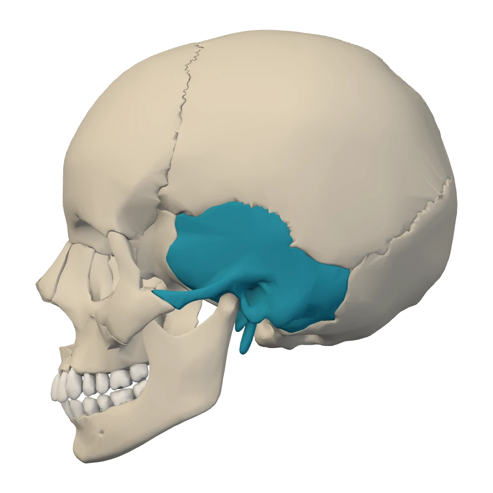
Left lateral view. The patient's left side: the face points to your left.
In this left lateral view one bone is highlighted. Which bone is it?
- Q2Landmark to bone: the cribriform plate and the ethmoidOpenStaxNetter p.19–20
Which bone carries the cribriform plate, the perforated strip in the midline of the anterior cranial fossa that forms the roof of the nasal cavity?
- Q3Articulation of the skull with the atlas: the occipital condylesOpenStaxNetter p.17–18
The whole skull rests on the vertebral column at two small joint surfaces. Where are they?
- Q4Recognize the palatine bones in the hard palateBodyParts3DOpenStaxNetter p.17–18
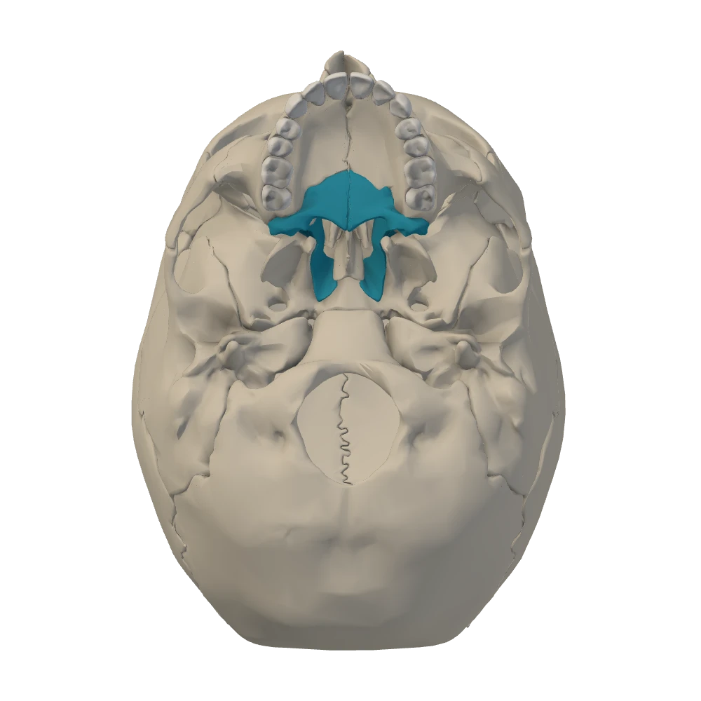
Inferior (external base) view. From below, mandible removed: the face is toward the top, the patient's left is on your right.
This is the inferior view with the lower jaw removed. A pair of bones is highlighted. Which bones are they?
- Q5Left and right in the anterior view: the nasal bonesBodyParts3DOpenStax
In the anterior view, a small bone at the bridge of the nose is selected. It lies just to the right of the midline as you look at the picture. Which bone is it?
- Q6Three conchae, two bonesOpenStaxNetter p.15–16
Each side wall of the nasal cavity carries three curled shelves: the superior, middle and inferior nasal conchae. To which bones do they belong?
- Q7Coronoid process versus condylar processOpenStaxNetter p.21–22
At the top of each ramus of the mandible there are two processes with a notch between them. Which statement is correct?
- Q8Mandibular foramen, mandibular canal and mental foramenOpenStaxNetter p.23–24
Before filling a lower molar, a dentist injects local anesthetic beside the opening where the inferior alveolar nerve enters the mandible. All the lower teeth of that side go numb, together with the chin and the lower lip.
Where is that opening?
- Q9Temporomandibular joint: forward glide, articular tubercle and anterior dislocationNetter p.25–26OpenStax
A student yawns widely and then finds that she cannot close her mouth. Her chin juts forward, and there is a hollow in front of each ear.
Where are the heads of her mandible?
- Q10Which suture joins which bones: the lambdoid suture and the lambdaBodyParts3DOpenStaxNetter p.11–12
In the posterior view you select the occipital bone. Its upper edge is a seam shaped like an inverted V. Name the suture, the bones on the other side of it, and the point at the tip of the V.
- Q11The four bones of the pterionOpenStaxNetter p.11–12
Which four bones meet at the pterion?
- Q12Pterion, middle meningeal artery and extradural hematomaNetter p.11–12OpenStax
A cyclist without a helmet falls and strikes his temple on the curb. He is dazed for a moment and then talks normally. Two hours later he is drowsy and confused. A scan shows a fracture at the pterion with a collection of blood beneath it.
Which vessel is most likely torn, and where has the blood collected?