The cranial base and the spaces of the skull
Lift off the skull cap and the floor under the brain turns out to be three steps going down from front to back. In front of and below those steps lie the orbits, the nasal cavity and the palate. This section teaches each space by its walls: which bone forms each wall, and what lies on the other side of it.
Three steps down: the cranial fossae
OpenStax · Cranial fossaeNetter p.19–20Switch the specimen to the internal cranial base view. The skull cap (calvaria) is removed and you are looking straight down at the floor the brain rests on. The face is toward the top of the screen and, because you are looking from above, the patient's right is on your right. This floor is the cranial base. What you see now is its upper, internal surface. Its lower, external surface is the underside of the skull, which you will turn to later in this section.
The floor is not flat. It drops in three steps from front to back, and each step is a cranial fossa (fossa means a trench or hollow). The anterior cranial fossa is the highest and shallowest. The middle cranial fossa is lower and shaped like a butterfly. The posterior cranial fossa is the lowest, deepest and largest. Each is molded to the part of the brain that lies in it, so once you can name the fossa you also know what rests there.
Specimen still
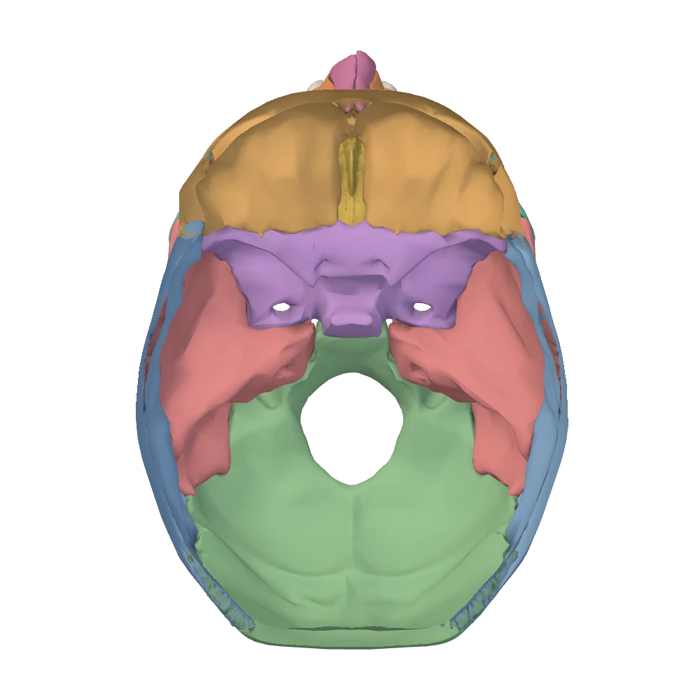
Internal cranial base view. Skull cap removed, looking down into the cranial cavity: the face is toward the top, the patient's right is on your right.
Three fossae at a glanceOpenStax · Cranial fossae
| Fossa | Floor formed by | What rests in it | Midline landmark | Fastest way to recognize it |
|---|---|---|---|---|
| Anterior | Orbital plates of the frontal bone at the sides; cribriform plate of the ethmoid in the middle; lesser wings and the front of the body of the sphenoid behind | Frontal lobes of the cerebrum; the olfactory bulbs lie on the cribriform plate | Crista galli | A shallow shelf that is also the roof of the orbits, with a narrow sieve on each side of a midline crest |
| Middle | Body of the sphenoid in the center; greater wings of the sphenoid, squamous part and front slope of the petrous part of each temporal bone at the sides | Temporal lobes at the sides; the pituitary gland in the center | Sella turcica | A butterfly: a narrow raised body between two deep hollows. Most of the small openings are here |
| Posterior | Occipital bone almost throughout; back slope of the petrous part and the mastoid part of each temporal bone in front and at the sides; the top of the clivus comes from the sphenoid | Cerebellum, pons and medulla oblongata | Clivus, sloping down to the foramen magnum | The deepest bowl, with the largest opening of the skull in its floor |
Anterior
- Floor formed by
- Orbital plates of the frontal bone at the sides; cribriform plate of the ethmoid in the middle; lesser wings and the front of the body of the sphenoid behind
- What rests in it
- Frontal lobes of the cerebrum; the olfactory bulbs lie on the cribriform plate
- Midline landmark
- Crista galli
- Fastest way to recognize it
- A shallow shelf that is also the roof of the orbits, with a narrow sieve on each side of a midline crest
Middle
- Floor formed by
- Body of the sphenoid in the center; greater wings of the sphenoid, squamous part and front slope of the petrous part of each temporal bone at the sides
- What rests in it
- Temporal lobes at the sides; the pituitary gland in the center
- Midline landmark
- Sella turcica
- Fastest way to recognize it
- A butterfly: a narrow raised body between two deep hollows. Most of the small openings are here
Posterior
- Floor formed by
- Occipital bone almost throughout; back slope of the petrous part and the mastoid part of each temporal bone in front and at the sides; the top of the clivus comes from the sphenoid
- What rests in it
- Cerebellum, pons and medulla oblongata
- Midline landmark
- Clivus, sloping down to the foramen magnum
- Fastest way to recognize it
- The deepest bowl, with the largest opening of the skull in its floor
Two ridges do the dividing, and both are easy to feel on a real skull. The front ridge is the sharp posterior edge of the lesser wings of the sphenoid; everything in front of it is anterior fossa. The back ridge is the thick upper border of the petrous part of the temporal bone on each side (the petrous ridge), which runs obliquely forward and toward the midline; everything behind it is posterior fossa. What lies between the two ridges is middle fossa. The next subtopic follows each boundary to the midline.
- If the surface you are looking at is directly above an eye socket, you are in the anterior fossa.
- If you can see the saddle of the sella turcica, or a deep hollow beside it, you are in the middle fossa.
- If you can see the foramen magnum, or the smooth bowl behind a petrous ridge, you are in the posterior fossa.
Where the boundaries run: sphenoid, petrous ridges, occipital bone
OpenStax · Sphenoid bone; Temporal bone; Cranial fossaeNetter p.13–14, 17–20The three fossae are separated by parts of three bones. Learn the boundaries as lines you could trace with a fingertip from the side wall to the midline, because the same lines tell you which fossa an opening belongs to.
Specimen still
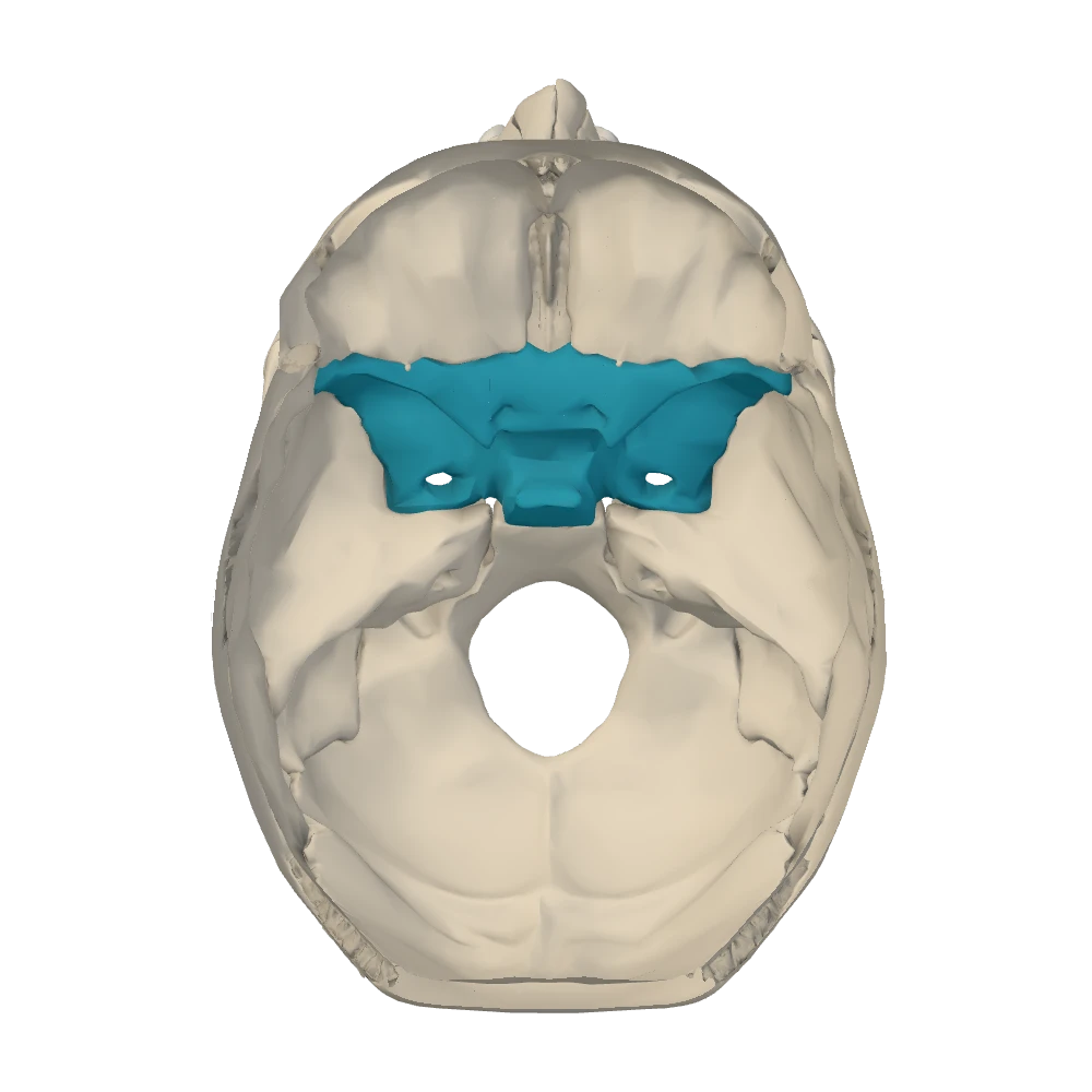
Internal cranial base view. Skull cap removed, looking down into the cranial cavity: the face is toward the top, the patient's right is on your right.
Anterior to middle: the lesser wings. Each lesser wing of the sphenoid is a thin, pointed plate that ends behind in a sharp free edge. Follow that edge toward the midline and it thickens into a small knob, the anterior clinoid process, which overhangs the opening of the optic canal. Between the two optic canals a low ridge on the body of the sphenoid completes the line. In front of this line you are over the orbits; behind it the floor drops into the middle fossa.
The center of the middle fossa: the sella turcica. The body of the sphenoid rises in the midline as the sella turcica (Latin for “Turkish saddle”). From front to back it has a low front bump (the tuberculum sellae), a seat (the hypophyseal fossa, which holds the pituitary gland) and a tall back (the dorsum sellae), whose upper corners are the posterior clinoid processes. The air-filled sphenoidal sinus lies inside the body just below the seat, which you can confirm in the midsagittal view.
Specimen still
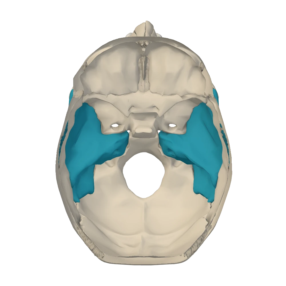
Internal cranial base view. Skull cap removed, looking down into the cranial cavity: the face is toward the top, the patient's right is on your right.
Middle to posterior: the petrous ridges. The petrous part of the temporal bone (petrous means rock-like; it houses the middle and inner ear) lies on the floor like a pyramid on its side. Its base is at the side wall near the ear. Its tip, the petrous apex, points forward and medially toward the body of the sphenoid and stops just short of it, at the ragged gap called the foramen lacerum. The upper border of the pyramid, the petrous ridge, is the boundary. Its front slope faces the middle fossa. Its back slope faces the posterior fossa and carries the internal acoustic meatus. In the midline the dorsum sellae joins the right and left ridges.
The posterior fossa: the occipital bone. Behind the petrous ridges almost everything is occipital bone. Its basilar part climbs forward from the foramen magnum to meet the body of the sphenoid; the smooth slope the two make together is the clivus, on which the pons and medulla rest. Its lateral parts flank the foramen magnum and carry the occipital condyles on their under-surface. Its broad squamous part, the occipital squama, forms the bowl for the cerebellum. Where the petrous part of the temporal bone meets the occipital bone, a large irregular gap is left on each side: the jugular foramen.
Walk the midline from front to back
- Crista galli (ethmoid)
- Cribriform plate on each side of it
- Flat top of the sphenoid body, between the lesser wings
- Ridge between the two optic canals: anterior fossa ends
- Tuberculum sellae
- Hypophyseal fossa (pituitary gland)
- Dorsum sellae: middle fossa ends
- Clivus (sphenoid above, occipital below)
- Foramen magnum
Two ridges that students swap
Edge of the lesser wing (sphenoid)
- Thin and sharp, like the edge of a shelf
- Runs almost straight from side to side
- Ends medially at the anterior clinoid process, beside the optic canal
- Separates anterior from middle fossa
Petrous ridge (temporal)
- Thick and rounded, the top of a solid block of bone
- Runs obliquely: backward and outward from the sella toward the ear
- Ends medially at the petrous apex, beside the foramen lacerum
- Separates middle from posterior fossa
The external base in outline
Netter p.17–18OpenStax · Temporal bone; Occipital bone; Sphenoid boneNow turn the skull over: choose the inferior view. The mandible is hidden so that it does not cover the base. The face is again toward the top of the screen, but you are looking up from below, so the sides are mirrored: the patient's left is on your right, exactly as in the anterior view and the opposite of the internal view you have just left.
The underside looks crowded, but it sorts into three bands from front to back.
The external base in three bandsNetter p.17–18
| Band | Runs from … to | What you see | Bones |
|---|---|---|---|
| Front | Upper teeth to the back edge of the hard palate | The hard palate inside the arch of upper teeth | Maxillae and palatine bones |
| Middle | Back edge of the palate to the front edge of the foramen magnum | The two posterior openings of the nasal cavity (choanae) with the vomer between them; the pterygoid processes at their sides; further out, the joint surface for the jaw, the styloid process and most of the openings of the base | Sphenoid, vomer, temporal bones, basilar part of the occipital bone |
| Back | Foramen magnum to the external occipital protuberance | The foramen magnum with an occipital condyle on each side; the mastoid processes laterally; the rough area for the neck muscles, up to the superior nuchal lines and the external occipital protuberance | Occipital bone, mastoid parts of the temporal bones |
Front
- Runs from … to
- Upper teeth to the back edge of the hard palate
- What you see
- The hard palate inside the arch of upper teeth
- Bones
- Maxillae and palatine bones
Middle
- Runs from … to
- Back edge of the palate to the front edge of the foramen magnum
- What you see
- The two posterior openings of the nasal cavity (choanae) with the vomer between them; the pterygoid processes at their sides; further out, the joint surface for the jaw, the styloid process and most of the openings of the base
- Bones
- Sphenoid, vomer, temporal bones, basilar part of the occipital bone
Back
- Runs from … to
- Foramen magnum to the external occipital protuberance
- What you see
- The foramen magnum with an occipital condyle on each side; the mastoid processes laterally; the rough area for the neck muscles, up to the superior nuchal lines and the external occipital protuberance
- Bones
- Occipital bone, mastoid parts of the temporal bones
Specimen still
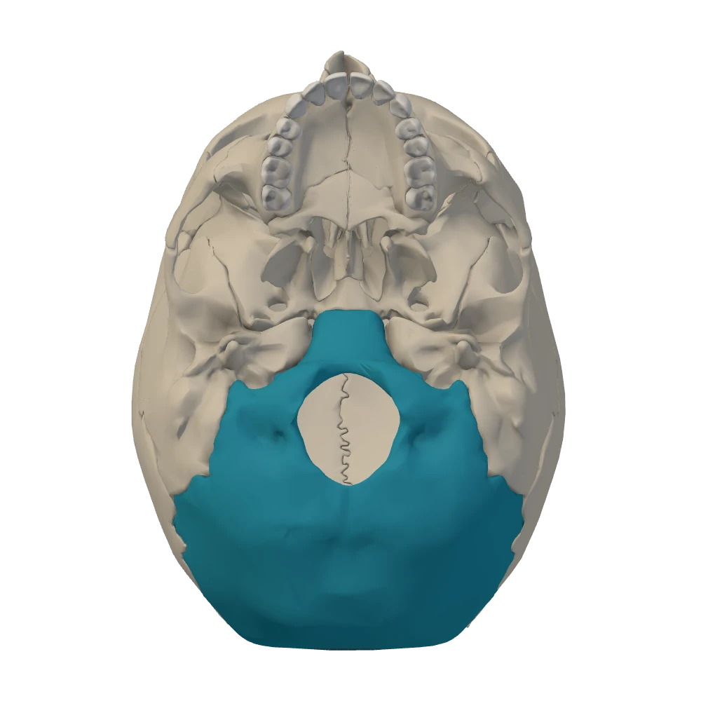
Inferior (external base) view. From below, mandible removed: the face is toward the top, the patient's left is on your right.
The middle band holds the detail. Find its landmarks in order, from the midline outward.
- Vomer and choanae. The back edge of the vomer is a thin vertical blade in the midline. The opening on each side of it is a choana (posterior nasal aperture), the way from the nasal cavity to the pharynx.
- Pterygoid process. Lateral to each choana a process of the sphenoid hangs down and splits into a medial and a lateral pterygoid plate. The medial plate is the side wall of the choana. The lateral plate gives attachment to chewing muscles.
- Under-surface of the greater wing. Behind and lateral to the root of the pterygoid process is the flat under-surface of the greater wing of the sphenoid, which roofs the infratemporal fossa. The foramen ovale and foramen spinosum open here.
- Joint surface for the jaw. Further laterally, on the temporal bone, lie the mandibular fossa and, in front of it, the articular tubercle: the temporal half of the temporomandibular joint.
- Styloid and mastoid. Behind the mandibular fossa the thin styloid process points down and forward. Behind and lateral to it is the blunt mastoid process. The stylomastoid foramen lies between the two.
- Carotid and jugular openings. Medial to the styloid process, the round lower opening of the carotid canal lies in front and the larger jugular foramen behind it.
The orbit: seven bones, four walls and an apex
OpenStax · The orbitNetter p.9–10Return to the anterior view and look into an eye socket. Each orbit is a pyramid lying on its side. Its base is the opening you are looking into, ringed by a thick margin. Its tip, the apex, points backward and medially, toward the center of the skull. Between base and apex are four walls: a roof, a floor, a medial wall and a lateral wall. The medial walls of the two orbits are nearly parallel, with only the nasal cavity between them. The lateral walls splay outward at about 45 degrees, which widens the field of vision to the side.
Seven bones line each orbit: frontal, sphenoid, zygomatic, maxilla, ethmoid, lacrimal and palatine. The useful way to learn them is wall by wall, together with what lies on the other side of each wall, because that tells you where a fracture or an infection will go.
Specimen still
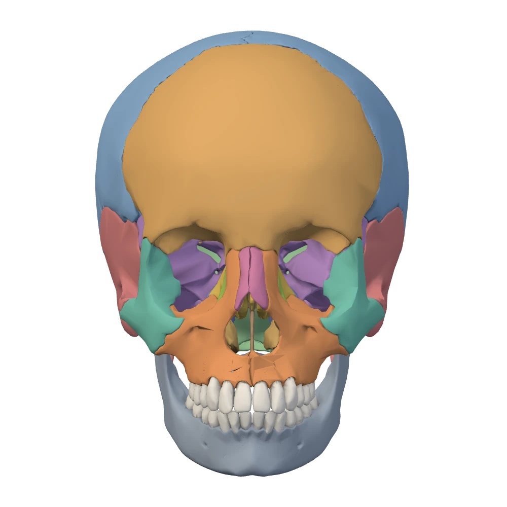
Anterior view. Facing the skull: the patient's left is on your right.
The walls of the orbitOpenStax · The orbit
| Wall | Bones, front to back | On the other side | Worth knowing |
|---|---|---|---|
| Roof | Orbital plate of the frontal bone; the lesser wing of the sphenoid at the very back | Anterior cranial fossa and the frontal lobe | The same thin plate is the floor of the anterior fossa |
| Lateral wall | Zygomatic bone in front; greater wing of the sphenoid behind | Temporal fossa in front; middle cranial fossa behind | The thickest wall, and the one most exposed to a blow from the side |
| Floor | Maxilla (most of it); zygomatic bone at the front and side; the small orbital process of the palatine bone at the back | Maxillary sinus | Thin; grooved by the infraorbital nerve on its way to the infraorbital foramen |
| Medial wall | Frontal process of the maxilla; lacrimal bone; orbital plate of the ethmoid (the largest part); a little of the body of the sphenoid | Nasal cavity and the ethmoidal air cells | Paper-thin; the lacrimal groove for the tear sac is at its front |
| Apex | Sphenoid | Middle cranial fossa | The optic canal opens here, with the superior orbital fissure just lateral to it |
Roof
- Bones, front to back
- Orbital plate of the frontal bone; the lesser wing of the sphenoid at the very back
- On the other side
- Anterior cranial fossa and the frontal lobe
- Worth knowing
- The same thin plate is the floor of the anterior fossa
Lateral wall
- Bones, front to back
- Zygomatic bone in front; greater wing of the sphenoid behind
- On the other side
- Temporal fossa in front; middle cranial fossa behind
- Worth knowing
- The thickest wall, and the one most exposed to a blow from the side
Floor
- Bones, front to back
- Maxilla (most of it); zygomatic bone at the front and side; the small orbital process of the palatine bone at the back
- On the other side
- Maxillary sinus
- Worth knowing
- Thin; grooved by the infraorbital nerve on its way to the infraorbital foramen
Medial wall
- Bones, front to back
- Frontal process of the maxilla; lacrimal bone; orbital plate of the ethmoid (the largest part); a little of the body of the sphenoid
- On the other side
- Nasal cavity and the ethmoidal air cells
- Worth knowing
- Paper-thin; the lacrimal groove for the tear sac is at its front
Apex
- Bones, front to back
- Sphenoid
- On the other side
- Middle cranial fossa
- Worth knowing
- The optic canal opens here, with the superior orbital fissure just lateral to it
OpenStax gives a shorter list for each wall; the fuller list here follows standard regional anatomy (see the source notes for this unit).
Notice which bones appear more than once. The sphenoid closes the back of the orbit and reaches three walls (roof, lateral, medial). The maxilla forms most of the floor and the front of the medial wall. The zygomatic bone turns the corner between the lateral wall and the floor. The frontal bone has the roof almost to itself. The ethmoid, lacrimal and palatine bones each appear once.
The rim is built from only three bones. The frontal bone forms the supraorbital margin, which carries the supraorbital notch. The zygomatic bone forms the lateral margin and the outer half of the infraorbital margin. The maxilla forms the inner half of the infraorbital margin and, with its frontal process, the medial margin.
How the orbit connects to its neighbors. At the apex, the optic canal and the superior orbital fissure both lead back into the middle cranial fossa. Along the junction of the lateral wall and the floor runs a second long gap, the inferior orbital fissure, which opens backward into the pterygopalatine fossa (a small space behind the maxilla) and, further forward, into the infratemporal fossa. At the front of the medial wall the lacrimal groove leads down into the nasolacrimal canal, which carries tears to the nasal cavity, below the inferior concha. The groove for the infraorbital nerve in the floor becomes a canal and opens on the face at the infraorbital foramen.
The nasal cavity: septum, conchae and meatuses
Netter p.13–16OpenStax · The nasal septum and nasal conchae; Ethmoid boneThe nasal cavity is the tall, narrow space behind the nose, between the two orbits and above the palate. In the anterior view you look into it through the pear-shaped front opening of the bony nose (the piriform aperture). It runs back to the two choanae you found on the external base. A midline partition, the nasal septum, divides it into right and left halves. Each half is narrow at the top and wide at the bottom, and has a roof, a floor, a medial wall (the septum) and a lateral wall.
The four walls of each half of the nasal cavityNetter p.13–16OpenStax · Ethmoid bone
| Wall | Bony parts | On the other side |
|---|---|---|
| Roof | Nasal bone and frontal bone in front; cribriform plate of the ethmoid in the middle; body of the sphenoid behind | Anterior cranial fossa above the cribriform plate; sphenoidal sinus behind |
| Floor | Palatine process of the maxilla in front; horizontal plate of the palatine bone behind | The mouth: this floor is the upper surface of the hard palate |
| Medial wall (septum) | Perpendicular plate of the ethmoid above; vomer below and behind | The other half of the nasal cavity |
| Lateral wall | Maxilla, lacrimal bone, ethmoid (with the superior and middle conchae), inferior nasal concha, perpendicular plate of the palatine bone, medial pterygoid plate of the sphenoid | Orbit and ethmoidal air cells above; maxillary sinus below |
Roof
- Bony parts
- Nasal bone and frontal bone in front; cribriform plate of the ethmoid in the middle; body of the sphenoid behind
- On the other side
- Anterior cranial fossa above the cribriform plate; sphenoidal sinus behind
Floor
- Bony parts
- Palatine process of the maxilla in front; horizontal plate of the palatine bone behind
- On the other side
- The mouth: this floor is the upper surface of the hard palate
Medial wall (septum)
- Bony parts
- Perpendicular plate of the ethmoid above; vomer below and behind
- On the other side
- The other half of the nasal cavity
Lateral wall
- Bony parts
- Maxilla, lacrimal bone, ethmoid (with the superior and middle conchae), inferior nasal concha, perpendicular plate of the palatine bone, medial pterygoid plate of the sphenoid
- On the other side
- Orbit and ethmoidal air cells above; maxillary sinus below
The bony septum. Open the midsagittal view: the left half of the skull is removed, the face points to your left, and the septum is the thin vertical plate filling the nasal region. It has two bony parts. The perpendicular plate of the ethmoid hangs down from the cribriform plate and forms the upper part. The vomer, a separate bone shaped like a plowshare, forms the lower and back part and reaches the body of the sphenoid behind; its free back edge is the blade you saw between the choanae. Along the floor, the vomer sits on a low crest raised by the maxillae and the palatine bones. In front, the two bony parts leave a wide angle that is filled in life by the septal cartilage, which continues forward into the external nose. A dry skull has lost that cartilage, so the front of the septum looks open.
Specimen still
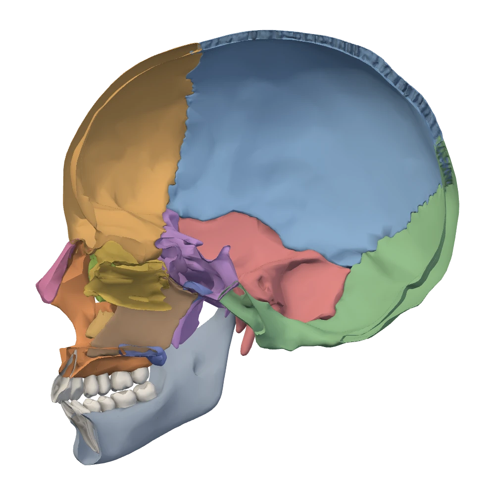
Midsagittal section view. Left half removed: you see the inside of the right half, and the face points to your left.
Conchae. The lateral wall is not flat. Three curved shelves of bone, the nasal conchae (singular concha, a shell), project from it and curl downward. The superior and middle nasal conchae are parts of the ethmoid. The inferior nasal concha is the largest and is a separate bone, one of the fourteen facial bones, fixed to the lateral wall. Covered with mucosa in life, the conchae (then called turbinates) enlarge the surface that warms, moistens and cleans the air. From the front only the inferior and middle conchae can be seen; the small superior concha is hidden high up and far back.
Meatuses. The air channel beneath each concha is a meatus (a passage), and it takes the name of the concha that roofs it. So the inferior meatus lies under the inferior concha, between it and the floor. The middle meatus lies under the middle concha. The superior meatus lies under the superior concha. The small pocket above and behind the superior concha, against the body of the sphenoid, is the sphenoethmoidal recess.
What opens where on the lateral wallOpenStax · Lacrimal bone; Paranasal sinuses
| Channel | Position | What opens into it |
|---|---|---|
| Sphenoethmoidal recess | Above and behind the superior concha | Sphenoidal sinus |
| Superior meatus | Under the superior concha | Posterior ethmoidal air cells |
| Middle meatus | Under the middle concha | Frontal sinus, maxillary sinus, anterior and middle ethmoidal air cells |
| Inferior meatus | Under the inferior concha | Nasolacrimal canal (tears from the orbit) |
Sphenoethmoidal recess
- Position
- Above and behind the superior concha
- What opens into it
- Sphenoidal sinus
Superior meatus
- Position
- Under the superior concha
- What opens into it
- Posterior ethmoidal air cells
Middle meatus
- Position
- Under the middle concha
- What opens into it
- Frontal sinus, maxillary sinus, anterior and middle ethmoidal air cells
Inferior meatus
- Position
- Under the inferior concha
- What opens into it
- Nasolacrimal canal (tears from the orbit)
The paranasal sinuses are kept brief in this unit. OpenStax supports that every sinus opens into the nasal cavity and that tears reach it beneath the inferior concha; the meatus for each sinus follows standard regional anatomy.
How the nasal cavity connects to its neighbors. Forward it opens on the face through the piriform aperture, and backward into the pharynx through the choanae. Upward, the many small holes of the cribriform plate link it to the anterior cranial fossa. From the orbit it receives the nasolacrimal canal. Through its lateral wall, high up and far back near the end of the middle concha, the sphenopalatine foramen leads to the pterygopalatine fossa. Through its floor, just behind the incisor teeth, the incisive canals lead down to the roof of the mouth. And every paranasal sinus opens into it.
The hard palate: floor of the nose, roof of the mouth
Netter p.15–18OpenStax · Maxillary bone; Palatine boneThe hard palate is one plate with two jobs: its upper surface is the floor of the nasal cavity and its lower surface is the roof of the mouth. In the inferior view it fills the horseshoe of the upper teeth.
Four pieces of bone make it. In front, the palatine process of each maxilla grows in from the tooth-bearing alveolar process and meets its partner in the midline; together the two form the larger front part of the palate, roughly two-thirds to three-quarters of it. Behind, the horizontal plate of each palatine bone completes it. The seams between the four pieces make a cross: a midline suture from front to back, and a transverse suture where the maxillae meet the palatine bones. The back edge of the hard palate is free; the soft palate hangs from it in life.
Specimen still
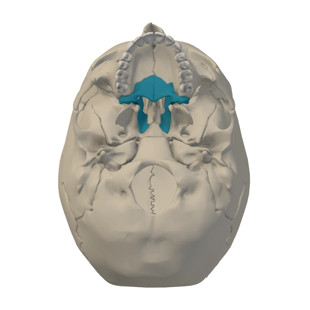
Inferior (external base) view. From below, mandible removed: the face is toward the top, the patient's left is on your right.
- The incisive fossa: a single pit in the midline just behind the central incisor teeth. The incisive canals from the nasal cavity open into it.
- The greater palatine foramen: one on each side, at the back outer corner of the palate, close to the last molar tooth, at the edge of the horizontal plate.
- The lesser palatine foramina: one or more small holes just behind each greater palatine foramen.
These three sets of openings mark the palate. What each one carries is in Section 5: Openings of the face, jaw and palate.
How the spaces connect: a map of doorways
OpenStax · Cranial fossae; The orbitNetter p.19–20You now have the main spaces of the skull: the cranial cavity with its three fossae, the two orbits, the nasal cavity and, below the palate, the mouth. Three smaller spaces on the outside of the base will keep appearing as destinations. The infratemporal fossa lies below the zygomatic arch, deep to the ramus of the mandible. The pterygopalatine fossa is a narrow cleft behind the maxilla and in front of the pterygoid process. The neck lies below the base. Every opening in the next section is a doorway between two of these.
Which doorway joins which spacesOpenStax · Cranial fossae; The orbitNetter p.19–20
| From | To | Doorway |
|---|---|---|
| Anterior cranial fossa | Nasal cavity | Foramina of the cribriform plate |
| Middle cranial fossa | Orbit | Optic canal; superior orbital fissure |
| Middle cranial fossa | Pterygopalatine fossa | Foramen rotundum |
| Middle cranial fossa | Infratemporal fossa | Foramen ovale; foramen spinosum |
| Neck | Middle cranial fossa | Carotid canal, through the petrous part of the temporal bone |
| Posterior cranial fossa | Inside of the petrous part (inner ear, facial canal) | Internal acoustic meatus |
| Posterior cranial fossa | Neck | Jugular foramen; hypoglossal canal |
| Posterior cranial fossa | Vertebral canal | Foramen magnum |
| Orbit | Pterygopalatine and infratemporal fossae | Inferior orbital fissure |
| Orbit | Nasal cavity | Nasolacrimal canal |
| Nasal cavity | Pharynx | Choanae |
| Nasal cavity | Pterygopalatine fossa | Sphenopalatine foramen |
| Nasal cavity | Mouth (hard palate) | Incisive canals, opening at the incisive fossa |
| Pterygopalatine fossa | Mouth (palate) | Palatine canals, opening at the greater and lesser palatine foramina |
Anterior cranial fossa
- To
- Nasal cavity
- Doorway
- Foramina of the cribriform plate
Middle cranial fossa
- To
- Orbit
- Doorway
- Optic canal; superior orbital fissure
Middle cranial fossa
- To
- Pterygopalatine fossa
- Doorway
- Foramen rotundum
Middle cranial fossa
- To
- Infratemporal fossa
- Doorway
- Foramen ovale; foramen spinosum
Neck
- To
- Middle cranial fossa
- Doorway
- Carotid canal, through the petrous part of the temporal bone
Posterior cranial fossa
- To
- Inside of the petrous part (inner ear, facial canal)
- Doorway
- Internal acoustic meatus
Posterior cranial fossa
- To
- Neck
- Doorway
- Jugular foramen; hypoglossal canal
Posterior cranial fossa
- To
- Vertebral canal
- Doorway
- Foramen magnum
Orbit
- To
- Pterygopalatine and infratemporal fossae
- Doorway
- Inferior orbital fissure
Orbit
- To
- Nasal cavity
- Doorway
- Nasolacrimal canal
Nasal cavity
- To
- Pharynx
- Doorway
- Choanae
Nasal cavity
- To
- Pterygopalatine fossa
- Doorway
- Sphenopalatine foramen
Nasal cavity
- To
- Mouth (hard palate)
- Doorway
- Incisive canals, opening at the incisive fossa
Pterygopalatine fossa
- To
- Mouth (palate)
- Doorway
- Palatine canals, opening at the greater and lesser palatine foramina
Rows that involve the pterygopalatine fossa, the sphenopalatine foramen and the palatine canals follow standard regional anatomy; those spaces are named here only as destinations.
Worked case · Trace the leak
After a fall onto the forehead, a patient notices that a clear, watery fluid drips from one nostril when he leans forward, and that he can no longer smell his coffee.
Which single plate of bone, if cracked, explains both findings? Name the two spaces it separates.
Netter p.19–20OpenStax · Ethmoid boneKnowledge check · 4
Answer, read why, and try again whenever you like. Answers are kept in both study modes.
- Q1Which fossa: middle cranial fossa and the temporal lobeOpenStax · Middle cranial fossa
In the internal cranial base view you select a point that lies behind the sharp edge of the lesser wing and in front of the petrous ridge, in the deep hollow to one side of the sella turcica.
Which fossa is this, and what rests in it?
- Q2Boundary: petrous ridge between middle and posterior fossaeOpenStax · Cranial fossae
Which structure separates the middle cranial fossa from the posterior cranial fossa on each side?
- Q3Sphenoid in all three fossaeOpenStax · Sphenoid bone; Cranial fossae
Which single bone contributes to the floor of all three cranial fossae?
- Q4Orbit: lateral wallOpenStax · The orbit
A hockey puck strikes the outer side of a player's eye socket and breaks its lateral wall.
Which two bones form that wall?
- Q5Orbit: floor and the maxillary sinus
After a punch to the eye, a CT scan shows orbital fat bulging downward through a break in the floor of the orbit. The patient's cheek and upper lip are numb on that side.
Into which space has the fat herniated, and which bone forms most of the broken wall?
- Q6Nasal septum: vomerNetter p.13–14OpenStax · The nasal septum and nasal conchae
In the midsagittal view, which bone forms the lower and posterior part of the bony nasal septum and ends behind as the free edge between the two choanae?
- Q7Inferior meatus and the inferior nasal conchaOpenStax · Lacrimal bone; Inferior nasal conchae
Tears drain from the orbit through the nasolacrimal canal. Under which shelf of bone does the canal open, and what kind of bone is that shelf?
- Q8Left and right: internal base versus inferior viewBodyParts3D
You are studying the patient's left foramen ovale. In the internal cranial base view (skull cap removed, face toward the top) it lies on the left half of the screen. You now switch to the inferior view of the external base, again with the face toward the top.
Where is the left foramen ovale now?